Ultrasensitive, highly selective and naked eye colorimetric recognition of D-penicillamine in aqueous media by CTAB capped AgNPs: applications to pharmaceutical and biomedical analysis

Laxman S. Walekara, Uttam R. Kondekara, Anil H. Gorea, Samadhan P. Pawara, V. Sudarsanb, Prashant V. Anbhulea, Shivajirao R. Patila and Govind B. Kolekar*a
aFluorescence Spectroscopy Research Laboratory, Department of Chemistry, Shivaji University, Kolhapur-416 004, Maharashtra, India. E-mail: gbkolekar@yahoo.co.in; Fax: +91 0231 2692333
bChemistry Division, Bhabha Atomic Research Centre, Trombay, Mumbai-400 085, Maharashtra, India

Received 14th June 2014 , Accepted 15th October 2014

First published on 17th October 2014


Abstract

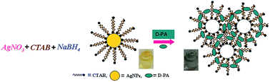
Herein, we are going to report a straightforward, highly selective and ultra sensitive naked eye colorimetric probe for the detection of D-penicillamine (D-PA) in aqueous solution using cetyltrimethyl ammonium bromide (CTAB) capped colloidal silver nanoparticles (AgNPs) based on induced aggregation. The synthesized CTAB-AgNPs and their interaction with D-PA were characterized by different analytical techniques such as UV-Vis absorption spectroscopy, transmission electron microscopy (TEM), dynamic light scattering (DLS) measurements and zeta potential measurements. The color of the CTAB-AgNPs solution changed from yellowish brown to colorless within short period of time after the successive addition of D-PA, resulting in a blue shift with quenching in the absorption spectra. Under the optimal conditions, a calibration plot of (A0A) against concentration of D-PA was linear in the range of 0.1–0.6 μg mL−1 with a correlation coefficient of 0.9901. The concentration of D-PA was quantitatively determined using an UV-Vis spectrophotometer with a limit of detection (LOD) of 0.056 μg mL−1 (56 ng mL−1). In addition, the method shows an excellent selectivity and sensitivity towards D-PA over the other interfering biomolecules and cations tested. The accuracy and reliability of the method were further ascertained from the detection of D-PA from pharmaceutical and biomedical samples via a standard addition method, with percentage recoveries in the range of 98.32–102.94%. A plausible reason for the observed color changes is also discussed. The proposed method is simple, rapid, specific and highly selective and sensitive with good precision.


Introduction

D-Penicillamine (2-amino-3-mercapto-3-methylbutanoic acid) abbreviated as D-PA is a naturally occurring amino acid and has been used as a vital medicinal active thiol compound. D-PA is a sulfur-containing amino acid, which belongs to the family of aminothiols where the hydrogen atom on the beta-carbon atom of cysteine is replaced by the methyl group. The D-PA molecule can exist in D and L enantiomeric forms; however, only the D type is pharmaceutically and clinically useful, whereas the L form is harmful due to its excessive toxicity.1 More recently, D-PA has been applied as a chelating agent for the treatment of a number of diseases, particularly in Wilson's disease, an autosomal recessive disorder of copper transport,2 as a antifibrotic agent to treat scleroderma,3 and as antirheumatic drug to treat patients with active rheumatoid arthritis.4

D-PA has received considerable importance in the field of medical applications, but possesses some adverse effects on human beings. About 50% of patients experience one or more adverse effect on treatment with D-PA such as an anorexia, loss of taste, oral ulceration, skin rashes, hematological effects, glomerulonephritis and nephrotic syndrome.5 Among these, nephrotic syndrome is general and most harmful to the health of patients,6 making the monitoring of D-PA in urine and plasma very important. The first symptoms of penicillamine-induced nephropathy are the proteinuria, which may lead to nephrotic syndrome in some patients. D-PA is the hydrolytic degradation product of penicillin having various modes of action.7–9 It reacts with collagen and elastin and also reduces disulphide groups in tissues to form mixed disulphides with other substances containing thiol groups.

Till date, several analytical methods have been reported for the determination of D-PA in both pharmaceutical preparations and biological samples such as high performance liquid chromatography,10 fluorimetry,11 spectrophotometry (the absence of a chromophore and/or auxochrome groups means direct spectrophotometric method cannot be used for its analysis),12 chemiluminescence,13 capillary electrophoresis,14 electrochemistry15 and colorimetry.16 However, these methods have their own limitations like expensive instruments, time consuming, requires complex mathematical calculations, robust sampling handling, requiring a large quantity of sample and so on. In order to overcome all these limitations, it is necessary to develop a simple, sensitive, rapid and cost effective method for the determination of D-PA in aqueous media. Nowadays, nanomaterial based colorimetric assays have attracted growing attention due to their simplicity, rapidity, high selectivity, color change under different conditions and ease of measurement.17,18 When compared to other assays, colorimetric sensors allow the direct and on-site analysis of samples using naked eye for clarification. Metal nanoparticles (MNPs) possess a very high molar extinction coefficient compared to most common dyes, and according to the Beer–Lambert law, MNPs can reach a considerably lower limit of detection (LOD) than common dyes.19 Besides novel optical properties, easy chemical modification also provides great convenience for the wide use of MNPs-based assays.

Among these, AgNPs-based colorimetric sensors have many advantages over gold nanoparticles-based ones, as they have higher extinction coefficients and lower prices, allowing direct detection with minimal consumption of material.20 Methods based on the color change of MNPs have been applied to determine targets such as proteins,21 cells,22 DNA,23 metal ions24 and many more substances.25 Liu et al. have demonstrated the visual detection of ATP molecules based on the redispersion of AuNPs upon the addition of ATP molecules to the assembled AuNPs.26 However, there are very few reports describing the colorimetric detection of pharmaceuticals using nanoparticles. The D-PA molecule lacks the presence of a chromophore and auxochrome groups, and hence direct spectrophotometric methods cannot be used for its analysis. In our proposed approach, we have developed a new colorimetric method for the direct detection of D-PA in an aqueous solution using CTAB capped AgNPs as a nanosensor based on inducing aggregation. The absorbance of CTAB-AgNPs is extremely quenched by D-PA with a blue shift in the λmax and a color change from yellowish brown to colorless, which is easily recognized by the naked eye. Furthermore, the proposed method was successfully applied for the determination of D-PA from pharmaceutical and biological samples by a standard addition method. This probe offers the advantages of simplicity, rapidity, selectivity, sensitivity and good stability, which makes it a valuable pathway for analytical purpose.

Experimental section

Equipment

All the spectroscopic analysis was carried out using a stable dispersion of nanoparticles. The absorption spectrum was recorded at room temperature using a UV 3600 Shimadzu UV-VIS-NIR spectrophotometer with a 1.0 cm quartz cell. The particle size distribution of the AgNPs in an aqueous suspension was measured using dynamic light scattering with a Zetasizer Nano ZS (Malvern Instruments Ltd., UK). Transmission electron microscopy (TEM) images were recorded on a Tecnai™ transmission electron microscope (TEM, FEI Tecnai 300). The images of the samples were taken using Canon digital camera.

Reagents

All chemical reagents were of analytical reagent grade and used as received without further purification. All aqueous solutions were prepared with doubly distilled water. Silver nitrate (AgNO3), cetyltrimethyl ammonium bromide (CTAB), and sodium borohydride (NaBH4) were purchased from S d fine-chem Ltd (Mumbai, India). D-PA was purchased from Sigma-Aldrich Ltd (Mumbai, India). A stock solution of D-PA was prepared by dissolving D-penicillamine (D-PA) in double distilled water. All the metal salts and biomolecules solutions used for the interference study were purchased from SD Fine-Chem Ltd (Mumbai, India).

Synthesis of AgNPs

The water-soluble CTAB capped AgNPs were synthesized by a simple chemical reduction method at room temperature. Appropriate amounts of AgNO3, CTAB and NaBH4 were first dissolved in doubly distilled water separately. Here, AgNO3 was used as a source of metal precursor, CTAB as a capping agent and NaBH4 the reducing agent. In a typical procedure, firstly, 5.0 mL of 0.1 mol L−1 AgNO3 solution was added slowly and dropwisely to 5.0 mL of 0.1 mol L−1 CTAB solution with continuous stirring until the reaction mixture becomes nearly colorless. Then, 10 mL of 0.1 mol L−1 NaBH4 was added dropwisely into the flask containing a mixture of AgNO3 and CTAB and the reaction mixture turns from colorless to yellowish brown, which indicates the complete reduction and formation of AgNPs. After stirring overnight (12 h), resultant aliquot was diluted up to 100 mL with water and stored in a freezer at 4 °C for further use. The concentration of the resultant AgNPs was 0.005 mol L−1 (calculated using the concentration of AgNO3 added) and used for further study. Here AgNO3, CTAB and NaBH4 were used in the ratio of 1[thin space (1/6-em)]:[thin space (1/6-em)]0.5[thin space (1/6-em)]:[thin space (1/6-em)]2, respectively, for the synthesis of the CTAB-AgNPs.

Colorimetric detection of D-PA with CTAB-AgNPs

The colorimetric recognition of D-PA in aqueous solution was performed at room temperature. A stock solution of D-PA was prepared by dissolving D-PA in doubly distilled water. The solutions for all metal ions and biomolecules were prepared by dissolving their respective salts/molecules in doubly distilled water and stored at room temperature. For a typical colorimetric analysis of D-PA with CTAB-AgNPs, to a 10 mL standard flask, solutions were added according to the following order: 2.0 mL of CTAB-AgNPs stock solution (5 × 10⁻⁴ mol L−1) and a known volume of standard metal ion solution. Then, the solutions were diluted with water up to the mark (i.e. 10 mL), mixed thoroughly, and maintained at room temperature for 5.0 min, and then, transferred separately into quartz cuvettes. Their absorption bands were recorded using an UV-Vis absorption spectrophotometer. In addition, the colorimetric detection of D-PA was also observed by the naked eye with respect to the color change from yellowish brown to colorless upon increasing concentrations of D-PA, whereas the colorimetric probe (i.e. CTAB-AgNPs) does not show any response in the form of color change or change in the absorbance towards other metal ions and biomolecules. In brief, it shows a color change and a drastic change in its absorbance for only the D-PA molecule. The photographic images of all samples were taken using a Canon digital camera.

Results and discussion

Characterization of CTAB-AgNPs

The CTAB capped AgNPs were firstly characterized using a UV-Vis absorption spectrophotometer and shows a maximum absorbance at a wavelength of λmax = 408 nm with an absorbance of A = 0.32 (as shown in Fig. 1) and extinction coefficient ε = 3200 M−1 cm−1, which had a concentration of 1 × 10⁻⁴ mol L−1. The absorbance peak at λmax = 408 confirms the formation of colloidal CTAB capped AgNPs. To measure the size of the nanoparticles, we performed TEM experiments to acquire the TEM image of the probe, as shown in Fig. 2. Fig. 2(a) shows the TEM image of CTAB-AgNPs and it was concluded that CPB-AgNPs are well dispersed in aqueous solution, with a slight variation in particle size ranging from 3.0–5.0 nm.
image file: c4ra05741d-f1.tif
Fig. 1 UV-visible absorption spectrum at λmax = 408 nm of the CTAB-AgNPs (1 × 10⁻⁴ mol L−1) (ε = 3200 M−1 cm−1) in aqueous colloidal solution.

image file: c4ra05741d-f2.tif
Fig. 2 TEM images of the (a) CTAB-AgNPs and (b) after 0.3 μg mL−1 D-PA added to the CTAB-AgNPs colloidal solution.

In addition, the measurement of particle size and size distribution of CTAB-AgNPs was also carried out by performing dynamic light scattering (DLS) experiment.27 Fig. 3 shows the typical size and size distribution of the synthesized CTAB-AgNPs measured by DLS. The average hydrodynamic diameter of the well-dispersed CTAB-AgNPs determined using DLS was 8.56 nm and 98% of the particle size distribution was found within the range of 6.78–13.56 nm.


image file: c4ra05741d-f3.tif
Fig. 3 Particle size distribution measured by DLS of the CTAB-AgNPs colloidal solution (1 × 10⁻⁴ mol L−1). The average size is 8.56 nm, 98% of the particles are between 6.78–13.56 nm.

Zeta (ζ) potential measurements were also performed to study the effectiveness of the capping agent on the surface of the AgNPs, to characterize the surface charge of the AgNPs and their stability.28 The CTAB is a cationic surfactant and hence the AgNPs are supposed to acquire a positive zeta potential. The zeta potential for the CTAB-AgNPs was found to be +40.1 mV.

Reason for the color change of CTAB-AgNPs with D-PA

To explore the reason for the color change of CTAB-AgNPs with D-PA, various experiments were performed consecutively at room temperature. Firstly, the cationic surfactant (i.e. CTAB) was capped on the surface of the AgNPs in order to make a stable colloidal suspension of nanoparticles during the synthesis (as shown in Scheme 1), and it was conceived that the CTAB was plainly adsorbed on the surface of the AgNPs, resulting in the formation of a CTAB monolayer around the AgNPs via self-assembly to form spherically shaped nanoparticles. It was supposed that the CTAB did not permanently bond with the AgNPs and was adsorbed on the surface of the AgNPs with its hydrophobic tails and hydrophilic heads via hydrophobic interactions, which forms a monolayer on the surface of AgNPs via self-assembly. In other words, it could be said that the CTAB has adsorbed on the surface of AgNPs, which makes the positive zeta potential (+40.1 mV) and spherical shape for the AgNPs. The freshly synthesized CTAB-AgNPs exhibits a yellowish brown color in aqueous solution due to excitation of the surface plasmon resonance vibrations (SPR band),29 and gives a maximum absorption band at λmax = 408 nm. Preceding the addition of D-PA to the colloidal solution of nanoparticles, the CTAB-AgNPs have a homogeneous and well dispersed nature in the colloidal suspension. In the presence of D-PA, the CTAB-AgNPs tend to aggregate, which results in the color change from yellowish brown to colorless in a short period of time with a blue shift in the wavelength of absorbance (λmax) and it was supported by TEM and DLS experiments. The change in color of solution after the addition of D-PA was easily detected by the naked eye.
image file: c4ra05741d-s1.tif
Scheme 1 Plausible schematic illustration for the color change of CTAB-AgNPs with D-PA.

It was expected that the D-PA molecule is mainly adsorbed on the surface of the CTAB-AgNPs and remains responsible for the aggregation of the nanoparticles. The interaction of D-PA with CTAB-AgNPs was tested by UV-Vis absorption spectroscopy. The change in absorbance of CTAB-AgNPs with respect to the concentration of D-PA is shown in Fig. 4. As indicated in Fig. 4, the absorbance of the CTAB-AgNPs decreases upon the successive addition of an aqueous solution of D-PA over a selected range at room temperature. From Fig. 4, it is noteworthy that the increased concentration of D-PA induces a blue shift with quenching in the absorption spectra, which could be attributed to an increase in particle size and confirms the aggregation of the CTAB-AgNPs. In this work, it was observed that D-PA has a strong interaction with the surface of the CTAB-AgNPs, which could be responsible for the aggregation of CTAB-AgNPs with a blue shift in the surface plasmon absorption band, which results in the color change of the solution from yellowish brown to colorless.


image file: c4ra05741d-f4.tif
Fig. 4 The UV-vis absorption spectra of the CTAB-AgNPs (1 × 10⁻⁴ mol L−1) solution with various concentrations of D-PA. The concentration of D-PA from top to bottom is 0.0, 0.1, 0.2, 0.3, 0.4, 0.5, 0.6 and 0.7 μg mL−1.

image file: c4ra05741d-f5.tif
Fig. 5 Photographic images of the CTAB-AgNPs (1 × 10⁻⁴ mol L−1) solution with various concentrations of D-PA. The concentration of D-PA from left to right is 0.0, 0.1, 0.2, 0.3, 0.4, 0.5, 0.6 and 0.7 μg mL−1.

To confirm the aggregation of these CTAB-AgNPs, we carried out a TEM study with and without the addition of designed concentrations of D-PA to the CTAB-AgNPs solution. Fig. 2(a) shows the TEM image of CTAB-AgNPs without D-PA and (b) upon the addition of 0.3 μg mL−1 D-PA. From Fig. 2(a) and (b) it was clearly observed that the CTAB-AgNPs have a great affinity to aggregate with each other upon the addition of D-PA.

Further, to evaluate the aggregation behavior of CTAB-AgNPs with D-PA, DLS experiments were carried out before and after the addition of D-PA to the colloidal suspension of nanoparticles. The particle sizes of the CTAB-AgNPs before and after the addition of D-PA are graphically represented in Fig. 9. The average particle size of CTAB-AgNPs was found to be 8.56 nm with 98% of particles in the range in between the 6.78–13.56 nm. Upon addition of 0.3 μg mL−1 D-PA, the average particle size was increased to 143.76 nm, which is clearly seen in Fig. 9(b), showing the tendency towards the formation of aggregates. The formation of an aggregated assembly between CTAB-AgNPs and D-PA are in accordance with the change in color of CTAB-AgNPs from yellowish brown to colorless (Fig. 5). From the above mentioned observations, it is reasonable to conclude that the aggregation of CTAB-AgNPs should be attributed to the addition of D-PA and thus aggregation is the main reason for the color change in the CTAB-AgNPs solution.

Calibration curve and limit of detection for the method

Under the most favorable conditions, the absorption spectra of CTAB-AgNPs with increasing amounts of D-PA were recorded. The results are shown in Fig. 4. The experimental data for D-PA detection were plotted to obtain a linear relationship. Here, the developed probe shows a good linearity for the calibration graph of (A0A) for CTAB-AgNPs at a chosen wavelength against the concentration of D-PA in the range of 0.1–0.6 μg mL−1 with a correlation coefficient of 0.9901 shown in Fig. 6. As shown in Fig. 7, no further linearity was observed after 0.6 μg mL−1 of D-PA had been added to the CTAB-AgNPs solution, indicating that the saturable concentration of D-PA on the surface of CTAB-AgNPs was reached. Interestingly, the plots of (A0A) versus D-PA concentration (Fig. 6) fit a conventional linear Beer–Lambert equation. The limit of detection (LOD) of the method is 0.056 μg mL−1 (56 ng mL−1), which is calculated by the equation LOD = (3σ/k), where σ is the standard deviation of the y-intercept of the regression lines and k is the slope of the calibration graph.30 The limit of detection value (LOD) of our proposed method was compared with other various published methods. The comparison chart with their values is shown in Table 1. The obtained value of method shows a greater sensitivity with low detection limit as compared to other methods (as shown in Table 1).
image file: c4ra05741d-f6.tif
Fig. 6 The graph shows the dependence (linearity) of the (A0A) values of CTAB-AgNPs solution (1 × 10⁻⁴ mol L−1) at 406 nm upon increasing the concentration of D-PA between 0.0, 0.1, 0.2, 0.3, 0.4, 0.5, and 0.6 μg mL−1.

image file: c4ra05741d-f7.tif
Fig. 7 The equilibrium curves between (A0A) of the CTAB-AgNPs solution (1 × 10⁻⁴ mol L−1) at 406 nm and the D-PA concentration. The concentration of D-PA ranges from 0.0, 0.1, 0.2, 0.3, 0.4, 0.5, 0.6 and 0.7 μg mL−1.
Table 1 Comparison of the proposed method with other various methods for the determination of D-PA
Sr. no. Methods used Linear range Limit of detection (LOD) Ref.
1 Colorimetry 4–20 μg mL−1 0.15 μg mL−1 31
2 Electrochemical sensor 10–480 μmol L−1 3.5 μmol L−1 or 0.52 μg mL−1 32
3 Sequential injection (SI) method (chemiluminescence) 0.2–24 μg mL−1 0.1 μg mL−1 33
4 Capillary electrophoresis 8.5 to 1.71 × 10⁻³ μg mL−1 2.58 μg mL−1 34
5 Vapor-phase generation (VPG) and Fourier transform infrared (FTIR) spectrometry 4–380 μg mL−1 0.5 μg mL−1 35
6 Cyclic voltammetry 0.5–10 mM 25 μMol or 3.73 μg mL−1 36
7 Kinetic analytical method 14.92–149.21 μg mL−1 3–5 μg mL−1 37
8 Ion-pairing reversed-phase liquid chromatography separation 1–200 μmol L−1 0.5 μmol L−1 or 0.074 μg mL−1 38
9 Normal-phase thin layer chromatography (TLC) Not mentioned 0.12 μg mL−1 39
10 Proposed method 0.1–0.6 μg mL−1 0.056 μg mL−1


Selectivity

A high selectivity and sensitivity towards the analyte is a characteristic property of excellent sensors. In our proposed approach, the optical response of CTAB-AgNPs were extended to other biomolecules as well as cations in aqueous solution (each 5.0 μg mL−1), which are seven times more concentrated than D-PA. Fig. 8a, b and c (correspond to the absorption spectra, bar graph and photographic images, respectively) show the selectivity of the CTAB-AgNPs as an optical sensor. As shown in Fig. 8a, only the addition of D-PA lead to the drastic absorbance of the CTAB-AgNPs, and no significant absorbance and color change were observed upon addition of the other biomolecules and cations, such as sucrose, SDS, tyrosine, starch, dextrose, L-tryptophan, NH4+, glucose, lactose, Ca2+, Cu2+, maltose, cysteine, glutathione, cysteamine hydrochloride (Cys. HCl) and thiourea, indicating that only D-PA shows a strong interaction with the CTAB-AgNPs, resulting in a color change (Fig. 8c). The sensing capability of CTAB-AgNPs towards D-PA and other biomolecules as well as cations was investigated by means of UV-Vis absorption spectroscopy. The absorption spectra (Fig. 8a), of CTAB-AgNPs with D-PA shows a dramatic decrease in the absorbance with a blue shift, whereas it did not show any change in absorbance with the other biomolecules and cations tested without any shift in wavelength. This absorption maxima of CTAB-AgNPs with D-PA corresponds to a distinct color change of the solution from yellowish brown to colorless upon increasing the concentration of D-PA (Fig. 8c), which supported the strong interactions between CTAB-AgNPs and D-PA.
image file: c4ra05741d-f8.tif
Fig. 8 (a) Absorption intensity of the CTAB-AgNPs (1 × 10⁻⁴ mol L−1) solution shows the specificity test for D-PA (0.7 μg mL−1) with other interfering metal ions (5 μg mL−1). (b) Red bars denote the response of individual metal ions (5.0 μg mL−1 each) and D-PA concentration (0.7 μg mL−1) and (c) the photographic images of the CTAB-AgNPs (1 × 10⁻⁴ mol L−1) solutions corresponding to the various interfering moieties.

image file: c4ra05741d-f9.tif
Fig. 9 Demonstrating how the size distribution measured by DLS varies in response to the addition of D-PA to CTAB-AgNPs (1 × 10⁻⁴ mol L−1). (a) In the absence of D-PA, and (b) in the presence of 0.3 μg mL−1 of D-PA.

Determination of D-PA from pharmaceutical and biomedical samples

The potential use of our colorimetric probe (i.e. CTAB-AgNPs) for the analysis of pharmaceutical preparations and biomedical sample was investigated. The applications of the proposed method were evaluated for the determination of D-PA from pharmaceutical capsules containing D-penicillamine markedly available under the name, Cilamin-250, which contains 250 mg of D-PA. The Cilamin-250 capsules contain a white granular powder. Firstly, the Cilamin-250 capsules were dissolved in a specific volume to make a stock solution of particular concentration. Then, constant volumes (0.1 mL) of the prepared capsule solution containing D-PA were spiked with the standard D-PA solution at three different concentration levels using a standard addition method; then the samples were diluted within the working linear range and analyzed using the proposed method via a standard addition method. For biomedical samples, firstly, we have collected the urine and blood samples of ourselves. The urine samples were filtered through a Whatman filter paper no. 41. The resulting filtrate (10 mL) was diluted to 100 mL. For blood samples, firstly it was centrifuged at 4000 rpm for 15 minutes, and then 1 mL of serum was diluted to the 10 mL. These diluted samples were used for the determination of D-PA. Here, we have spiked the known concentration of standard D-PA at three different concentration levels within the urine and blood samples by a standard addition method, and then the samples were diluted within the linear working range and analyzed with the proposed method by a standard addition method. The accuracy and reliability of the method were further ascertained by recovery studies via a standard addition method with percentage recoveries in the range of 98–102%. The results are summarized in Table 2 and show good output with the found values. These resulting values demonstrate that the designed probe is applicable for naked eye D-PA detection from pharmaceutical and biomedical samples.
Table 2 Determination of D-PA in pharmaceutical and biomedical samples (urine & blood) using a standard addition method (n = 3)
Samples studied Amount of D-PA in tablet (μg mL−1) Amount of standard D-PA added (μg mL−1) Total D-PA found (μg mL−1) (n = 3) Recovery of pure D-PA added (%) (n = 3) RSD (%) Relative error (%)
a Samples for analysis were collected from the Primary Health Centre (PHC), Shivaji University, Kolhapur (M.S., India).b Panacea Biotec Ltd., Malapur, Baddi, Tehsil Nalagarh (H.P., India).
Urine sample (PHC, SUKa) 0.3 0.29 98.70 3.30 −1.29
0.4 0.41 102.72 2.03 3.23
0.5 0.49 99.22 1.07 −0.78
Blood serum sample (PHC, SUKa) 0.3 0.31 102.67 0.55 2.67
0.4 0.41 102.97 0.61 4.20
0.5 0.50 100.31 0.62 0.31
Cilamin-250 mg capsuleb 0.1 0.2 0.31 100.21 1.56 0.31
0.1 0.3 0.39 99.57 0.76 0.39
0.1 0.4 0.52 100.86 0.74 0.52


Conclusions

The synthesis of CTAB capped AgNPs was carried out at room temperature using a simple chemical reduction method and have the ability to selectively and sensitively detect D-PA tested over other biomolecules as well as cations in an aqueous solution. Upon the successive addition of an increasing amount of D-PA, the CTAB-AgNPs aggregate with each other resulting in a considerable decrease in absorbance and a visible color change from yellowish brown to colourless, which proves the highly selective and sensitive detection of D-PA. The evident color change induced by D-PA can be easily observed by the naked eye. This study reports a highly selective and sensitive method for the recognition of D-PA in aqueous solutions using a simple route. As compared to traditional methods, this method was specifically characterized by its straightforwardness, sensitivity, selectivity, simplicity, and rapidity and was easy to handle. Under the optimal conditions, the developed probe shows a good linearity for the calibration graph (A0A) against concentration of D-PA in the range from 0.1 μg mL−1 to 0.6 μg mL−1 with a correlation coefficient of 0.9901. The limit of detection (LOD) for this probe is 0.056 μg mL−1 (56 ng mL−1). This method might offer a new cost-effective, rapid and most simple solution to the inspection of D-PA in pharmaceutical samples.

Acknowledgements

We gratefully acknowledge the DAE-BRNS, Mumbai (no. 2011/37C/01/BRNS/0081), DST-FIST & UGC-SAP for providing funds to our department.

References

  1. R. Munro and H. A. Capell, Br. J. Rheumatol., 1997, 36, 104–109 CrossRef CAS.
  2. A. Członkowska, J. Gajda and M. Rodo, J. Neurol., 1996, 243, 269–273 CrossRef.
  3. E. C. Leroy, M. Trojanowska and E. A. Smith, Clin. Exp. Rheumatol., 1991, 9, 173–177 CAS.
  4. H. A. Kim and Y. W. Song, Rheumatol. Int., 1997, 17, 5–9 CrossRef CAS PubMed.
  5. M. A. Saracino, C. Cannistraci, F. Bugamelli, E. Morganti, I. Neri, R. Balestri, A. Patrizi and M. A. Raggi, Talanta, 2013, 103, 355–360 CrossRef CAS PubMed.
  6. G. S. Habib, W. Saliba, M. Nashashibi and Z. Armali, Eur. J. Intern. Med., 2006, 17, 343–348 CrossRef CAS PubMed.
  7. A. G. Mowal and E. G. Huskisson, Clin. Rheum. Dis., 1975, 1, 319 Search PubMed.
  8. New Aspects of Rheumatoid Inflammation, ed. H. Vapaatalo, I. B. Linden Parantainen and J. O. Lovgren, Astra-Syntex Scandinavia AB, Sodertalje, 1980, vol. 17, p. 307 Search PubMed.
  9. J. C. Crawhall, D. Lecavaler and P. Ryan, Biopharm. Drug Dispos., 1979, 1, 73–95 CrossRef CAS PubMed.
  10. R. Saetre and D. L. Rabenstein, Anal. Chem., 1978, 50, 276–280 CrossRef CAS.
  11. V. Cavrini, R. Gatti, P. Roveri and M. R. Cesaroni, Analyst, 1988, 113, 1447–1452 RSC.
  12. F. E. O. Suliman, H. A. J. Al-Lawati, S. M. Z. Al-Kindy, I. E. M. Nour and S. B. Salama, Talanta, 2003, 61, 221–231 CrossRef CAS PubMed.
  13. Z. D. Zhang, W. R. G. Baeyens, X. R. Zhang and G. VanderWeken, Analyst, 1996, 121, 1569–1572 RSC.
  14. X. Yang, H. Yuan, C. Wang, X. Su, L. Hu and D. Xiao, J. Pharm. Biomed. Anal., 2007, 45, 362–366 CrossRef CAS PubMed.
  15. A. A. J. Torriero, H. D. Piola, N. A. Martinez, N. V. Panini, J. Raba and J. J. Silber, Talanta, 2007, 71, 1198–1204 CrossRef CAS PubMed.
  16. P. B. Issopoulos and S. E. Salta, Farmaco, 1997, 52, 113–118 CAS.
  17. J. Wang and X. Qu, Nanoscale, 2013, 5, 3589–3600 RSC.
  18. K. Saha, S. Agasti, C. Kim, X. Li and V. Rotello, Chem. Rev., 2012, 112, 2739–2779 CrossRef CAS PubMed.
  19. S. K. Ghosh and T. Pal, Chem. Rev., 2007, 107, 4797–4862 CrossRef CAS PubMed.
  20. J. S. Lee, A. K. R. L. Jean, S. J. Hurst and C. A. Mirkin, Nano Lett., 2007, 7, 2112–2115 CrossRef CAS PubMed.
  21. Y. Y. Chen, C. W. Tseng, H. Y. Chang, Y. L. Hung and C. C. Huang, Biosens. Bioelectron., 2011, 26, 3160–3166 CrossRef CAS PubMed; C. S. Tsai, T. B. Yu and C. T. Chen, Chem. Commun., 2005, 14, 4273–4275 RSC; H. Wei, B. Li, J. Li, E. Wang and S. Dong, Chem. Commun., 2007, 168, 3735–3737 RSC.
  22. W. Lu, R. Arumugam, D. Senapati, A. K. Singh, T. Arbneshi, S. A. Khan, H. Yu and P. C. Ray, ACS Nano, 2010, 4, 1739–1749 CrossRef CAS PubMed; C. D. Medley, J. E. Smith, Z. Tang, Y. Wu, S. Bamrungsap and W. Tan, Anal. Chem., 2008, 80, 1067–1072 CrossRef PubMed.
  23. M. S. Han, A. K. R. Lytton-Jean, K. B. Oh, J. Heo and C. A. Mirkin, Angew. Chem., Int. Ed., 2006, 45, 1807–1810 CrossRef CAS PubMed; S. J. Hurst, M. S. Han, A. K. R. Lytton-Jean and C. A. Mirkin, Anal. Chem., 2007, 79, 7201–7205 CrossRef PubMed.
  24. Y. Wang, F. Yang and X. Yang, Biosens. Bioelectron., 2010, 25, 1994–1998 CrossRef CAS PubMed; G. Wang, Y. Wang, L. Chen and J. Choo, Biosens. Bioelectron., 2010, 25, 1859–1868 CrossRef PubMed.
  25. M. C. Daniel and D. Astruc, Chem. Rev., 2004, 104, 293–346 CrossRef CAS PubMed; T. R. Jensen, M. Malinsky, C. L. Haynes and R. P. Van Duyne, J. Phys. Chem. B, 2000, 104, 10549–10556 CrossRef; D. Y. Lin, H. L. Liu, K. Qian, X. Zhou, L. B. Yang and J. H. Liu, Anal. Chim. Acta, 2012, 744, 92–98 CrossRef PubMed; X. Zhang, F. Zhao, H. Y. Xue, Z. J. Wu, Y. Zhang, Y. J. He, X. J. Li and Z. B. Yuan, Biosens. Bioelectron., 2012, 34, 112–117 CrossRef PubMed; W. Li, Z. Nie, K. Y. He, X. H. Xu, Y. Li, Y. Huang and S. Z. Yao, Chem. Commun., 2011, 47, 4412–4414 RSC; Y. Jiang, H. Zhao, Y. Q. Lin, N. N. Zhu, Y. R. Ma and L. Q. Mao, Angew. Chem., Int. Ed., 2010, 49, 4800–4804 CrossRef PubMed; Y. L. Pan, M. L. Z. Guo, Y. Q. Nie, Y. Huang, A. F. Peng, M. Liu, S. Qing and Z. Yao, Chem. Commun., 2012, 48, 997–999 RSC; Q. Cao, H. Zhao, Y. J. He, X. J. Li, L. X. Zeng, N. Ding, J. Wang, J. Yang and G. W. Wang, Biosens. Bioelectron., 2010, 25, 2680–2685 CrossRef PubMed; Z. J. Wu, H. Zhao, Y. Xue, Q. Cao, J. Yang, Y. J. He, X. J. Li and Z. B. Yuan, Biosens. Bioelectron., 2011, 26, 2574–2578 CrossRef PubMed.
  26. J. W. Liu and Y. Lu, Angew. Chem., Int. Ed., 2006, 45, 90–94 CrossRef CAS PubMed.
  27. Dynamic Light Scattering, Application Note by Malvern Instruments Ltd., Worcestershire, U. K., March 30, 2012, http://www.malvern.co.uk Search PubMed.
  28. Measuring Zeta Potential, Application Note by Malvern Instruments Ltd., Worcestershire, U. K., March 30, 2012, http://www.malvern.co.uk Search PubMed.
  29. M. Forough and K. Farhadi, Turk. J. Eng. Environ. Sci., 2010, 34, 281–287 CAS.
  30. International Conference on Harmonization (ICH) of Technical Requirements for Registration of Pharmaceuticals for Human Use, Topic Q2 (R1): Validation of Analytical Procedures: Text and Methodology, 2005, http://www.ich.org Search PubMed.
  31. A. A. Al-Majed, J. Pharm. Biomed. Anal., 1999, 21, 827–833 CrossRef CAS PubMed.
  32. L. L. Bang, H. L. Jun, Q. L. Hong and B. L. Nian, Sens. Actuators, B, 2013, 186, 96–102 CrossRef.
  33. E. O. S. Fakhr, M. Mohammed Al-Hinai, M. Z. Salma Al-Kindy and S. B. Salama, Talanta, 2008, 74, 1256–1264 CrossRef PubMed.
  34. S. Lijuan, G. Zhenpeng and C. Yi, Electrophoresis, 2012, 33, 2056–2063 CrossRef PubMed.
  35. Z. Mohsen, R. G. Mohammad, N. Parviz and R. M. Seyyed, Talanta, 2009, 78, 584–589 CrossRef PubMed.
  36. N. Wangfuengkanagul and O. Chailapakul, Talanta, 2002, 58, 1213–1219 CrossRef CAS PubMed.
  37. R. M. Naik, S. Prasad, B. Kumar and V. Chand, Microchem. J., 2013, 111, 97–102 CrossRef CAS.
  38. K. Krzysztof and B. Edward, Anal. Chim. Acta, 2007, 590, 132–137 CrossRef PubMed.
  39. R. Bhushan and A. Charu, Biomed. Chromatogr., 2008, 22, 1237–1242 CrossRef CAS PubMed.

This journal is © The Royal Society of Chemistry 2014
Click here to see how this site uses Cookies. View our privacy policy here.