Processing of plasma-modified and polymer-grafted hydrophilic PET surfaces, and study of their aging and bioadhesive properties

Maria Jesus Perez-Roldan , Dominique Debarnot and Fabienne Poncin-Epaillard*
LUNAM Université, UMR Université du Maine – CNRS no 6283, Institut des Molécules et Matériaux du Mans – Département Polymères, Colloïdes et Interfaces, Avenue Olivier Messiaen, 72085 Le Mans Cedex, France. E-mail: fabienne.poncin-epaillard@univ-lemans.fr

Received 13th June 2014 , Accepted 3rd July 2014

First published on 8th July 2014


Abstract

In this work, poly(ethylene terephthalate) (PET) films were treated by oxygen and helium plasmas and their chemistry and morphology were studied. Samples were characterized by X-ray photoelectron spectroscopy (XPS), atomic force microscopy (AFM), scanning electron microscopy (SEM) and water contact angle (WCA) measurements. The aging of plasma-treated PET films was studied in different media (air and water) by WCA. The anti-fouling properties of the plasma treated surfaces were evaluated by confocal microscopy. Both oxygen and helium plasma-treatments produced hydrophilic and nano-structured surfaces that presented a remarkable reduction of the bioadhesive character. Besides, the grafting of plasma treated surfaces was explored using Pluronic F108 in order to improve the anti-fouling properties of the plasma treated surfaces.
Introduction

The improvement of polymer surface properties has been the aim of numerous research groups in the last few years.1–5 Among the different techniques used for modifying the polymer surface properties6–8 plasma modification has been broadly used due to its ability to alter the surface without modification of the bulk characteristics.2,9–12 The different species present in the plasma can interact with the polymer surface and produce free radicals inducing degradation of the polymer chains, functionalization or cross-linking.13 These reactions can change the properties of the polymers such as wettability and adhesion and they can be controlled by the gas selected to form the plasma and the plasma parameters, i.e., power (P), gas flow (F) and treatment time (t). Particularly, the wettability of polymeric surfaces can be improved by the use of gases that allows the grafting of polar groups on the surface.1,14,15

The use of reactive gases like O2 has been extendedly applied in order to increase the oxygen-containing groups on the surface and produce an increment of the hydrophilic character of the surface. But inert gases, like He, that produce a surface cross-linking, can also increase the surface wettability.16,17 Apart from the chemical modification, plasma treatments can change the roughness of the surface and create nano-structured surfaces.2,18 Both factors, roughness and chemical composition, are considered of relevant importance on the bioadhesive properties of the surfaces.19–21

A characteristic of the plasma-treated surfaces is the aging effect that produces alterations on the polymeric surface chemistry over the storage period. Such changes affect the wettability and the bioadhesive properties of plasma-treated surfaces. Aging is assigned to two rearrangements: a post-plasma oxidation of radicals formed during the plasma treatment and a surface adaptation due to the motion of the polymeric chains from the surface into the bulk.22

In this work, we have modified by means of He and O2 plasma-treatments a polymer commonly used in biomedical applications, the polyethylene terephthalate (PET). The chemical composition of plasma-treated surfaces was characterized by X-ray photoelectron spectroscopy (XPS), meanwhile the changes produced in the morphology of the PET surfaces were studied by atomic force microscopy (AFM) and scanning electron microscopy (SEM). The wettability of plasma-treated surfaces was studied by water contact angle (WCA) measurements, making a special effort in evaluating the aging of the treated surfaces stored at two different conditions, air and water.

Besides, the grafting of polymers on plasma-activated surfaces was explored. This technique, grafting following the plasma-treatment, allows the functionalization of the surface with the properties of the chosen polymer.22,23 Here, two model polymers were selected Pluronic, an amphiphilic triblock copolymer, well-known by its protein repulsive properties that it is commonly used to confer antifouling properties to the surfaces.24,25 Particularly, the adhesion of Pluronic F108 (PEO132-PPO50-PEO132) on He and O2 plasma-treated surfaces and its stability in water was evaluated by contact angle measurements. The bioadhesive properties conferred to the PET substrates by the post-grafting of Pluronic F108 were analyzed by confocal microscopy.

Experimental

Chemical and materials

Poly(ethylene terephthalate) films (PET-G Veralite 200 - VPC Display) cut in pieces of approximately 2 × 2 cm2, were cleaned by ultrasonication in ethanol (5 min) and then rinsed with distillated water and dried under compressed air. Poly(ethylene glycol)-block-poly(propylene glycol)-block-poly(ethylene glycol), so-called Pluronic F108 (PF108), was purchased from Sigma Aldrich and diluted in distilled water at a concentration of 0.5 mg ml−1 by overnight stirring. Immnunoglobulin G from rabbit serum (IgG), anti-rabbit IgG FITC conjugate (Ab-IgG-FITC) developed in goat, ovoalbumine and phosphate buffered saline (PBS) were purchased from Sigma Aldrich. All protein solutions were prepared in PBS.

Plasma treatment

Experiments were performed in a home-made radiofrequency (RF) plasma reactor. The system was pumped with a turbomolecular pump with the nominal pumping speed of 900 m3 h−1 backed with a two-stage oil rotary pump with a pumping speed of 25 m3 h−1. The discharge chamber is made of stainless steel (30 × 15 × 10 cm3). In the upper part of the chamber a flat rectangular electrode is powered by a RF generator via a matching network that is in turn connected to a 13.56 MHz RF generator. Gases are leaked into the discharge chamber through a precise flow controller. Commercially available, highly purified helium (purity > 99%, Air Liquide) and oxygen were used in the experiments. Plasma treatments were carried out at a fixed power of P = 75 W and a flow rate of F = 10 sccm and 50 sccm for He and O2 respectively. The pressure of the chamber before gas injection was of 6.4 × 10⁻⁵ mbar. After injection of gases the pressure reached 3.3 × 10⁻⁴ mbar and 1.3 × 10⁻² mbar for He and O2, respectively.

Grafting procedure

Plasma treated PET samples were immersed 3 min in Pluronic F108 solution (0.5 mg ml−1 in distilled water) immediately after the plasma treatment. Then, the samples were extensively rinsed with distilled water to remove not well anchored polymer on the surface and softly dried under compressed air. Then, the grafted samples were kept under the laminar flux hood (1 h) for longer drying. Samples grafted for bio-characterization experiments were kept under the fume hood (4 h) for longer drying before protein immobilization procedure.

Surface characterization

Contact angles (CA) were measured using a goniometer (Ramé-Hart 100-00) at room temperature. For each measurement, a 3 μl drop of ultra-pure water was placed on the surface. Mapping of contact angle distribution water drops on the treated surfaces was carried out using a picoliter dosing system DSA100M (Krüss GmbH, Hamburg). An 8 × 8 matrix of 5 × 5 μm2 area was designed using DSA Mapping Editor software (Krüss GmbH, Hamburg) to obtain a contact angle map of the surface. The CA value of every drop (64 drops in total) was measured by the DSA100M software (4 measurements per second). The average of the CA values was used to draw the map using the Matlab software.

Atomic force microscopy (AFM) measurements were performed in air at room temperature, in tapping mode using a Scanning Probe Microscope (Innova, Bruker, Santa Barbara CA) with the software version Nanodrive v8.02 (Bruker) for data acquisition. Phosphorus-doped Silicon cantilever MPP-11100-Tap 300 (Bruker) was use at a frequency of 309.274 kHz. AFM images were processed with the free software Gwyddion using the second-order flatten option, then the average surface roughness (Ra) was calculated from each image. To obtain representative results Ra was obtained from three different images of 10 × 10 μm2 area each.

X-ray photoelectron spectroscopy (XPS) measurements were performed with an AXIS NOVA Spectrometer (KRATOS Analytical, UK). The samples were irradiated with monochromatic AlK a X-rays ( = 1486.6 eV) using X-ray spot size of 100 μm diameter and a take off angle of 90° with respect to the sample surface. The charge compensation system was used on all samples and all spectra were corrected by setting the C1s hydrocarbon component to 285.00 eV binding energy. For each sample, a survey spectrum (0–1350 eV) was recorded at pass energy of 160 eV. Sample compositions were obtained from the survey spectra after shirley background subtraction and using the RSF (Relative Sensitivity Factors) 0.78 for O and 0.278 for C. In addition one set of high-resolution spectra was recorded on each sample at pass energy of 20 eV. The data were processed using Casa-XPS v2.3.16 (Casa software, UK). The core level envelopes were fitted with Gaussian–Lorentzian function (G/L = 30) and variable full width half maximum previous background subtraction. Measurements were performed on samples stored for a period of 10 days at laboratory conditions.

Scanning electron microscopy (SEM) was used to study the morphology of treated surfaces. A JEOL-6700F microscope operating at a working distance of 8 mm and an acceleration voltage of 3 keV was used. Each sample was covered by a thin platinum layer to improve image resolution. SEM images of plasma-treated PET were analyzed with ImageJ using the “analyze particles” tool in order to quantify the fraction of nano-structuration on the surface.

Bio-characterization

The bioadhesive character of plasma-treated (10 min of He plasma and 20 min of O2 plasma) and pristine surfaces was studied. In order to compare the bioactivity of treated and non-treated surfaces, half area of every sample was covered with a mask during the plasma treatment to avoid the plasma exposure. In that way, half sample was plasma-treated and half sample remained untreated. Samples were incubated in IgG (20 μg ml−1) at 4° overnight. After a generous washing with PBS to remove not well adhered proteins, samples were incubated with ovoalbumin (1[thin space (1/6-em)]:[thin space (1/6-em)]100) at room temperature for 1 hour in static mode. Then, washed quite a lot with PBS and incubated with Ab-IgG-FITC (10 μg ml−1) at room temperature for 1 hour in static mode. Finally, samples were carefully cleaned with PBS. For each treatment, a total of four samples were analyzed. From each sample, five confocal images were taking from plasma treated and non treated areas and the mean intensity of the five images was averaged. Then, the average intensity from treated surfaces was divided by the average intensity of the non plasma treated areas to obtain their intensity ratio. Imaging of protein coated samples was performed by confocal microscopy, Leica TCS-SP2 (Leica Microsystems Heidelberg, Germany), working with FITC filter (488 nm), beam intensity at 50% and the PMT1 set at 600 V for every image.

Results and discussion

Plasma modification of PET surfaces and characterization results

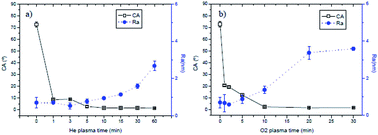
PET is a common plastic material, however its use in bio-application is rather limited because of its surface properties. Therefore, its surface was exposed to different plasma treatments in order to modify its hydrophobic character and enhance the anti-fouling properties. Two types of plasma phases were selected, the first (O2 plasma) should strongly oxidize the PET surface while the second one (He plasma) should induce a weaker surface modification. Plasma-treated surfaces were characterized studying their roughness, wettability, chemical composition and aging. WCA measurements performed on pristine PET substrates showed a CA value of 72°. The values obtained immediately after plasma treatments presented a remarkable drop (Fig. 1). With He plasma a value of 15° after 30 s of treatment was measured and for plasma times of 10 min or higher the CA value decreases to almost 0°, conferring highly hydrophilic character to the PET substrate. O2 plasma-treatment produces a significant decrease. Hence, both plasma treatments increase the hydrophilic character of PET surfaces and in particular, highly hydrophilic surfaces are obtained with plasma times around the 10 minutes.
image file: c4ra05691d-f1.tif
Fig. 1 Contact angle (empty squares) and Ra values (filled circles) of He (a) and O2 (b) plasma-treated PET versus the duration.

The surface topography of plasma-treated substrates (Fig. 1) showed an increment of the average surface roughness (Ra) with plasma time: from 0.7 nm (pristine PET) to 1.6 and 3.6 nm after 30 min of treatment, for He and O2 plasma respectively.

From the height AFM images (Fig. 2), it can be observed that pristine PET film is a smooth surface that presents conical structures of different sizes not uniformly distributed. After plasma-treatment, the smooth area on the upper surface is removed giving place to a nanostructurated surface. Features ranging 30–40 nm of diameter were obtained with He plasma-treatments of at least 5 min, meanwhile structures ranging 20–35 nm were found with O2 plasma-treatments. However, for oxygen plasma-treatments of 30 min structures with dimensions around 120 nm were observed, which could be associated to a bundler formation of the nanostructures as has been reported by Fernandez-Blazquez et al.26


image file: c4ra05691d-f2.tif
Fig. 2 2D and 3D height images of virgin PET (a), He plasma-treated PET 10 min (b), and O2 plasma-treated PET 10 min (g). Images have a scan area of 2 × 2 μm except for non-treated PET image that an area of 10 × 10 μm is showed.

SEM images of He plasma-treated PET showed a 42% and 36% of nano-structuration with treatments of 10 and 30 min, respectively. No nano-structuration was produced by 3 min of treatment. O2 plasma-treatment presented a nano-structured area of 35%, 29% and 19% for 10, 20 and 30 min respectively. Results indicate a reduction of the nano-structured area increasing the plasma-treatment time. Such effect increases applying O2 plasma, 19% of nano-structuration against a 36% with He plasma after 30 min of treatment. Moreover, the shape of the nano-structuration differs on the applied plasma-treatment. A granular structuration is obtained with the plasma-oxidation meanwhile a tubular shaped structuration results from He plasma-treatment, as can be observed in the Fig. 3. Therefore, both plasma treatments confer to the PET a nanostructured topography but their roughness and distribution differ depending on the gas used on the plasma treatment. Hence, by selecting the proper plasma parameters, surfaces with similar structuration and different chemistry can be produced to study their bioadhesive properties.


image file: c4ra05691d-f3.tif
Fig. 3 SEM images of surfaces treated for 30 min with He plasma (a) and with O2 plasma.

The surface functionalization of the PET samples was studied by XPS. From the survey spectra, the relative atomic concentration of C and O elements was obtained. Results (Table 1) showed an increment of the oxygen groups on the treated surfaces for both He and O2 plasma plasma-treated surfaces. Regarding to the O/C ratio versus treatment time, a gradual increment in the O/C ratio is observed on He plasma-treated samples; meanwhile, practically no change is observed with the O2 plasma treatment. Both plasma-treatments induce the bond scission and radical formation depending on duration. But, the formed radicals are immediately oxidized in O2 plasma phase while they are only quenched after the He plasma-treatment through post-oxidation in air, giving place to the formation of different chemical groups. This surface reaction is associated to the aging.

Table 1 O/C ratio and proportions (%) of decomposed peaks from the C1s spectra of pristine and plasma-treated PET at different durations
  O/C C1 C–C/C–H C2 C–O C3 O–C[double bond, length as m-dash]O C4 arom. C5 C[double bond, length as m-dash]O
  0.26 71.1 15.81 12.67 0.45
 
He plasma-treated PET
5 min 0.38 70.1 18.7 5.3 0.7 5.2
10 min 0.45 72.8 18.7 6.0 0.5 2
30 min 0.64 59.3 28.6 6.6 2 3.5
 
O2 plasma-treated PET
5 min 0.42 60.3 23.1 14.2 2.4
10 min 0.42 56.0 26.2 15.2 2.6
20 min 0.45 62.0 21.4 14.6 2.0


The high-resolution C1s spectrum of the pristine PET surface was decomposed into three main peaks, C1 at 285.0 eV attributed to the aliphatic carbon (C–C and C–H), C2 at 286.7 eV assigned to the ether carbon (C–O) and C3 at 289.1 eV corresponding to the carboxylic carbon (O–C[double bond, length as m-dash]O).27 Besides, a shake-up peak is observed at 291.4 eV (peak C4) indicating the presence of aromatic rings. After He plasma-treatment, a new functional group appears around 287.8 eV (peak C5) attributed to the C[double bond, length as m-dash]O groups,28–30 meanwhile no new groups were added to the surface by O2 plasma treatments. The chemical change produced on the PET surface by the He plasma (Table 1) is remarkable with long treatment times, were a reduction of 16% and 48% can be observed on the aliphatic and carboxylic contents respectively (peaks C1 and C3) and a increase of 81% in ether is obtained (peak C2). Regarding to the O2 plasma (Table 1) similar changes are observed for the different treatment times. After 20 min of treatment the PET surface shows a decrease of 12% of the aliphatic carbon and an increase of both ether and carboxylic groups of 35% and 15%, respectively. This oxidation plateau may be assigned to the competition between functionalization and degradation, since longer treatment times can emphasize the surface etching. Therefore, the different plasma treatments produce a similar O/C ratio, around 0.45, but O2 plasma-treated surfaces seem to be more oxidized as they are mostly composed of ester groups. Remark also the significant change in the carboxylic content of the PET surfaces, that is reduced by He plasma-treatments and increased by O2 plasma-treatments. Such changes in the chemical composition are the main responsible of the improved wettability observed in the plasma-treated PET films. Particularly, the key factor is the increment of the oxygen containing groups achieved by both He and O2 plasma treatments.1 Moreover, the shake-up satellite peak did not disappear completely with the plasma treatments. With a similar O/C ratio, around 0.45, the He or O2 plasma-treated surfaces showed different substructures. Indeed, the latter seemed to be more oxidized as mostly composed of ester groups.

Study of the aging of the plasma-modified PET surfaces

The surface functionalities and the wettability of plasma-treated surfaces can be lost due to aging processes1,14,26 that can be attributed to external influences like atmospheric adsorption or oxidation, and to internal restructuring processes.31 The aging effect on plasma-treated PET films was studied by water contact angle measurements. Fig. 4 shows the variation of the CA values of PET films stored in closed Petri dishes at laboratory conditions. A loss on the hydrophilic character versus time is observed in all the samples.
image file: c4ra05691d-f4.tif
Fig. 4 Contact angle versus aging-time of PET surfaces plasma-treated at different durations, (a) He and (b) O2 plasmas.

The increase in the CA values shows three different regimes regarding to the He plasma treatment time. At short treatment times, 3–5 min, the CA increases quickly during the first days after the plasma exposure, then a week after the plasma-treatment a slower increment is observed, reaching values around the CA of the pristine PET film. The following regime is observed for treatments of 10–15 min, where a high increment on the CA is observed during the first two weeks of storage and then a slower increment takes place. In the last regime, for treatments of 30–60 min, a slow variation of the CA values is shown during the first two weeks after the plasma treatment and then increases remarkably. PET films treated with O2 plasma during 1–10 min present the same trend than substrates treated with short helium treatment times. With oxygen plasma times of 20–30 min a slower recovery takes place the first days after the treatment. Then, a fast increase in the CA values is observed, reaching the same plateau values than substrates treated for 30–60 min with He plasma but in shorter time, 20 days and 40 days for O2 and He plasmas, respectively. Hence, the hydrophilic character of plasma-treated PET is reduced with aging time. Nevertheless, for long He treatment times the hydrophilic character is kept even two weeks after the plasma-treatment.

Since the aging behavior depends on different factors, such as temperature, humidity or aging medium,32,33 two different storage conditions (in distilled water and in air) were selected in order to study the storage effect on the surface wettability. After 28 days considering as the time needed to reach a stable surface, the storage conditions were changed, that means, samples immersed in water were kept into a closed Petri dish and samples stored in air were immersed in distilled water. Samples were kept in that conditions for 14 days and then, the storage conditions were changed again for a total of 4 times. In that way, a period of three months was covered, the first one with fixed storage conditions and the following ones with cycles of 14 days. After 28 days of the plasma treatment, higher CA values were obtained for samples initially stored in air, as can be seen in Fig. 5. Nevertheless, the samples presented a hydrophilic recovery when they were kept in water, showing all of them similar CA values independently of the plasma treatment time; an averaged value of 21° ± 1 and 33° ± 0 for He and O2 plasma-treatments respectively. Samples initially stored in water present lower CA values even after aging in air (Petri dish storage) and their hydrophilic recovery after immersion in distilled water is similar to the samples initially stored in air; an averaged value of 18° ± 2 and 33° ± 1 for He and O2 plasma-treatments respectively. Moreover, a plasma-treatment time dependency is observed on the aging of samples treated with helium and stored initially in water, increasing the hydrophilic character with a reduction of the plasma treatment time, 13° ± 3 for 1 min treatment versus 24° ± 4 for 10 min of treatment.


image file: c4ra05691d-f5.tif
Fig. 5 Contact angle of PET films treated with (a) He and (b) O2 plasmas after switching the storage conditions (water or air).

Such results indicate that not all the hydrophilic character obtained by plasma-treatments is lost during the aging process, since independently of the storage conditions and the plasma-treatment time, a hydrophilic recovery is observed for all the samples after immersion in distilled water. Therefore, keeping the treated samples into a hydrophilic environment helps to recover the surface wettability that was initially lost by atmospheric surface adsorption processes. Hence, the highest wettability can be achieved with short He plasma-treatments and storing the sample into a hydrophilic environment.

Alternative preparation of stable hydrophilic PET surfaces

As described above, the aging of plasma-modified PET depends on the plasma parameters and on the storage conditions. Minimizing the interfacial energy between the treated surface and the surrounding medium, here hydrophilic surface in contact of highly polar phase such water, stabilizes the surface and allows a partial recovery of the attached functional groups. In order to enhance the stability, a thin protective layer of Pluronic F108 was deposited onto the plasma-treated PET. Then, the samples were immersed in water 24 hours to check the stability of the grafting in water. As reference, samples non-grafted and treated at the same plasma conditions were also immersed in water for the same period of time. Then, after drying under compressed air, the CA was measured (Fig. 6).
image file: c4ra05691d-f6.tif
Fig. 6 Contact angle of PET films plasma-treated and grafted with Pluronic F108 versus plasma parameters duration; (a) He and (b) O2 plasmas.

No difference in the CA values from non plasma treated PET (grafted and non-grafted) was obtained, indicating the solubility in water of the surfactant grafted on the pristine PET surface. However, for all He and O2 plasma treatment times, higher CA values were obtained from grafted samples showing the presence of the surfactant on the surface after overnight in distilled water. Samples modified with different plasma treatments (10 min of He plasma and 20 min of O2 plasma) were grafted with Pluronic F108 and the CA distribution of ultra-pure water picodrops on an area of 5 × 5 μm2 is showed in the Fig. 7.


image file: c4ra05691d-f7.tif
Fig. 7 Contact angle mapping of (a) He and (b) O2 plasma-treated surfaces grafted with Pluronic F108.

Grafted He plasma-treated surfaces showed an average contact angle of 11.4° ± 3.9, meanwhile grafted O2 plasma-treated surfaces presented slightly higher CA, 20.4° ± 5.6, as well as higher variation on the surfactant distribution as indicates the increase in the standard deviation values. The increment on the CA values on grafted samples regarding to the values obtained from non grafted surfaces indicates a good and uniform coverage of the surface by the surfactant Pluronic F108.

The stability in water of plasma-treated surfaces grafted with Pluronic was studied by CA measurements. For reference values, plasma-treated surfaces were immersed in distilled water. Initially samples grafted with the Pluronic F108 presented higher contact angle than non grafted surfaces with a difference around 5 and 11 degrees for He and O2 plasma-treated surfaces, respectively. After two weeks immersed in distilled water, the contact angle differences between grafted and non grafted samples were maintained, 5 and 7.5° for He and O2 plasmas respectively, indicating the stability in water of the grafted Pluronic on the surface (Fig. 8).


image file: c4ra05691d-f8.tif
Fig. 8 Contact angle of modified PET versus immersion time in water.

Besides, grafted and non grafted surfaces exposed to the same plasma treatment show the same trend of aging indicating no influence of grafting on reducing the aging effect, the chemical nature of the medium has a stronger influence and therefore, samples have to be kept in water.

Study of the anti-fouling character of the relating highly hydrophilic PET surfaces

As an example, the bioadhesive character of plasma-treated (10 min of He and 20 min of O2 plasma-treatments) and pristine surfaces was studied for the adsorption of ovoalbumin protein. For such purpose, the bioadhesive character of plasma-treated and pristine surfaces was studied by biorecognition of Ab-IgG-FITC on surfaces previously functionalized with IgG antibodies. The complete functionalization process is described in the experimental part. For such purpose, half of the sample was masked during the plasma treatment in order to have a non exposed area as reference in each measurement.

The intensity measured from confocal images showed higher values on pristine surfaces, indicating a higher adhesion of proteins on non plasma-treated PET. The intensity ratios between treated and non-treated surfaces for O2 and He plasma-treatments were respectively 0.54 ± 0.1 and 0.52 ± 0.1. Hence, almost half adhesion of proteins was observed on plasma-treated surfaces. In Fig. 9, one can be observed the higher intensity signal on pristine surfaces (upper part of the image) and the drastic signal reduction achieved by the O2 or He plasma-treatments (low part of the image). Regarding to surfaces grafted with Pluronic F108, an enhancement on the antifouling behavior was also achieved, since the intensity ratio of O2 or He plasma-treated and grafted with Pluronic F108 were respectively 0.36 ± 0.04 and 0.45 ± 0.06. Bioadhesion is dependent on the different surface parameters, such as the roughness and the chemical affinity (hydrophilicity and charge effect).34,35 However, these plasma-treated surfaces can be considered as smooth ones since their roughness (Ra) was shown to be lower than 1 nm (0.72 ± 0.03 nm).


image file: c4ra05691d-f9.tif
Fig. 9 Confocal microscopy image of ovoalbumin adsorption onto a PET surface where the lower part of the sample is treated with (a) He and (b) O2 plasma (scale: 158 × 158 μm).

Therefore, a similar reduction of the bioadhesive character is obtained with the different plasma-treatments. However, the anti-fouling character is improved grafting the plasma-treated surfaces with an anti-fouling copolymer.

Conclusions

In this study, we have explored the plasma-treatment of PET surfaces with two different gases, helium and oxygen, to enhance their surface wettability. After plasma treatment nanostructured hydrophilic surfaces were obtained by the proper selection of the plasma parameters. The increase in the wettability of the surfaces was achieved by increasing the oxygen-containing groups on the surface. For both treatments, aliphatic carbon contents are reduced and the ether groups are increased. However, the carboxylic carbon content is increased by oxygen treatments and reduced with helium treatments. Besides, a new functional group, C[double bond, length as m-dash]O, is anchored on the surface by the helium plasma.

The dependence on the surrounding media in the aging of plasma treated surfaces was studied. Indicating a hydrophilic recovery of plasma-treated surfaces after immersion into a hydrophilic medium like water. Furthermore, He plasma-treated samples presented a hydrophilic recovery dependent on the initial storage conditions. Samples immersed in water had a better hydrophilic recovery than samples exposed to air. Morphology studies by SEM and AFM techniques showed a nanostructuration of plasma treated surfaces the shape of the nanostructures obtained was highly influenced by the gas used during the plasma treatment. Roughness measurements indicated a higher modification of the surface by O2 plasma treatments. Plasma-treated surfaces presented a good grafting of Pluronic F108 on them as well as the stability of the grafted polymer after immersion in water.

Confocal microscopy confirmed an improvement in the antifouling properties of plasma-treated surfaces, showing a 50% less adhesion of proteins than in pristine samples. Furthermore, plasma treated surfaces were grafted with a well know non-bioadhesive polymer, Pluronic F108, in order to tailor the anti-fouling properties of the plasma-treated surface. Results showed a remarkable reduction of the protein adhesion on grafted surfaces, 64% and 55% for oxygen and helium plasma-treated and grafted surfaces respectively.

Acknowledgements

This work was supported by ANR French agency in the frame of SANBACT project.

References

  1. D. Hegemann, H. Brunner and Ch. Oehr, Nucl. Instrum. Methods Phys. Res., Sect. B, 2003, 208, 281–286 CrossRef CAS.
  2. M. Collaud Coen, R. Lehmann, P. Groening and L. Schlapbach, Appl. Surf. Sci., 2003, 207, 276–286 CrossRef CAS.
  3. H. Dong and T. Bell, Surf. Coat. Technol., 1999, 111, 29–40 CrossRef CAS.
  4. Y. Ikada, Biomaterials, 1994, 15(10), 725–736 CrossRef CAS.
  5. B. D. Ratner, Biosens. Bioelectron., 1995, 10(9–10), 797–804 CrossRef CAS.
  6. S. Burkert, M. Kuntzsch, C. Bellmann, P. Uhlmann and M. Stamm, Appl. Surf. Sci., 2009, 255(12), 6256–6261 CrossRef CAS PubMed.
  7. S. Han, Y. Lee, H. Kim, G. H. Kim, J. Lee, J. H. Yoon and G. Kim, Surf. Coat. Technol., 1997, 93(2–3), 261–264 CrossRef CAS.
  8. J. Friedrich, L. Wigant, W. Unger, A. Lippitz and H. Wittrich, Surf. Coat. Technol., 1998, 98(1–3), 879–885 CrossRef CAS.
  9. G. Poletti, F. Orsini, A. Raffaele-Addamo, C. Riccardi and E. Selli, Appl. Surf. Sci., 2003, 219(3–4), 311–316 CrossRef CAS.
  10. M. M. Hossain, D. Hegemann, A. S. Herrmann and P. Chabrecek, J. Appl. Polym. Sci., 2006, 102(2), 1452–1458 CrossRef CAS.
  11. N. De Geyter, R. Morent, C. Leys, L. Gengembre and E. Payen, Surf. Coat. Technol., 2007, 201(16–17), 7066–7075 CrossRef CAS PubMed.
  12. S. M. Pelagade, N. L. Singh, A. Qureshi, R. S. Rane, S. Mukherjee, U. P. Deshpande, V. Ganesan and T. Shripathi, Nucl. Instrum. Methods Phys. Res., Sect. B, 2012, 289(2012), 34–38 CrossRef CAS PubMed.
  13. A. Friedman, in Plasma chemistry, Cambrodge University Press, 2008 Search PubMed.
  14. K. N. Pandiyaraj, V. Selvarajan, R. R. Deshmukh and M. Bousmina, Surf. Coat. Technol., 2008, 202(17), 4218–4226 CrossRef CAS PubMed.
  15. N. K. Pandiyaraj, V. Selvarajan, R. R. Deshmukh and C. Gao, Vacuum, 2008, 83(2), 332–339 CrossRef PubMed.
  16. M. Gheorghiu, F. Arefi, J. Amouroux, G. Placinta, G. Popa and M. Tatoulian, Plasma Sources Sci. Technol., 1997, 6(1), 8–19 CrossRef CAS.
  17. D. Papakonstantinou, E. Amanatides, D. Mataras, V. Ioannidis and P. Nikolopoulos, Plasma Processes Polym., 2007, 4(S1), S1057–S1062 CrossRef.
  18. E. Wohlfart, J. P. Fernández-Blázquez, E. Knoche, A. Bello, E. Pérez, E. Arzt and A. del Campo, Macromolecules, 2010, 43(23), 9908–9917 CrossRef CAS.
  19. G. Legeay, A. Coudreuse, F. Poncin-Epaillard, J. M. Herry and M. N. Bellon-Fontaine, J. Adhes. Sci. Technol., 2010, 24(13–14), 2301–2322 CrossRef CAS PubMed.
  20. P. Koegler, A. Clayton, H. Thissen, G. N. C. Santos and P. Kingshott, Adv. Drug Delivery Rev., 2012, 64(15), 1820–1839 CrossRef CAS PubMed.
  21. G. Speranza, G. Gottardi, C. Pederzolli, L. Lunelli, R. Canteri, L. Pasquardini, E. Carli, A. Lui, D. Maniglio, M. Brugnara and M. Anderle, Biomaterials, 2004, 25(11), 2029–2037 CrossRef CAS PubMed.
  22. K. S. Siow, L. Britcher, S. Kumar and H. J. Griesser, Plasma Processes Polym., 2006, 3(6–7), 392–418 CrossRef CAS.
  23. T. Vrlinic, D. Debarnot, M. Mozetic, A. Vesel, J. Kovac, A. Coudreuse, G. Legeay and F. Poncin-Epaillard, J. Colloid Interface Sci., 2011, 362(2), 300–310 CrossRef CAS PubMed.
  24. V. M. De Cupere, J. Van Wetter and P. G. Rouxhet, Langmuir, 2003, 19(17), 6957–6967 CrossRef CAS.
  25. Y. B. Liou and R. Y. Tsay, J. Taiwan Inst. Chem. Eng., 2011, 42(3), 533–540 CrossRef CAS PubMed.
  26. J. P. Fernández-Blázquez, D. Fell, E. Bonaccurso and A. del Campo, J. Colloid Interface Sci., 2011, 357(1), 234–238 CrossRef PubMed.
  27. H. Hantsche, High resolution XPS of organic polymers, the scienta ESCA300 database, ed. G. Beamson and D. Briggs, Wiley, Chichester 1992, p. 295 Search PubMed.
  28. J. R. Friedrich, I. Loeschcke, H. Frommelt, H. D. Reiner, H. Zimmermann and P. Lutgen, Polym. Degrad. Stab., 1991, 31(1), 97–114 CrossRef CAS.
  29. Y. Ren, C. Wang and Y. Qiu, Surf. Coat. Technol., 2008, 202(12), 2670–2676 CrossRef CAS PubMed.
  30. P. A. Charpentier, A. Maguire and W. K. Wan, Appl. Surf. Sci., 2006, 252(18), 6360–6367 CrossRef CAS PubMed.
  31. M. Morra, E. Occhiello, R. Marola, F. Garbassi, P. Humphrey and D. Johnson, J. Colloid Interface Sci., 1990, 137(1), 11–24 CrossRef CAS.
  32. C. C. Dupont-Gillain, Y. Adriaensen, S. Derclaye and P. G. Rouxhet, Langmuir, 2000, 16(21), 8194–8200 CrossRef CAS.
  33. J. Nakamatsu, L. F. Delgado-Aparicio, R. Da Silva and F. Soberon, J. Adhes. Sci. Technol., 1999, 13(7), 753–761 CrossRef CAS PubMed.
  34. X. Zhang, L. Wang and E. Levaren, RSC Adv., 2013, 3, 12003–12020 RSC.
  35. N. J. Shirtcliffe, Methods Mol. Biol., 2013, 949, 269–281 CAS.

Footnote

Present address: CIC NanoGUNE Consolider, Tolosa Hiribidea 76, 20018 Donostia-San Sebastian, Spain.

This journal is © The Royal Society of Chemistry 2014
Click here to see how this site uses Cookies. View our privacy policy here.