Mild temperature synthesis of gold nanoplates using polyethyleneimine and their improved surface enhanced Raman signal

Taekyung Yu, Zhaohui Wu and Woo-Sik Kim*
Department of Chemical Engineering, College of Engineering, Kyung Hee University, Youngin, 446-701, Korea. E-mail: wskim@khu.ac.kr

Received 10th June 2014 , Accepted 5th August 2014

First published on 6th August 2014


Abstract

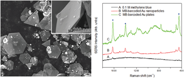
This study explores the facile controlled-synthesis and localized surface plasmonic properties of Au nanoplates. Au nanoplates were synthesized via a reaction of HAuCl4 with branched polyethyleneimine (BPEI) in the presence of urea at a reaction temperature as low as 30 °C. Structural characterizations revealed that the synthesized Au nanoplates had an average lateral size of around 2–8 μm and a thickness of around 40 nm, plus they were highly crystalline in spite of the low reaction temperature. Studies of the growth factors for Au nanoplates showed that the PEI morphology, molar ratio of BPEI/HAuCl4, pH of the reaction solution, and concentration of urea all played important roles in the formation of thin Au nanoplates. With a low BPEI/HAuCl4 ratio or under acidic conditions, thick Au plates were synthesized, whereas Au nanoparticles around 10–20 nm were observed with a high BPEI/HAuCl4 ratio or under basic conditions. When the synthesis was conducted in the absence of urea, thick Au nanoplates were synthesized, indicating that urea can serve as a capping agent for thin Au nanoplates. The resulting Au nanoplates also exhibited an enhanced surface-enhanced Raman scattering (SERS) signal when compared with Au nanoparticles due to their sharp corners and edges.


Introduction

Gold (Au) nanostructures have attracted considerable attention due to their widespread use in catalysis, photonics, electronics, optoelectronics, biological labeling, imaging, sensing, and surface-enhanced Raman scattering (SERS).1–9 When compared with their size, composition, and crystallinity, the morphological control of Au nanostructures is especially important as it is one of the most sensitive knobs for tuning the optical properties of Au nanostructures. For example, the position of the localized surface plasmon resonance (LSPR) peaks of Au nanostructures has been found to be strongly dependent on the morphology.10,11 In particular, two-dimensional Au nanostructures including nanoplates, microplates, and nanosheets have drawn increasing interest due to their unique optical properties and potential use in various sensing applications.12–15 Moreover, the sharp corners and edges of these metal plates make them useful as possible substrates for SERS.16 To date, various synthetic routes involving chemical or photochemical reduction methods have been developed to generate Au nanoplates.17–26 For example, Sastry and co-workers synthesized Au nanoplates using a lemongrass plant extract as the reducing agent.17 Au nanoplates have also been synthesized by reducing Au salts in the presence of capping agents and stabilizers, plus certain capping agents have been proposed to play a critical role in the formation of nanoplates by adsorption onto the {111} facets of the developing nanoplates.18–26 Yet, despite such successful experimental demonstrations, these methods are invariably difficult to scale-up due to expensive capping agents, tedious synthetic steps, and high reaction temperatures of more than 90 °C. Therefore, developing a simple and reliable Au nanoplate production route for industrial application remains a challenge.

Accordingly, this study investigated the synthesis of Au nanoplates when reacting HAuCl4 with branched polyethyleneimine (BPEI) in the presence of urea in an aqueous solution at a reaction temperature as low as 30 °C. In this synthesis, the morphology of the PEI, molar ratio of BPEI/HAuCl4, pH of the reacting solution, and concentration of urea were all found to play an important role in the formation of the Au nanoplates. With a low BPEI/HAuCl4 ratio or under acidic conditions, thick Au plates were synthesized, whereas Au nanoparticles around 10–20 nm were observed with a high BPEI/HAuCl4 ratio or under basic conditions, confirming that the reduction rate of the Au ions and stabilizing the nanoparticles in an aqueous solution are both important for the formation of thin Au nanoplates. When the synthesis was conducted in the absence of urea, thick Au nanoplates were synthesized, indicating that urea can serve as a capping agent for thin Au nanoplates. Furthermore, the Au nanoplates were found to exhibit an enhanced SERS signal when compared with Au nanoparticles. Therefore, the present results represent a good example of the anisotropic growth of Au nanostructures and advance the current understanding of the growth processes involved in the aqueous-phase synthesis of Au nanoplates.

Experimental section

Materials

The branched polyethyleneimine (BPEI, MW = 25[thin space (1/6-em)]000 and 750[thin space (1/6-em)]000) and linear polyethyleneimine (LPEI, MW = 25[thin space (1/6-em)]000) were purchased from Polysciences. The urea was purchased from Aldrich and used without further purification. The chloroauric acid (HAuCl4·nH2O (n = 3.6)) was purchased from Kojima, and the methylene blue (certified by the BSC, FW = 373.9) obtained from Sigma-Aldrich.

Synthesis of Au nanoplates

For the typical synthesis of Au nanoplates, 2.5 mg of PEI and 300 mg of urea were dissolved in 5 mL of deionized water and heated to 30 °C. 10 μL of an aqueous HAuCl4 solution (1 M) was then added to the reaction solution using a pipette. After maintaining the reaction solution at 30 °C for 24 h under magnetic stirring, the reaction mixture was cooled to room temperature.

Synthesis of Au nanoparticles

20 mg of BPEI (MW ∼ 750[thin space (1/6-em)]000) was dissolved in 5 mL of deionized water and heated to 50 °C. Next, 10 μL of an aqueous HAuCl4 solution (1 M) was added to the reaction solution. After maintaining the reaction solution at 50 °C for 12 h under magnetic stirring, the reaction mixture was then cooled to room temperature.

SERS measurements

The methylene blue (MB) was completely dissolved in 4 mL of deionized water to form a 0.1 M homogenous solution. The original solutions (5 mL) of Au plates and Au nanoparticles were precipitated by centrifugation, and then 2 mL of the MB solution was added to the Au plates and Au nanoparticles to form a suspension. After sonication, the suspensions were kept for 12 h at room temperature. Finally, 100 μL of the suspensions were dropped onto a load fragment to measure the SERS spectrum.

Characterization

The scanning electron microscopy (SEM) images were obtained using a Jeol JSM-6701F microscope operated at 15 kV. The transmission electron microscopy (TEM) images were captured using a JEM-2100F microscope operated at 200 kV. The UV-vis spectra were recorded using a Jasco UV-vis spectrophotometer within a range of 400–1300 nm. The powder X-ray diffraction (XRD) patterns were obtained using a Rigaku D-MAX/A diffractometer at 35 kV and 35 mA. The SERS spectra were obtained using a FT-Raman spectrometer (Renishaw, RENISHAW plc, U.K.) with He–Ne 785 nm as the laser source.

Results and discussion

The Au nanoplate synthesis was conducted in an aqueous solution containing chloroauric acid trihydrate (HAuCl4·3H2O), branched polyethyleneimine (BPEI, MW ∼750[thin space (1/6-em)]000), and urea (1 M) at a low reaction temperature of 30 °C for 24 h. The BPEI served as a mild reductant and capping agent, similar to the mechanism of conventional BPEI-based Au nanoparticle synthesis, as reported previously.27 Several hours into the reaction, the solution gradually changed from light yellow to shiny golden, indicating the formation of Au nanoplates. Fig. 1(a) and (b), are typical scanning electron microscopy (SEM) images of the product, showing hexagonal and triangular nanoplates with lateral dimensions of 2–8 μm and a thickness of approximately 40 nm. The transmission electron microscopy (TEM) and corresponding electron diffraction (ED) patterns taken by directing the electron beam perpendicular to the flat faces of a hexagonal Au nanoplate exhibited diffraction spots with a six-fold rotational symmetry, indicating that the top and bottom faces of the Au nanoplates were enclosed by {111} planes (Fig. 1(c)). The spots circled and squared in the ED patterns can be indexed to the {220} and forbidden 1/3{422} reflections, respectively, in which the latter indicates the presence of planar defects such as stacking faults in the {111} planes.28 The powder X-ray diffraction (XRD) peaks recorded from the sample were assigned to diffraction from (111), (200), (220), and (311) planes of face-centered cubic (fcc) gold (Fig. 1(d), Fm3m, a = 4.078 Å, Joint Committee on Powder Diffraction Standards (JCPDS) file number 04-0784). It is worth noting that the intensity ratio between the (111) and (200) diffraction peaks was higher than the literature value of JCPDS (0.67 versus 0.4), indicating that the present nanoplates grew in the [111] directions of an fcc structure. The UV-vis extinction spectrum taken from an aqueous suspension of the as-prepared Au nanoplates revealed a broad band at around 1300 nm, matching well with previous reports on Au nanoplates with a similar side dimension (Fig. S1 in ESI).12 Au nanoparticles with sizes of around 10–20 nm were also synthesized using a similar method with the current Au nanoplates and exhibited a strong peak at around 520 nm, showing the importance of morphology control for the optical properties of Au nanostructures.
image file: c4ra05528d-f1.tif
Fig. 1 (a) and (b) SEM images, (c) TEM image, and (d) XRD patterns of Au nanoplates synthesized by reducing HAuCl4 with BPEI (MW ∼750[thin space (1/6-em)]000) in presence of urea in aqueous solution.

In the present synthesis, PEI serves as both a stabilizer to protect the Au nanoplates from agglomerization and a reducing agent due to its primary and secondary amine groups. When the synthesis was conducted using linear polyethyleneimine (LPEI) instead of BPEI, small Au nanoplates with a lateral dimension of around 2 μm were synthesized (Fig. 2). Typically, the amine group in LPEI is mostly secondary amines (∼99%), whereas the amine group in BPEI is composed of 25% primary amines, 50% secondary amines, and 25% tertiary amines. Thus, the significant portion of secondary amines in LPEI gives it a higher reducing power than BPEI. As a result, when using LPEI as the reagent, a large number of seeds would seem to be produced during the early stage, leading to the formation of small Au nanoplates. When the synthesis was conducted using BPEI having smaller molecular weight (MW ∼25[thin space (1/6-em)]000), Au nanoplates with a lateral dimension of around 5 μm were observed, supporting the importance of reducing power rather than chain length of PEI in this synthesis (Fig. S2 in ESI). During the growth of nanoparticles, the selective chemisorptions of the capping agent can control the growth rate of specific crystallographic planes, which determines the final morphology of the nanoparticles. In the present synthesis, urea seemed to act as a capping agent for the formation of Au nanoplates. When the synthesis was conducted in the absence of urea and the other experimental parameters were the same as in Fig. 1, the product consisted of a mixture of nanostructures, including small Au nanoparticles, large aggregates, and a small portion of thick Au nanoplates (Fig. 3(a)). Thus, to elucidate the role of urea in controlling the morphology of Au nanoplates, a set of experiments was conducted with different urea concentrations. When using a low concentration of urea (0.5 M), the resultant Au nanoplates had small lateral dimensions of 1–2 μm (Fig. 3(b)). However, when increasing the urea concentration to 1 M, the lateral dimensions of the Au nanoplates exhibited the dramatic growth to 2–5 μm, demonstrating that urea can act as a capping agent for the formation of Au nanoplates with a large portion of {111} facets on the surface (Fig. 3(c)). It is also interesting to note that the lateral dimensions of the Au nanoplates decreased to less than 2 μm with a high urea concentration of 2.0 M (Fig. 3(d)). Therefore, it would seem that low selective chemisorptions occurred with the high urea concentration, leading to the formation of relatively small Au nanoplates.


image file: c4ra05528d-f2.tif
Fig. 2 TEM image of a sample prepared under same conditions as those in Fig. 1, except synthesis was conducted in presence of LPEI instead of BPEI.

image file: c4ra05528d-f3.tif
Fig. 3 TEM images of samples prepared under same conditions as those in Fig. 1, except synthesis was conducted with different urea concentrations: (a) 0, (b) 0.5, (c) 1.0, and (d) 2.0 M, respectively.

The effect of the BPEI/HAuCl4 ratio on the formation of Au nanoplates was also systematically investigated. A previous report already confirmed that manipulating the concentration of the reducing agent and/or stabilizer can vary the size of the resulting Au nanoparticles.27 Thus, when the synthesis was conducted with a high molar ratio of BPEI/HAuCl4 at around 8, small Au nanoparticles with sizes of around 10–20 nm were synthesized (Fig. 4(a)). Plus, with a high molar ratio of BPEI/HAuCl4, the plentiful amine groups induced a high supersaturation, leading to the formation of a huge number of small Au nanoparticles. Meanwhile, with an intermediate BPEI/HAuCl4 ratio of 2, the relatively low supersaturation formed a small portion of Au nuclei and large portion of Au–polymer complexes in the early stage, thereby promoting the 2-D growth of nanoplates (Fig. 1(a)). However, when the BPEI concentration respective to the Au concentration was too low (BPEI/HAuCl4 ratio of 1), thick Au plates were synthesized due to a low reducing rate and insufficiency of stabilizers (Fig. 4(b)). Interestingly, small Au nanoparticles with sizes of around 10 nm were also observed with a low BPEI/HAuCl4 ratio (inset of Fig. 4(b)). Thus, it is would seem that urea is a relatively weak reducing agent and stabilizer in the synthesis of Au nanoparticles. Fig. S3 in ESI is a TEM image of the product synthesized when reacting HAuCl4 and urea in the absence of BPEI at 30 °C for 24 h, showing the formation of Au nanoparticles with sizes of around 10 nm and aggregates containing small Au nanoparticles. Notwithstanding, a complete understanding of the role of urea in this synthesis will require further studies based on both experiments and simulations. Fig. 5 briefly summarizes the different reaction conditions used in this study and the shapes of the Au nanoparticles according to each reaction condition.


image file: c4ra05528d-f4.tif
Fig. 4 TEM images of samples prepared under same conditions as those in Fig. 1, except synthesis was conducted with different molar ratios of BPEI/HAuCl4: (a) 8 and (b) 1, respectively.

image file: c4ra05528d-f5.tif
Fig. 5 Schematic illustration of reaction pathways leading to Au nanoparticles with various morphologies.

In previous research on the synthesis of Au nanoparticles using BPEI as a reacting agent, the present authors found that varying the pH of the reaction solution could control the ratio of the protonated amine group and the amine group, allowing manipulation of the size and stability of the Au nanoparticles.27,29–31 In the present study, the Au nanoplates were synthesized under weak acidic conditions at pH 3.0. However, when the synthesis was conducted under strong acidic conditions at pH 1.8, thick Au nanoplates with a thickness of around 120 nm were synthesized, demonstrating that the protonated amine group had a weaker ability in terms of reducing the Au ions to Au metal and capping the Au nanoplates (Fig. 6(a) and (b)). When increasing the pH value of the reaction solution (pH 7.0), small Au nanoplates with lateral dimensions of less than 3 μm were obtained (Fig. 6(c)). Plus, under strong basic conditions of pH 11.0, small Au nanoparticles with sizes of around 10 nm were synthesized due to the high reduction, capping, and stabilization activities of the amine groups in the BPEI (Fig. 6(d)). Therefore, these results demonstrate that adjusting the ratio between the protonated amine group and the amine group by controlling the pH value of the reaction solution is a key factor for the synthesis of thin Au nanoplates.


image file: c4ra05528d-f6.tif
Fig. 6 TEM images of samples prepared under same conditions as those in Fig. 1, except synthesis was conducted at various initial pH values: 1.8 (a) and (b), 7.0 (c), and 11.0 (d), respectively.

SERS is a technique that enhances the Raman scattering cross-sections of molecules in an environment of metal nanostructures.32 Metal nanostructures are essential for SERS as their localized surface plasmon resonances can increase the Raman signals by many orders of magnitude.33,34 In this study, methylene blue (MB) was used as the probe molecule since it has a large scattering cross section. Thus, Fig. 7 compares the SERS activities of thin films cast from MB and two types of MB-modified Au nanostructures, Au nanoplates, and spherical Au nanoparticles. The Au nanoplates have lateral dimensions of 2–8 μm and a thickness of approximately 40 nm. The spherical Au nanoparticles with sizes of around 9 nm were synthesized by reacting HAuCl4 with BPEI in an aqueous solution, as reported previously (Fig. S4 in ESI).27 All the samples exhibited SPR peaks at 450 nm, 1400 nm, and 1620 nm and the Au nanoplates exhibited well-defined and relatively strong SERS signals, whereas the signals from the Au nanoparticles were quite weak. Because all experiments used same concentration of Au nanoplates and nanoparticles, we can conclude that morphology difference between nanoplates and nanoparticles play a key role in the improvement of Raman signal.16 Although the current SERS results are purely qualitative, the enhanced Raman signal obtained with the Au nanoplates demonstrates the importance of controlling the morphology of Au nanostructures in optical applications.


image file: c4ra05528d-f7.tif
Fig. 7 SERS spectra taken from (a) methylene blue (MB), (b) MB-barcoded Au nanoparticles, and (c) MB-barcoded Au nanoplates.

Conclusions

This study explored the controlled synthesis and localized surface plasmonic properties of Au nanoplates with an average lateral size of around 2–5 μm and thickness of around 40 nm using a simple route based on the reduction of HAuCl4 with BPEI in the presence of urea at a low reaction temperature of 30 °C. In this synthesis, the morphology of the PEI, molar ratio of BPEI/HAuCl4, pH of the reaction solution, and urea concentration all played important roles in the formation of the Au nanoplates. Furthermore, it was also found that the Au nanoplates exhibited an enhanced SERS signal compared with Au nanoparticles due to their sharp corners and edges. The present results represent a good example of the anisotropic growth of Au nanostructures and advance the current understanding of the growth processes involved in the aqueous-phase synthesis of Au nanoplates. Moreover, the present synthetic strategy could also be extended to the synthesis of other anisotropic metal nanostructures.

Acknowledgements

This work was supported through a Mid-Career Researcher based on a National Research Foundation grant funded by the Korean Ministry of Education, Science and Technology (2010-0017993). This work was also supported by the Engineering Research Center of Excellence Program of Korea Ministry of Science, ICT & Future Planning (MSIP)/National Research Foundation of Korea (NRF) (Grant NRF-2014-009799).

References

  1. L. N. Lewis, Chem. Rev., 1993, 93, 2693–2730 CrossRef CAS.
  2. S. Nie and S. R. Emory, Science, 1997, 275, 1102–1106 CrossRef CAS PubMed.
  3. T. A. Taton, C. A. Mirkin and R. L. Letsinger, Science, 2000, 289, 1757–1760 CrossRef CAS.
  4. S. A. Maier, M. L. Brongersma, P. G. Kik, S. Meltzer, A. A. G. Requicha and H. A. Atwater, Adv. Mater., 2001, 13, 1501–1505 CrossRef CAS.
  5. M. A. El-Sayed, Acc. Chem. Res., 2001, 34, 257–264 CrossRef CAS PubMed.
  6. S. Chen and Y. Yang, J. Am. Chem. Soc., 2002, 124, 5280–5281 CrossRef CAS PubMed.
  7. Y. C. Cao, R. Jin and C. A. Mirkin, Science, 2002, 297, 1536–1540 CrossRef CAS PubMed.
  8. A. G. Tkachenko, H. Xie, D. Coleman, W. Glomm, J. Ryan, M. F. Anderson, S. Franzen and D. L. Feldheim, J. Am. Chem. Soc., 2003, 125, 4700–4701 CrossRef CAS PubMed.
  9. J. Chen, F. Saeki, B. J. Wiley, H. Cang, M. J. Cobb, Z.-Y. Li, L. Au, H. Zhang, M. B. Kimmey, X. Li and Y. Xia, Nano Lett., 2005, 5, 473–477 CrossRef CAS PubMed.
  10. T. R. Jensen, L. Kelly, A. Lazarides and G. C. Schatz, J. Cluster Sci., 1999, 10, 295–317 CrossRef CAS.
  11. I. O. Sosa, C. Noguez and R. G. Barrera, J. Phys. Chem. B, 2003, 107, 6269–6275 CrossRef CAS.
  12. J. E. Millstone, S. Park, K. L. Shuford, L. Qin, G. C. Schatz and C. A. Mirkin, J. Am. Chem. Soc., 2005, 127, 5312–5313 CrossRef CAS PubMed.
  13. S. S. Shankar, A. Rai, A. Ahmad and M. Sastry, Chem. Mater., 2005, 17, 566–572 CrossRef CAS.
  14. C. S. Ah, Y. J. Yun, H. J. Park, W.-J. Kim, D. H. Ha and W. S. Yun, Chem. Mater., 2005, 17, 5558–5566 CrossRef CAS.
  15. J. E. Millstone, G. S. Métraux and C. A. Mirkin, Adv. Funct. Mater., 2006, 16, 1209–1214 CrossRef CAS PubMed.
  16. Y. Xiong, J. M. McLellan, J. Chen, Y. Yin, Z.-Y. Li and Y. Xia, J. Am. Chem. Soc., 2005, 127, 17118–17127 CrossRef CAS PubMed.
  17. S. S. Shankar, A. Rai, B. Ankamwar, A. Singh, A. Ahmad and M. Sastry, Nat. Mater., 2004, 3, 482–488 CrossRef CAS PubMed.
  18. J.-U. Kim, S.-H. Cha, K. Shin, J. Y. Jho and J.-C. Lee, Adv. Mater., 2004, 16, 459 CrossRef CAS PubMed.
  19. Y. Shao, Y. Jin and S. Dong, Chem. Commun., 2004, 1104–1105 RSC.
  20. L. Wang, X. Chen, J. Zhan, Y. Chai, C. Yang, L. Xu, W. Zhuang and B. Jing, J. Phys. Chem. B, 2005, 109, 3189–3194 CrossRef CAS PubMed.
  21. B. Liu, J. Xie, J. Y. Lee, Y. P. Ting and J. P. Chen, J. Phys. Chem. B, 2005, 109, 15256–15263 CrossRef CAS PubMed.
  22. M. Tsuji, M. Hashimoto, Y. Nishizawa, M. Kubokawa and T. Tsuji, Chem. –Eur. J., 2005, 11, 440–452 CrossRef CAS PubMed.
  23. C. Kan, X. Zhu and G. Wang, J. Phys. Chem. B, 2006, 110, 4651–4656 CrossRef CAS PubMed.
  24. H.-C. Chu, C.-H. Kuo and M. H. Huang, Inorg. Chem., 2006, 45, 808–813 CrossRef CAS PubMed.
  25. W.-L. Huang, C.-H. Chen and M. H. Huang, J. Phys. Chem. C, 2007, 111, 2533–2538 CAS.
  26. B. Lim, P. H. C. Camargo and Y. Xia, Langmuir, 2008, 24, 10437–10442 CrossRef CAS PubMed.
  27. R. Kim, H. S. Park, T. Yu, J. Yi and W.-S. Kim, Chem. Phys. Lett., 2013, 575, 71–75 CrossRef CAS PubMed.
  28. C. Lofton and W. Sigmund, Adv. Funct. Mater., 2005, 15, 1197–1208 CrossRef CAS PubMed.
  29. W. Maketon, C. Z. Zenner and K. L. Ogden, Environ. Sci. Technol., 2008, 42, 2124–2129 CrossRef CAS.
  30. K. Kim, J. W. Lee, J.-Y. Choi and K. S. Shin, Langmuir, 2010, 26, 19163–19169 CrossRef CAS PubMed.
  31. T. Yu, R. Kim, H. Park, J. Yi and W.-S. Kim, Chem. Phys. Lett., 2014, 592, 765 Search PubMed.
  32. D. L. Jeanmaire and R. P. Van Duyne, J. Electroanal. Chem., 1977, 84, 1–20 CrossRef CAS.
  33. K. Kneipp, H. Kneipp, I. Itzkan, R. Dasari and M. Feld, Chem. Rev., 1999, 99, 2957–2976 CrossRef CAS PubMed.
  34. M. Rycenga, M. H. Kim, P. H. C. Camargo, C. Cobley, Z.-Y. Li and Y. Xia, J. Phys. Chem. A, 2009, 113, 3932–3939 CrossRef CAS PubMed.

Footnote

Electronic supplementary information (ESI) available: UV-vis spectra and additional TEM images of Au nanoparticles. See DOI: 10.1039/c4ra05528d

This journal is © The Royal Society of Chemistry 2014
Click here to see how this site uses Cookies. View our privacy policy here.