Toxicity of ionic liquids toward microorganisms interesting to the food industry

A. G. Santos*a, B. D. Ribeiroa, D. S. Alvianob and M. A. Z. Coelhoa
aEscola de Quíımica, Universidade Federal do Rio de Janeiro, Centro de Tecnologia, Bl.E, Lab. 103, Cidade Universitária, 21949-900, Rio de Janeiro, RJ, Brazil. E-mail: ariane_gs@hotmail.com
bInstituto de Microbiologia Paulo Góes, Universidade Federal do Rio de Janeiro, 21941-590 Rio de Janeiro, RJ, Brazil

Received 4th June 2014 , Accepted 4th August 2014

First published on 5th August 2014


Abstract

Considering the potential applications of ionic liquids (ILs) as solvents in biotechnological processes such as food production via microbial synthesis, the work presented here aimed to evaluate the toxicity of these new solvents to microorganisms of interest in the food industry. Following the international standard method of the CLSI (Clinical and Laboratory Standards Institute), the maximum non-toxic concentration (MNTC) was determined for nine ILs (containing an imidazolium, cholinium or phosphonium cation) toward nine microorganisms (the bacteria Bacillus subtilis, Lactobacillus delbrueckii subs. delbrueckii, Pseudomonas aeruginosa, the actinobacteria Streptomyces drozdowiczii, the yeasts Saccharomyces cerevisiae, Yarrowia lipolytica, Kluyveromyces marxianus, and the filamentous fungi Aspergillus brasiliensis and Rhizopus oryzae). Among the bacteria, B. subtilis and P. aeruginosa were more tolerant to hydrophilic imidazolium ILs with [C2mim] cations combined with [EtSO4], [EtSO3] and [Cl] anions. In the presence of hydrophilic choline and phosphonium based-ILs, the Gram-negative bacterium P. aeruginosa was more resistant than others. The same effect was observed for the [NTf2]-based ILs, in which only P. aeruginosa could grow. Regarding the fungi, A. brasiliensis and R. oryzae tolerated high concentrations of ILs. Among the yeasts, only Y. lipolytica was tolerant to all tested ILs. In general, ILs containing choline as the cationic moiety were more biocompatible since they allowed the growth of all the studied microorganisms.

Introduction

The term ionic liquid (IL) refers to a solvent composed only of ions, organic cations combined with organic or inorganic anions, with a melting point below 100 °C.1 Due to these and other interesting features, these liquid salts have emerged as alternative solvents in bioprocesses. Changing the cations and anions that comprise the ILs allows the tuning of their properties, such as viscosity, density and hydrophobicity, in order to satisfy the requirements of a particular process; this is the most attractive characteristic of ILs. Since 1990, studies related to the application of ILs in biotechnology have been carried out; however, this application still remains quite limited in the literature, and is restricted to enzymatic2–4 and whole-cell process2 involving biotransformation reactions, mainly the asymmetric reduction of ketones to produce chiral alcohols.5–10 In whole-cell processes, water-immiscible ILs can be used in a biphasic system as a reservoir of substrate and products that can act as inhibitors. Other studies have also reported the use of water-miscible ILs for increasing the availability of insoluble substrates.

An important requirement to successfully apply a solvent as an extractor or additive in a whole-cell process is low toxicity towards microorganisms. Thus, the selection of an IL for bioprocess applications requires the screening of different cations and anions to identify the most biocompatible combination. Within the context of the toxicity of ILs, investigators have been searching for new compounds with efficient antimicrobial activities for use as biocides.11–16 In these studies, the toxic effects of ILs are evaluated toward a range of bacteria and fungi that are important for human health. These organisms include Micrococcus luteus, Staphylococcus aureus, Enterococcus faecalis, Escherichia coli, Klebsiella pneumoniae, Pseudomonas aeruginosa, Candida albicans, Rhodotorula rubra, and Bacillus subtilis, among others. Some toxicological studies have evaluated the susceptibility of microorganisms considered to be ecotoxicological models, such as the bacteria Vibrio fischeri.17,18 In the context of biotechnology, there are still few studies, and they are focused on more commonly used microorganisms such as E. coli and Saccharomyces cerevisiae and use mainly imidazolium-based ionic liquids such as [C4mim] and [C6mim] with [PF6], [BF4] and [NTf2] anions.2

In general, antimicrobial activity has been shown to be affected by the cation alkyl length; a longer alkyl chain is associated with stronger biological activity for microorganisms11–13,15,19 and others organisms including the nematode Caenorhabditis elegans,20 the freshwater snail Physa acuta,21 and the green algae Pseudokirchneriella subcapitata.22 This effect is related to an increase in the molecular lipophilicity through the QSAR concept (quantitative structure-activity relationship) that statistically relates the chemical structure, in this case lipophilicity parameters, to the toxic effect,11,23 suggesting that high molecular lipophilicity increases the interaction with cell membranes along with the toxic effect. Thus, the most biocompatible ILs seem to be those with short alkyl chains.

Although ILs have been deemed green solvents because of their negligible vapor pressures and non-flammability in contrast to molecular solvents, ILs can be very toxic for organisms in the environment and poorly biodegradable.24 The number of studies with ILs based on biocompatible ions, such as cholinium cation,16,25 the anions saccharinate and acesulfamate,26–28 and organic acids,29 has increased. With the aim of applying ILs to the biocatalytic processes of the food industry that use chemicals such as organic acids, enzymes, vitamins, polysaccharides, and others, the present work evaluated the tolerance to ILs of a range of microorganisms used in the food industry or having a potential use in this field. In this way, short alkyl chain ILs with more common cations including imidazolium and phosphonium and sustainable cations including choline associated with different anions were used to test both hydrophilic and hydrophobic ILs. The objective was to identify differences in tolerance between groups of microorganisms and identify the most biocompatible combinations of cations and anions with the potential to be used as solvents in biotechnology.

Results and discussion

Hydrophilic ILs

The growth inhibition effect of nine ILs toward nine microorganisms (bacteria, actinobacteria, yeast and filamentous fungi) was evaluated. The MNTC value was considered as the maximum concentration at which it was possible to observe microbial growth and viable cells; in other words, the higher the MNTC value, the lower the toxicity of the IL.

As the relationship between the cation alkyl chain length and IL toxic effect to a variety of organisms has been previously described in the literature, this work applied cations with short alkyl chains (between 2–4 carbons).

Hydrophilic imidazolium-based ILs (Table 1) exhibited higher toxicity to yeasts, especially S. cerevisiae and K. marxianus, which did not grow within the studied concentration range. Among yeasts, only Y. lipolytica showed any resistance to hydrophilic imidazolium-based ILs. The filamentous fungi A. brasiliensis and R. oryzae were quite tolerant to the hydrophilic ILs, exhibiting growth at the higher tested concentration. R. oryzae growth was not inhibited at 825.5 mM of [C2mim][Cl] (Table 1). Among bacteria, B. subtilis and P. aeruginosa were comparatively more resistant to hydrophilic imidazolium-based ILs. Apparently, this effect is not related to the membrane and cell wall compositions since these two bacteria differ in this regard.

Table 1 MNTC values relative to water-miscible imidazolium-based ionic liquids
Microorganisms MNTC (mM)
[C2mim][Cl] [C2mim][EtSO4] [C2mim][EtSO3]
Gram-positive bacteria
Bacillus subtilis 426 270 581
Lactobacillus delbrueckii subsp. delbrueckii 53 67.5 72
Actinobacteria
Streptomyces drozdowickii 27 34 36
Gram-negative bacteria
Pseudomonas aeruginosa 852.5 135 145
Yeasts
Saccharomyces cerevisiae <6.7 <4.2 <4.5
Yarrowia lipolytica 235 270 145
Kluyveromyces marxianus <6.7 <4.2 <4.5
Filamentousfungi
Aspergillus brasiliensis 426 >540 >581
Rhizopus oryzae >852.5 >540 290


It was not possible to observe a clear pattern between the results obtained for [Cl], [EtSO4] and [EtSO3] anions; in general, they demonstrating low toxicity toward the tested microorganisms. In others works with Clostridium butyricum30 and E. coli,31 alkylsulfate anions were considered non-toxic and promising for producing biocompatible and biodegradable ILs.

Ions based on natural products have been shown to be a good choice to form biocompatible and biodegradable ILs;32 the choline cation, for example, is an essential micronutrient of B complex vitamins.16 [Ch][Cl] (or vitamin B4) is a low-cost organic salt used, for example, as feed additive for chickens. Due to its high melting point (298–304 °C), this salt is not considered as an IL; however, it is used in choline-based ILs by anion substitution27 and applied in the synthesis of deep eutectic solvents (DES). DES are produced from a mixture of two substances in specific proportions that possesses a melting point lower than those of the separated components (e.g., the mixture of choline chloride and urea).33 Thus, it was interesting to study choline chloride as it presents a potential use as a component of DES.

Water-miscible ILs with choline cation (Table 2) were, in general, more biocompatible than the previously studied ILs with imidazoliumcation since they allowed the growth of all studied microbial strains.

Table 2 MNTC values relative to water-miscible choline- and phosphonium-based ionic liquids
Microorganisms MNTC (mM)
[Ch][CH3COO] [Ch][Cl] [P4441][MetSO4]
Gram-positive bacteria
Bacillus subtilis 24.5 28 <3.75
Lactobacillus delbrueckii subsp. delbrueckii 24.5 112 8
Actinobacteria
Streptomyces drozdowikzii 24 112 < 3.75
Gram-negative bacteria
Pseudomonas aeruginosa 98 448 15
Yeasts
Saccharomyces cerevisiae 98 224 1.95
Yarrowia lipolytica 196 448 7.8
Kluyveromyces marxianus 98 224 <3.75
Filamentous fungi
Aspergillus brasiliensis 196 448 8
Rhizopus oryzae 392 >896 8


Some studies involving other ILs containing tetralkylammonium non-cyclic cations reported that these solvents are less toxic than those with cyclic quaternarium ammonium such as alkylimidazolium.30,34 Furthermore, previous results showed that the introduction of hydroxyl in the alkyl chain substituent, as in choline(2-hydroxyethyltrimethylammonium), produces non-toxic salts both for microbial31 and mammalian cell strains.35

The Gram-positive bacteria, including S. drozdowikzii, showed low MNTC values to [Ch][CH3COO] and [Ch][Cl] in relation to the imidazolium-based ILs studied in this work.

Choline-based cations are structurally similar to quaternary ammonium compounds (quats), which are cationic surfactants with biocidal activities. These substances have the capacity to attract negatively-charged compounds such as proteins. They can also change the superficial tension, solubilize and denature proteins and disintegrate the cell membrane, making them more effective against Gram-positive bacteria than Gram-negative ones.36 Since P. aeruginosa exhibited a higher tolerance to choline-based ILs compared to Gram-positive bacteria, a similar mechanism of action is suggested for these compounds. In a previous work,23 the authors observed the same trend between P. aeruginosa and Gram-positive bacteria when evaluating the antimicrobial activity of quaternary ammonium choline-based compounds associated with chloride anion.

For filamentous fungi and yeasts, [Ch][CH3COO] and [Ch][Cl] exhibited high MNTCs and were less inhibitory to Y. lipolytica, A. brasiliensis and R oryzae. The cell walls of many fungi contain chitin, a polymer of N-acetylglucosamine that forms microfibrils with a crystalline structure. Due to its crystallinity, chitin is one of the most insoluble natural substances.37 The chitin contents in the cell walls of the fungi used in this study are estimated to be 1% to 3% for the yeasts K. marxianus and S. cerevisiae38 and approximately 15% for Y. lipolytica;39 reported literature values suggest a content ranging from 7% to 26% for Aspergillus species and 3% to 8% for Rhizopus species.40 The high contents of chitin in the cell walls of Y. lipolytica, A. brasiliensis and R. oryzae may make them more resistant to dissolution in ILs. In addition, some results about the ability of ILs to dissolve chitin have been reported for imidazolium cation associated with chloride, acetate41 and bromide anions42,43 for extreme temperature conditions (100–110 °C) and high IL concentration. ILs with chloride and bromide anions are generally not efficient in chitin dissolution,41,42 unlike acetates, which seem to be more effective in clear liquid forms.41,44 It has been suggested that weak acids characterized by basicity (in other words, those with stronger hydrogen bonding acceptability, e.g., acetate) interact with the H-bond of chitin, destroying its compact crystal structure.41 This was confirmed with choline-based ILs, for which a decrease in MNTC values was observed relative to [Ch][CH3COO] when compared to [Ch][Cl] in all cases. This result confirmed the possible association with the role of chitin in resistance to IL effects.

In another case, also was observed the more toxic effect of acetate anion, [C2mim][CH3COO], when compared with the same cation associated with chloride anion, [C2mim][Cl], on the growth of S. cerevisiae.45 Other authors have also observed that among seven anions in acetate- and chloride-containing pyridinium-based ionic ILs, acetate was more inhibitory than chloride for the growth of diverse microorganisms.46

Even though the phosphonium cation was less toxic to yeasts than imidazolium, it showed low MNTC values in all cases. After the addition of resazurin to the medium, an immediate color change was observed in the first four wells, even without growth. Since resazurin is a redox indicator, this color change can be attributed to oxidative stress, which may have contributed to growth inhibition.

Hydrophobic ILs

Hydrophobic ILs with [NTf2] anion were, in general, more toxic to microorganisms, highlighting the important role of the anion in the toxicity of ILs (Table 3). Both cations [C2mim] and [Ch] presented discrepant results when associated with different anions; for example, Gram-positive bacteria tolerated concentrations up to 200 mM when [C2mim] was associated with hydrophilic anions, while [C2mim][NTf2] inhibited their growth. Different results were observed for the Gram-negative bacterium P. aeruginosa, which was able to tolerate high concentrations of the [NTf2]-based ILs used in this work. The mechanism of action related to this result is unknown; however, Gram-negative bacteria, particularly species of the genus Pseudomonas, are known to employ diverse tolerance mechanisms to organic solvents,47 which could be active in this situation. In contrast to Gram-positive bacteria, Gram-negative bacteria have an outer membrane that constitutes a semipermeable barrier to the uptake of antibiotics and substrate molecules; this is especially true for P. aeruginosa.48 Furthermore, this bacterium also appears to have a low permeability toward hydrophobic solutes, as reported in the literature.49
Table 3 MNTC values relative to water-immiscible ionic liquids
Microorganisms MNTC (mM)
[C2mim][NTF2] [C4mim][NTF2] [Ch][NTF2]
Gram-positive bacteria
Bacillus subtilis <5 <4.7 10
Lactobacillus delbrueckii, subsp. delbrueckii <5 <4.7 <5
Actinobacteria
Streptomyces drozdowikzii <5 <4.7 <5
Gram-negative bacteria
Psedomonas aeruginosa 10 300 74
Yeasts
Saccharomyces cerevisiae <2.5 <2.5 10
Yarrowia lipolytica 10 5 37
Kluyveromyces marxianus <2.5 <2.5 <2.5
Filamentous fungi
Aspergillus brasiliensis 160 150 74
Rhizopus oryzae 20 >300 74


In agreement with data for hydrophilic ILs, the yeasts S. cerevisiae and K. marxianus did not grow in the presence of hydrophobic imidazolium-based ILs, suggesting that the imidazolium cation is quite toxic to these microorganisms. To support this hypothesis, S. cerevisiae in contact with [Ch][NTf2] exhibited growth, although in low concentration (10 mM). Some studies have asserted that the anion portion does not play a significant role in the toxicities of ILs.17,50 However, other works,30,45,51 in addiction to results obtained in this work, show that the toxic effect of the IL also depends on anion structure. In previous studies performed with S. cerevisiae,8 the IL [C4mim][NTf2] was considered to be biocompatible at 20% (v/v), in contrast with the results of the present work. However, the initial inoculum concentration (OD600 nm = 1) as well as the criteria of growth determination were different. As discussed in the literature, the contrasting results can be explained by different biomass concentrations.2

These hydrophobic salts mainly inhibited bacterial growth. This trend is confirmed by a study finding that the bacterium of [Cnmim][NTf2] (n = 2-8) toward the bacterium Escherichia coli could not grow in the presence of 2% of all these salts.31

The present work demonstrated a trend of increasing toxicity of [NTf2]-based ionic liquids when associated with short alkyl chains in the quaternary ammonium cation (i.e., more water miscible). This trend is the opposite of the idea of ‘the shorter the cation alkyl chain, the less toxic the IL’. The result suggests that the presence of the hydrophilic cation may facilitate the migration of the anion to the aqueous phase, increasing the toxic effect.31 To decrease error, dimethylsulfoxide (DMSO) was used as a detergent during the transfer of IL from the stock solution to the medium in order to homogenize the solution. Thus, the IL solubility was altered in the medium, although it was not possible to obtain a completely homogeneous solution. Therefore, the toxicity of water-immiscible ILs might be enhanced by the addition of DMSO since this substance facilitates the interaction of the [NTf2] anion with the aqueous medium as well as with the microorganisms. Another important observation was the formation of a precipitate in the assay with L. delbrueckii and the hydrophobic ILs. This might result from the interaction between the MRS medium, which was used only for this bacterium, and the ILs, causing the precipitation of medium components. More studies are needed to evaluate the influence of this medium.

The toxic effect of [NTf2] anion has also been reported in a study indicating that the inhibition intensities of anions to the bacterium Clostridium sp. were reduced in the following order: [NTf2] > [PF6] > [BF4] > trifluoroacetate [CF3COO] > methane sulfonate [OMS].52 Thus, [NTf2] was considered the most toxic anion this bacterium. This inhibitory effect was linearly related with the number of fluorine atoms presents in the anionic portion; [NTf2] and [PF6] have six fluorine atoms, [BF4] and [CF3COO] have three and four atoms, respectively, and [OMS] does not contain any fluorine atoms.52

Despite the toxic effects reported for this anion, filamentous fungi were quite tolerant to ILs with [NTf2], making them promising for future investigations in processes where the use of water-immiscible solvents is required (e.g., extractive fermentation). The use of filamentous fungi species as models for the investigation of the toxicity of ILs to eukaryotic organisms is quite recent. The first work to do so was published in 2009 and investigated the tolerance of ten Penicillium species to 16 ILs; among them were some used in the present study such as [C2mim][Cl], [C2mim][EtSO4], [Ch][Cl] and [Ch][NTf2].51 The authors used a fixed concentration of 50 mM, which was, according to them, a high value; thus, those species that grew were considered to be quite resistant. Comparing the some unit with the results described in Tables 1, 2 and 3, A. brasiliensis and R. oryzae were not inhibited at even higher concentrations in contact with ILs: [C2mim][Cl] (426 mM and 852.5 mM), [C2mim][EtSO4] (540 mM), [Ch][Cl] (448 mM and 896 mM) and [Ch][NTF2] (74 mM). This shows the potential for the application of ILs in processes involving this group of microorganisms.

The typically-studied hydrophobic ILs are those compounded by [NTf2] and [PF6]. The former is considered toxic to organisms, while the latter is known for hydrolyzing in water to generate hydrofluoric acid (HF). Due to this, the application of these types of ILs in bioprocesses may not be a good idea. A recent study53 sought to directly design ILs with low toxicity and high hydrophobicity and evaluated their effects on the growth of different trophic level organisms such as Vibrio fischeri (bacteria), P. subcapitata (marine algae) and D. magna (crustacea). The authors observed that it is possible to decrease the toxic effect of the anions by using more benign cations. In this case, the non-aromatic cyclic cation piperidinium and pyrrolidinium increased the hydrophobicity and decreased the toxicity.53 Choline cation also can be a good choice (Table 3), especially considering its biocompatibility and low cost.

Experimental

Ionic liquids

1-Ethyl-3-methylimidazolium chloride [C2mim][Cl], 1-ethyl-3-methylimidazolium ethylsulfate [C2mim][EtSO4], 1-ethyl-3-methylimidazolium ethylsulfonate [C2mim][EtSO3], choline acetate [Ch][CH3COO], choline chloride [Ch][Cl], tributylmethylphosphoniummethylsulfate [P4441][MetSO4], 1-ethyl-3-methylimidazolium bis(trifluoromethylsulfonyl)imide [C2mim] [NTf2], and 1-butyl-3-methylimidazolium bis(trifluoromethylsulfo-nyl)imide [C4mim][NTf2] were provided by Isabel M. Marrucho (ITQBq), Universidade Nova de Lisboa, Portugal. Choline bis(trifluoromethylsulfo-nyl)imide [Ch][NTf2] was purchased from Iolitec with purity >99%.

Strains and growth media

The following strains were used in this study: bacteria Pseudomonas aeruginosa ATCC 9027 and Bacillus subtilis ATCC 6633 were maintained in brain heart infusion (BHI) agar, lactic acid bacteria Lactobacillus delbrueckii subsp. delbrueckii ATCC 9649 was routinely cultured in deMan, Rogosa and Sharpe (MRS) agar, and actinobacteria Streptomyces drozdowiczii M7a was cultured in a yeast extract-malt extract medium.54 The fungal strains used were: Saccharomyces cerevisiae ATCC 2601, Yarrowia lipolytica IMUFRJ 506822 (Instituto de Microbiologia/Universidade Federal do Rio de Janeiro), Kluyveromyces marxianus IMUFRJ 508152, Aspergillus brasiliensis ATCC 16404, Rhizopus oryzae UCP 15063 (Universidade Católica de Pernambuco). Fungal strains were kept in Sabouraud agar.

Microdilution method

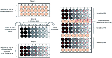
The microdilution method was used to determine the maximum non-toxic concentrations (MNTC) based on international standard methodology M27-A2 (yeast),55 M38-A (filamentous fungi),56 M7–A6 (bacteria),57 M24-A2 (actinobacteria),58 M45 A (fastidious bacteria)59 and CLSI/NCCLS (Clinical and Laboratory Standards Institute). The MNTC assay was undertaken in 96-well plates by eight successive dilutions (1[thin space (1/6-em)]:[thin space (1/6-em)]2) of a stock solution using 500 mg mL−1 of ionic liquids in 100 μL of culture medium: Müeller Hinton for bacteria and actinobacteria, MRS broth for lactobacilli and RPMI-MOPS (pH 7.2) for fungi. Wells were inoculated with 10 μL of bacterial suspension or 100 μL of fungal suspension. The microplates were incubated overnight at 37 °C for bacteria, at room temperature (28–30 °C) for actinobacteria, and at room temperature over 48 h for fungi. Pure medium was used as the negative control, and positive controls comprised inoculated growth medium. After incubation, the determination of MNTC was based on visual growth (turbidity) confirmed by 30 μL of resazurin (Sigma-Aldrich) added aseptically to the microplate wells and incubated at 37 °C for 1 h. MNTC values were expressed as the average of at least two independent assays performed in triplicate (Fig. 1).
image file: c4ra05295a-f1.tif
Fig. 1 Scheme of MNTC determination assay.

The hydrophobic ionic liquids ([C2mim][NTF2], [C4mim][NTF2], [Ch][NTF2]) were diluted in dimethyl sulfoxide (DMSO) to allow the pipetting of a homogenous aliquot from stock solution. The highest concentration of DMSO used in the first well of the microplate was 4% for bacteria and 2% for fungi. In order to assure the non-influence of this solvent on the obtained results, a control was set up with the concentration of DMSO used in the assays; the microorganisms showed growth in all cases under the applied concentrations.

Conclusions

By using the microdilution test, it was possible qualitatively and quantitatively determine the toxicity of some ionic liquids toward a range of microorganisms, some of them tested for the first time. Choline cation proved promising for use in processes involving microorganisms since it exhibited low toxicity to most microorganisms. The filamentous fungi tested herein were generally the most tolerant microorganisms to ionic liquids, followed by the yeast Yarrowia lipolytica. The anion bis(trifluoromethylsulfonyl)imide [NTf2] was quite toxic for the tested microorganisms. Thus, the study of other anions to obtain hydrophobic ionic liquids is needed to utilize biphasic systems in fermentation applications.

This study has revealed the promising application of biocompatible ionic liquids in processes involving microorganisms in biochemical production.

Acknowledgements

We wish to thank the Brazilian funding agencies CAPES and CNPq for scholarship and Isabel M. Marrucho (ITQBq), Universidade Nova de Lisboa, Portugal, for supplying some ionic liquids.

Notes and references

  1. R. D. Rogers and K. R. Seddon, Science, 2003, 302, 792–793 CrossRef PubMed.
  2. G. Quijano, A. Couvert and A. Amrane, Bioresour. Technol., 2010, 101, 8923 CrossRef CAS PubMed.
  3. Z. Yang and W. Pan, Enzyme Microb. Technol., 2005, 37, 19 CrossRef CAS PubMed.
  4. U. Kragl, M. Eckstein and N. Kaftzik, Curr. Opin. Biotechnol., 2002, 13, 565–571 CrossRef CAS.
  5. H. Pfruender, M. Amidjojo, U. Kragl and D. Weuster-botz, Angew. Chem., Int. Ed., 2004, 43, 4529 CrossRef CAS PubMed.
  6. H. Pfruender, R. Jones and D. Weuster-Botz, J. Biotechnol., 2006, 124, 182 CrossRef CAS PubMed.
  7. H. Pfruender, M. Amidjojo, F. Hang and D. Weuster-botz, Appl. Microbiol. Biotechnol., 2005, 67, 619 CrossRef CAS PubMed.
  8. M. Sendovski, N. Nir and A. Fishman, J. Agric. Food Chem., 2010, 58, 2260 CrossRef CAS PubMed.
  9. D. Dennewald, W.-R. Pitner and D. Weuster-Botz, Process Biochem., 2011, 46, 1132 CrossRef CAS PubMed.
  10. S. Bräutigam, S. Bringer-Meyer and D. Weuster-Botz, Tetrahedron: Asymmetry, 2007, 18, 1883 CrossRef PubMed.
  11. J. Pernak, J. Rogoza and I. Mirska, Eur. J. Med. Chem., 2001, 36, 313 CrossRef CAS.
  12. J. Pernak, K. Sobaszkiewicz and I. Mirska, Green Chem., 2003, 5, 52 RSC.
  13. J. Pernak, I. Goc and I. Mirska, Green Chem., 2004, 6, 323 RSC.
  14. J. Pernak and J. Feder-Kubis, Chem.–Eur. J., 2005, 11, 4441 CrossRef CAS PubMed.
  15. J. Pernak, M. Smiglak, S. T. Griffin, W. L. Hough, T. B. Wilson, A. Pernak, J. Zabielska-Matejuk, A. Fojutowski, K. Kita and R. D. Rogers, Green Chem., 2006, 8, 798 RSC.
  16. J. Pernak, A. Syguda, I. Mirska, A. Pernak, J. Nawrot, A. Pradzyńska, S. T. Griffin and R. D. Rogers, Chem.–Eur. J., 2007, 13, 6817 CrossRef CAS PubMed.
  17. J. Ranke, K. Mölter, F. Stock, U. Bottin-Weber, J. Poczobutt, J. Hoffmann, B. Ondruschka, J. Filser and B. Jastorff, Ecotoxicol. Environ. Saf., 2004, 58, 396 CrossRef CAS.
  18. K. M. Docherty and C. F. Kulpa Jr, Green Chem., 2005, 7, 185 RSC.
  19. A. Cornellas, L. Perez, F. Comelles, I. Ribosa, A. Manresa and M. T. Garcia, J. Colloid Interface Sci., 2011, 355, 164 CrossRef CAS PubMed.
  20. R. P. Swatloski, J. D. Holbrey, S. B. Memon, G. A. Caldwell, K. A. Caldwell and R. D. Rogers, Chem. Commun., 2004, 668–669 RSC.
  21. R. J. Bernot, E. E. Kennedy and G. a. Lamberti, Environ. Toxicol. Chem., 2005, 24, 1759 CrossRef CAS.
  22. T. P. T. Pham, C.-W. Cho, J. Min and Y.-S. Yeoung, J. Biosci. Bioeng., 2008, 105, 425 CrossRef CAS PubMed.
  23. J. Pernak and P. Chwała, Eur. J. Med. Chem., 2003, 38, 1035 CrossRef CAS PubMed.
  24. M. Petkovic, K. R. Seddon, L. P. N. Rebelo and C. Silva Pereira, Chem. Soc. Rev., 2011, 40, 1383–1403 RSC.
  25. M. Petkovic, J. L. Ferguson, H. Q. N. Gunaratne, R. Ferreira, M. C. Leit, K. R. Seddon, N. Rebelo and C. Silva, Green Chem., 2010, 12, 643–649 RSC.
  26. W. L. Hough-Troutman, M. Smiglak, S. Griffin, W. Matthew Reichert, I. Mirska, J. Jodynis-Liebert, T. Adamska, J. Nawrot, M. Stasiewicz, R. D. Rogers and J. Pernak, New J. Chem., 2009, 33, 26 RSC.
  27. P. Nockemann, B. Thijs, K. Driesen, C. R. Janssen, K. Van Hecke, L. Van Meervelt, S. Kossmann, B. Kirchner and K. Binnemans, J. Phys. Chem. B, 2007, 111, 5254–5263 CrossRef CAS PubMed.
  28. M. Stasiewicz, E. Mulkiewicz, R. Tomczak-Wandzel, J. Kumirska, E. M. Siedlecka, M. Gołebiowski, J. Gajdus, M. Czerwicka and P. Stepnowski, Ecotoxicol. Environ. Saf., 2008, 71, 157–165 CrossRef CAS PubMed.
  29. Y. Fukaya, Y. Iizuka, K. Sekikawa and H. Ohno, Green Chem., 2007, 9, 1155 RSC.
  30. M. Rebros, H. Q. N. Gunaratne, J. Ferguson, R. Seddon and G. Stephens, Green Chem., 2009, 11, 402 RSC.
  31. N. Wood, J. L. Ferguson, H. Q. N. Gunaratne, K. R. Seddon and G. M. Stephens, Green Chem., 2011, 13, 1843 RSC.
  32. J. Restolho, J. L. Mata and B. Saramago, Fluid Phase Equilib., 2012, 322–323, 142–147 CrossRef CAS PubMed.
  33. A. P. Abbott, G. Capper, D. L. Davies, R. K. Rasheed and V. Tambyrajah, Chem. Commun., 2003, 70–71 RSC.
  34. D. J. Couling, R. J. Bernot, K. M. Docherty, J. K. Dixon and E. J. Maginn, Green Chem., 2006, 8, 82 RSC.
  35. S. Stolte, J. Arning, U. Bottin-Weber, A. Müller, W.-R. Pitner, U. Welz-Biermann, B. Jastorff and J. Ranke, Green Chem., 2007, 9, 760 RSC.
  36. P. R. Massaguer, Microbiologia dos Processos Alimentares, Varela Editora e Livraria Ltda, São Paulo, 1st edn, 2005 Search PubMed.
  37. J. Ruiz-Herrera, Fungal Cell Wall: Structure, Synthesis, and Assembly, CRC Press, Boca Raton, 2nd edn, 2012 Search PubMed.
  38. T. H. Nguyen, G. H. Fleet and P. L. Rogers, Appl. Microbiol. Biotechnol., 1998, 50, 206 CrossRef CAS.
  39. R. Vega and A. Domínguez, Arch. Microbiol., 1986, 144, 124 CrossRef CAS.
  40. H. J. Blumenthal and S. Roseman, J. Bacteriol., 1957, 74, 222 CAS.
  41. Y. Wu, T. Sasaki, S. Irie and K. Sakurai, Polymer, 2008, 49, 2321 CrossRef CAS PubMed.
  42. K. Prasad, M. Murakami, Y. Kaneko, A. Takada, Y. Nakamura and J. Kadokawa, Int. J. Biol. Macromol., 2009, 45, 221 CrossRef CAS PubMed.
  43. T. Setoguchi, T. Kato, K. Yamamoto and J. Kadokawa, Int. J. Biol. Macromol., 2012, 50, 861 CrossRef CAS PubMed.
  44. Y. Qin, X. Lu, N. Sun and R. D. Rogers, Green Chem., 2010, 12, 968 RSC.
  45. M. Ouellet, S. Datta, D. C. Dibble, P. R. Tamrakar, P. I. Benke, C. Li, S. Singh, K. L. Sale, P. D. Adams, J. D. Keasling, B. A. Simmons, B. M. Holmes and A. Mukhopadhyay, Green Chem., 2011, 13, 2743 RSC.
  46. J. Pernak, J. Kalewska, H. Ksycinska and J. Cybulski, Eur. J. Med. Chem., 2001, 36, 899 CrossRef CAS.
  47. Y. Sardessai and S. Bhosle, Res. Microbiol., 2002, 153, 263–268 CrossRef CAS.
  48. R. E. W. Hancock, Clin. Infect. Dis., 1998, 27, 93–99 Search PubMed.
  49. H. Nikaido and R. E. W. Hancock, in The Bacteria, ed. J. Sokatch, Academic Press, Orlando, Florida, 1986, pp. 145–193 Search PubMed.
  50. A. Romero, A. Santos, J. Tojo and A. Rodríguez, J. Hazard. Mater., 2008, 151, 268–273 CrossRef CAS PubMed.
  51. M. Petkovic, J. Ferguson, A. Bohn, J. Trindade, I. Martins, M. B. Carvalho, M. C. Leit, C. Rodrigues, H. Garcia, R. Ferreira, K. R. Seddon, L. P. N. Rebelo and C. S. Pereira, Green Chem., 2009, 11, 889–894 RSC.
  52. H. Wang, S. V. Malhotra and A. J. Francis, Chemosphere, 2011, 82, 1597 CrossRef CAS PubMed.
  53. S. P. M. Ventura, A. M. M. Gonçalves, T. Sintra, J. L. Pereira, F. Gonçalves and J. a. P. Coutinho, Ecotoxicology, 2013, 22, 1–12 CrossRef CAS PubMed.
  54. E. B. Shirling and D. Gottlieb, Int. J. Syst. Bacteriol., 1966, 16, 313–340 CrossRef PubMed.
  55. CLSI, Reference method for broth dilution antifungal susceptibility testing of yeasts. Approved standard M27-A2, Wayne, Pensylvania, USA, 2nd edn, 2002, vol. 22 Search PubMed.
  56. CLSI, Reference method for broth dilution antifungal susceptibility testing of yeasts. Approved standard, M38-A, Wayne, Pensylvania, USA, 2002, vol. 22 Search PubMed.
  57. CLSI, Methods for Dilution Antimicrobial Susceptibility Tests for Bacteria That Grow Aerobically; Approved Standard, M7–A6, Wayne, Pensylvania, USA, 6th edn, 2003, vol. 23 Search PubMed.
  58. CLSI, Susceptibility Testing of Mycobacteria, Nocardiae, and Other Aerobic Actinomycetes; Approved Standard, M24-A, Wayne, Pensylvania, USA, 2003, vol. 23 Search PubMed.
  59. CLSI, Methods for Antimicrobial Dilution and Disk Susceptibility Testing of Infrequently Isolated or Fastidious Bacteria; Approved Guideline, M45-A, Wayne, Pensylvania, USA, 2006 Search PubMed.

This journal is © The Royal Society of Chemistry 2014
Click here to see how this site uses Cookies. View our privacy policy here.