In vivo cytotoxicity of MgO-doped nanobioactive glass particles and their anticorrosive coating on Ti–6Al–4V and SS304 implants for high load-bearing applications

M. Prabhua, R. Suriyaprabhaa, V. Rajendran*a, P. Kulandaivelub and S. Valiyaveettilc
aCentre for Nano Science and Technology, K. S. Rangasamy College of Technology, Tiruchengode-637215, Tamil Nadu, India. E-mail: veerajendran@gmail.com; Fax: +91-4288-274880 (direct), 274860; Tel: +91-4288-274741-4, 274880
bDepartment of Mechanical Engineering, K. S. Rangasamy College of Technology, Namakkal (Dt.), Tiruchengode-637215, Tamil Nadu, India
cDepartment of Chemistry, Faculty of Science, National University of Singapore, 3 Science Drive 3, 117543, Singapore

Received 23rd May 2014 , Accepted 9th September 2014

First published on 9th September 2014


Abstract

In this study, magnesium-doped nanobioactive glass (NBG) composites (SiO2–CaO–P2O5–MgO) were prepared by simple sol–gel method, which were characterized and coated on Ti alloy (Ti–6Al–4V) and stainless steel (SS304) implants by spin-coating technique. The prepared nanocomposite shows amorphous nature and spherical morphology with particle size of less than 100 nm. The adsorption and desorption isotherms showed the prepared nanocomposites to be in mesoporous range with a specific surface area of 104.1 m2 g−1. The coated implant was found to have a uniform structure without any cracks and pores. Magnesium-doped NBG-coated Ti implants show high corrosion resistance and hardness. In addition, formation of bone-like apatite layer on the coated implant was found to be high in magnesium-doped NBG particles. In addition, in vivo toxicity of the glasses was studied in zebrafish (Danio rerio) embryos, and the results confirmed significant toxicity at higher concentration. Hence, magnesium-doped NBG-coated implant is found to be a potential nanocomposite for high load-bearing applications with better anticorrosive property and long-term stability.

Introduction

Metallic implants such as pure titanium (Ti), titanium alloys (Ti–6Al–4V), and stainless steels (SS 316L) are widely for orthopedic and high load bearing applications.1–5 When an implant is inserted inside the human body, it may weaken the surrounding bone and cause failure between bone and implants due to microbial infections.4,5 To resolve this problem, most of the researchers are interested in developing calcium- and phosphate-based ceramic-coated implant to induce osteoconductive properties along with antimicrobial activity.6–8 A review of previous studies has shown that limited work has been conducted on the fabrication of a composite coating of bioactive glass (BG) and Ti–6Al–4V for biomedical implants.2,9–12 In this regard, many coating methodologies such as plasma spraying,13 pulsed laser deposition,14 and dip-coating1 have been adapted. However, the area of efficiency and durability of coated materials on implant is interesting and still to be explored. Spin coating of porous silicon- and zinc-doped hydroxyapatite (HAp) on SS implants may enhance their adhesiveness and long-term durability,15,16 which are favorable for bone implant applications.

In fact, for improved osteogenic properties, coating of antimicrobial and biocompatible nanobioactive glass (NBG) particles should be used on implants to expand the application of NBG for high load-bearing applications.17 HAp sprayed on the implant surface as a bioactive coating material has few drawbacks in terms of adherence to the metal substrate. For example, HAp coating on titanium implant surfaces may result in rough surface and increase osseointegration, but its adherence to the metal is not perfect.18 Bioglass may be known to act as a better osteoproductive, abrasive, and highly resorbable surface material for implants,17,19 but extensive investigations are required to explore the bioactive glass coating as an active biomaterial on clinical implants.

The NBGs are coated on glass films to explore higher bioactivity.20 However, it is found that chemical composition and microstructure of the glass particles have an important role in implant applications. Hence, the glass composition that possesses optimal physicochemical properties and better in vitro biocompatibility is used for the implant-coating applications. Our earlier investigations on the development of NBG composites prepared with Mg, Ag, and Zr show the in vitro bioactivity of the material for biomedical applications.21,22 Among the glass compositions tested, the one with 10% Mg was found to possess the required mechanical and physicochemical properties. Hence, the coating of Mg-doped NBG particles on implant materials led to desirable biomimetic characteristics for high load-bearing applications.

The screening of the in vivo toxicity of the optimized Mg-doped NBG (SiO2–CaO–P2O5–MgO) particles is mandatory for any implant coating. In this regard, a zebrafish model can be used to analyze the biocompatibility and in vivo cytotoxicity of the prepared glass compositions. As the genotype of zebrafish is closely similar to the human genotype,23 the analysis of toxicity in fish enables us to understand the human health risks of novel biomaterials. In addition, large quantity of zebrafish embryos can be developed rapidly at low cost, which serves as an inexpensive and faster in vivo assay to screen the biocompatibility, pharmacological efficacy, and toxicity of biomaterials.

This study aimed to examine the in vivo cytotoxicity of Mg-doped NBG (SiO2–CaO–P2O5–MgO) particles in zebrafish (Danio rerio) embryos and to coat these particles on implant materials such as Ti alloy (Ti–6Al–4V) and stainless steel (SS304) plates using spin-coating technique. In addition, this study aimed to explore the in vitro bioactivity using simulated body fluid (SBF), and to explore the mechanical and anticorrosive properties using artificial saliva on the coated implants.

Results and discussion

Characterization of prepared Mg-doped NBG particles

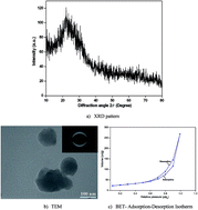
The XRD pattern of the synthesized Mg-doped NBG particles shows amorphous nature as no diffraction peaks are observed except a broad band between 10° and 35° (Fig. 1a). TEM images showed that Mg-doped NBG particles possess uniform spherical morphology with a particle size of 100 nm. However, the selected area electron diffraction pattern (inset in Fig. 1b) confirms the amorphous nature of the prepared Mg-doped NBG particles. The 10-point nitrogen adsorption–desorption isotherms of the prepared Mg-doped NBG particles are given in Fig. 1c. The observed results indicate that prepared particles are highly mesoporous (<50 nm), which follows type IV isotherm with H1 hysteresis loops in the mesoporous range.24 The SSA, average pore diameter, and total pore volume of the Mg-doped NBG particles are, 104.1 m2 g−1, 20.17 nm, and 0.53 cm3 g−1, respectively. Similarly, the base (without Mg) NBG particles also show an amorphous and agglomerated spherical morphology with a particle size in the range from 100 to 150 nm.25
image file: c4ra04892j-f1.tif
Fig. 1 Characterization of MgO-doped bioactive glass nanoparticles.

Characterization of nanocomposites coated on Ti–6Al–4V and SS304 implant

Fig. 2 shows the XRD pattern of pure titanium alloy (TIP), NBG-coated (TINBG), and Mg-doped NBG-coated (TIMgNBG) implant before in vitro study. The characteristic peaks observed at 35.9°, 39.1°, 40.9°, 53.7°, 64.2°, and 71.5° can be assigned, respectively, to (100), (002), (101), (102), (110), and (103) crystalline nature of Ti–6Al–4V (Fig. 2). Similarly, the diffraction peaks observed at 45.3° (111), 75.29° (200), and 51.16° (220) in Fig. 3 correspond to crystalline nature of stainless steel (SS304).
image file: c4ra04892j-f2.tif
Fig. 2 XRD patterns of NBG- and Mg-doped NBG-coated and un-coated titanium alloy (Ti–6Al–4V) implants.

image file: c4ra04892j-f3.tif
Fig. 3 XRD patterns of NBG- and Mg-doped NBG-coated and uncoated stainless steel (SS304) implants.

The amorphous NBG and Mg-doped NBG particles are successfully deposited onto Ti alloy and stainless steel after spin coating, and confirms through HR-SEM analysis (Fig. 4). HR-SEM images and corresponding EDX spectra of TIP, TINBG, and TIMgNBG implants before in vitro study are shown in Fig. 4. The polished TIP shows smooth surface without any scratches (Fig. 4a). The EDX pattern shows 100% purity (Fig. 4a). Similarly, the pure SS304 sample also show plain smooth surface without any impurities (Fig. 5a). The surfaces of the TINBG and TIMgNBG implants prepared using spin-coating technique are shown in Fig. 4b and c. From the observed results it is revealed that the deposition of uniform coating of particles is achieved and shows slight aggregation on the coated substrate which may due to the presence of excess PVP in the nanocomposite. The interface between the coating and alloy is dense, and no cracks are found at the interface between titanium alloy and coating. Controlling the ratio of nanobioactive glass and PVP, one can control the aggregation of bioactive glass particles in PVP matrix. The EDX pattern of TIMgNBG implant confirms the presence of elements such as Ti, Si, Ca, P, and Mg on the implant surface (Fig. 4c).


image file: c4ra04892j-f4.tif
Fig. 4 HR-SEM images of NBG- and Mg-doped NBG-coated and uncoated titanium alloy (Ti–6Al–4V) implants.

image file: c4ra04892j-f5.tif
Fig. 5 HR-SEM images of NBG- and Mg-doped NBG-coated and un-coated stainless steel (SS304) implants.

The surface morphology of the NBG- and Mg-doped NBG-coated SS304 implant also shows uniform coating and smooth surface without any cracks and pores (Fig. 5). These results indicate strong adherence of coating on the implant surface (Ti–6Al–4V and SS304). The surface morphology of the NBG and Mg-doped NBG implants became much smoother because of the PVP composite coating. The EDX pattern of Mg-doped NBG-coated stainless steel implant confirms the presence of elements such as Fe, Cr, Si, Ca, P, and Mg on the coated surface (Fig. 5b and c).

The elemental compositions of the uncoated and coated NBG particles are confirmed by XRF analysis (Table 1). The existence of elements such as Si, Ca, P, and Mg on the implant surface is confirmed in NBG particles and Mg-doped NBG composites. The remaining elements (Ti, V, Al, Fe, Ni, and Cr) are present in the substrate. From the observed results, it is concluded that the developed nanocomposite-coated implant shows negligible impurities. These results are in good agreement with the observed EDX pattern.

Table 1 Elemental composition and mechanical properties of magnesium doped nanobioactive glass coated implant confirmed through XRF analysis and Nano indentation techniques
Sample Element (wt%) Mechanical properties
Ti Al V Fe Cr Ni Si Ca P Mg Hardness (GPa) Young's modulus (GPa)
TIP 88.52 7.46 2.62 1.40 0.12 2.85
TINBG 84.18 2.96 0.17 8.68 2.40 1.59 0.56 15.72
TiMgNBG 82.91 3.50 8.37 3.57 1.41 0.24 1.65 12.78
SSP 70.42 19.54 07.56 01.59 0.18 4.48
SSNBG 56.52 16.03 5.90 12.67 6.63 2.19 0.17 5.37
SSMgNBG 56.66 15.71 6.22 11.18 7.03 2.10 0.94 0.75 4.57


AFM images of the Ti alloy (Ti–6Al–4V) and stainless steel (SS304) surface before and after coatings are given in Fig. 6 and 7. The two-dimensional (2D) and three-dimensional (3D) views of the AFM images of polished Ti alloy and SS304 surface are shown in Fig. 6a and 7a. The AFM images of the composite coated surfaces (Fig. 6a and 7a) show that the surface is uniform with no scratches. Fig. 6b and c and 7b and c show the 2D and 3D views of Ti alloy and SS304 sample after coating of NBG- and Mg-doped NBG-coated particles. Further, it confirms that an effective coating is achieved by spin-coating method. In addition, the surface of the implant subjected to a constant tip loading is seen in Fig. 6 and 7. It is reported that bioactive glass coating on metallic implant enhances the osteoconductivity and cellular differentiation.26 However, glass composition with enhanced mechanical properties promotes the stability of the implant for high load bearing applications.


image file: c4ra04892j-f6.tif
Fig. 6 AFM images of NBG- and Mg-doped NBG-coated and un-coated Ti–6Al–4V implants.

image file: c4ra04892j-f7.tif
Fig. 7 AFM images of NBG- and Mg-doped NBG-coated and uncoated stainless steel (SS304) implants.

Fig. 8 shows the potentiodynamic polarization curve of NBG particles coated on Ti–6Al–4V and SS304 to explore the corrosion resistance in artificial saliva. Using Tafel extrapolation method,27,28 the corrosion potential (Ecorr), corrosion current density (Icorr), anodic/cathodic Tafel (βα and βc), and polarization resistance (Rp) are obtained from polarization curves (Fig. 8). The values of Ecorr, Icorr, βα, βc, Rp, and corrosion rate per year for NBG- and Mg-doped NBG-coated particles on Ti–6Al–4V and SS304 implants are given in Table 2. TINBG implant shows higher Icorr value of 69.4 μA cm−2 whereas SSNBG implant shows higher Icorr value of 67.3 μA cm−2. Similarly, corrosion current densities of TIMgNBG and SSMgNBG implants were 13.5 and 9.55 μA cm−2, respectively. From the observed results, higher Icorr values are evident for TINBG and SSNBG when compared with those of TIMgNBG and SSMgNBG implants.


image file: c4ra04892j-f8.tif
Fig. 8 Potentiodynamic polarization curve of the NBG- and Mg-doped NBG-coated Ti–6Al–4V and SS304 implants in artificial saliva.
Table 2 Corrosion parameters from the potentiodynamic polarization test for NBG and Mg-NBG coated sample in artificial saliva
Sample Ecorr (V) Icorr (μA cm−2) βα (V per decade) βc (V per decade) Polarisation resistance (Rp) (Ω) Corrosion rate (mm per year)
TINBG −0.620 69.4 0.123 0.127 391.86 0.806
TIMgNBG −0.534 13.5 0.098 0.080 1415.5 0.157
SSNBG −0.635 67.3 0.119 0.126 396.6 0.782
SSMgNBG −0.614 9.55 0.038 0.028 754.99 0.111


It suggests that Mg-doped NBG-coated samples are passivated spontaneously with passive current density in an artificial solution. Higher polarization resistances of 1415.5 and 754.99 Ω are observed for TIMgNBG and SSMgNBG implants, respectively. In addition, Mg-doped NBG-coated samples show that the lower corrosion rates for Ti–6Al–4V and SS304 are 0.157 and 0.111 mm per year, respectively. These observed results are in good agreement with already reported studies.28,29 The presence of bioactive glass coating improves corrosion resistance of titanium alloy and stainless steel in artificial saliva solution.29 Finally, it is concluded that the presence of Mg in NBG particles improves corrosion resistance of titanium alloys and stainless steels while coating and they act as a barrier that extends the lifetime of the implant.

The typical load–displacement curves of nanoindentation experiments for uncoated and coated nanocomposites on Ti–6Al–4V and SS304 are shown in Fig. 9. From the obtained curves, the hardness (H) and Young's modulus (E) values are calculated and are given in Table 1. The hardness value of TIP is measured as 0.12 GPa whereas those of TINBG and TIMgNBG implants are measured as 0.56 and 1.65 GPa, respectively. Similarly, Young's moduli of TIP, TINBG, and TIMgNBG implants are found to be 2.85, 15.72, and 12.78 GPa, respectively. In addition, the hardness value of pure SS304 (SSP) is measured as 0.18 GPa whereas those of SSNBG and SSMgNBG implants are measured as 0.17 and 0.75 GPa, respectively.


image file: c4ra04892j-f9.tif
Fig. 9 Typical load–displacement curve of the nanocomposite-coated and uncoated Ti–6Al–4V and SS304 implants.

Moreover, Young's moduli of SSP, SSNBG, and SSMgNBG implants are observed to be 4.48, 5.37, and 4.57 GPa, respectively. From the observed results, it is seen that TIMgNBG and SSMgNBG implants show an increase in hardness and Young's moduli when compared with uncoated implants. The hardness of the materials determines the interfacial and intrinsic bonding of the materials for in vitro biological behavior.30 Generally, the surface roughness does not influence the immediate nucleation of calcium phosphate deposits onto Ti6Al4V surfaces.31 However, further growth and mechanical attachment of the final Ca–P deposition is favoured by a rougher topography rather than a smoother topography. Therefore, the Mg-doped NBG coated implants with an improved anticorrosion property along with mechanical stability are used for high load bearing implant applications in biomedical field.

In vitro bioactivity evaluation

The XRD patterns of the NBG- and Mg-doped NBG-coated particles on Ti–6Al–4V and SS304 substrates after 21 days of immersion in SBF are shown in Fig. 10. It is observed that after immersion, sharp diffraction peaks observed at 31.7° (JCPDS file no. 090432) correspond to formation of the apatite layers on the surface of the coated TIMgNBG and SSMgNBG implants. However, the diffraction peaks of apatite deposition on TINBG and SSNBG implants are found to be very weak.
image file: c4ra04892j-f10.tif
Fig. 10 XRD patterns of the nanocomposites-coated Ti–6Al–4V and SS304 substrates after 21 days of immersion in SBF.

The apatite layer formation on the prepared implant surface after immersion in SBF for 21 days is further confirmed by HR-SEM analysis and the results are shown in Fig. 11. From the observed results, it is seen that Mg-doped NBG coating provides a suitable surface for bone-like apatite formation. It is observed that the uncoated implant immersed in SBF is different from the coated implants before and after immersion in SBF. The image obtained after 21 days of SBF immersion of coated implant indicates the formation of white globular apatite-like structure on the surface of both Ti- and SS-coated implants. Polyvinylpyrrolidone (PVP) is known for its water-soluble and biocompatible polymer properties, used in the present study to induce the formation of apatite crystal.32 PVP can effectively combine with a larger amount of Ca2+ through a strong ion-polar interaction due to the presence of C–N and C[double bond, length as m-dash]O. The incorporated Ca2+ ions facilitate to combine with PO43− and CO32− ions in SBF solution, which inturn accelerates the nucleation of HAp crystals on the implant surface.33


image file: c4ra04892j-f11.tif
Fig. 11 HR-SEM images of the nanocomposites-coated Ti–6Al–4V and SS304 substrates after 21 days of immersion in SBF.

From the observed results, it is evident that the developed Mg-doped NBG-coated implants show better bioactivity and improved mechanical properties, which in turn become potential glass compositions for high load-bearing applications with better anticorrosion property and long-term stability. Similarly, previous observations of HAp-coated implants show significant bioactivity.16 However, the HR-SEM analysis of the Mg-doped NBG-coated implant shows better HAp layer formation on the implant surface.

From the in vivo cytotoxicity evaluation results, it is observed that hatching rate and touch response of the zebrafish are similar in control and in the NBG-treated samples (Table 3). No delay in hatching is observed for control and the NBG samples tested at 100 μg mL−1. However, the mortality rate is reduced by 20% in the NBG-treated samples than control while observing under a microscope (Fig. 12). The treatment of metal oxide nanoparticles at a concentration above 50 μg mL−1 causes a decrease in hatching rate as well as an increase in the percentage of mortality.23 The toxic response of MgO-doped NBG particles for in vitro cytotoxicity evaluation against human AGS cell line21 is closely related to that for in vivo analysis in terms of dosage effect. These observations convey that addition of optimized MgO (10%) to glass matrix does not enhance cytotoxicity when compared to base glass even though the addition of nanoparticles at 100 μg mL−1 causes stress to zebrafish, as observed from Fig. 12, due to surface functionalization of particles and their binding affinity for biological molecules.34,35 Thus, rapid in vivo cytotoxicity screening results indirectly point out the benefits associated with humans while applying the MgO-doped glass particles for implant and other clinical applications.

Table 3 In vivo toxicity of zebrafish embryos treated with bioactive glass samplesa
Stages of embryos (no.) Control NBG MgNBG
After 48 h After 72 h After 48 h After 72 h After 48 h After 72 h
a N – normal, Ab – abnormal.
Hatched embryos (N/Ab) 0/0 10/0 3/0 9/0 2/1 1/7
Unhatched embryos (N/Ab) 10/0 0 1/5 0 1/4 0
Dead embryos 0 0 1 1 2 2


image file: c4ra04892j-f12.tif
Fig. 12 In vivo analysis of the prepared nanocomposites using zebrafish (D. rerio) embryos.

Conclusions

Mg-doped NBG particles were prepared by a simple sol–gel process, mixed with PVP and deposited to achieve a uniform coating on a Ti alloy and a stainless steel substrate using spin-coating technique. The in vivo toxicity of Mg-doped NBG particles showed better biocompatibility against zebrafish embryos. Electron microscopic observations of Mg-doped NBG-coated Ti–6Al–4V and SS304 implants indicated uniform coating achieved without cracks or micropores on the surface. Compared with TIP and SS substrate, Mg-doped NBG-coated Ti implants showed an increase in mechanical properties in terms of hardness. In addition, the results of in vitro bioactivity studies gave a better HAp layer formation on the coated Ti–6Al–4V and SS304 implants. The electrochemical polarization of Mg-doped NBG-coated Ti implant led to higher corrosion resistance in artificial saliva when compared with base glass coating. From the observed results, it can be concluded that the prepared Mg-doped NBG-coated implants are better nanocomposites for high load-bearing applications with good anticorrosive property and long-term stability.

Materials and methods

Materials

The implant materials such as Ti–6Al–4V (20 mm × 20 mm × 1.5 mm) and SS304 (20 mm × 20 mm × 1.5 mm) were obtained as per the ASI standard. The reagents such as tetraethyl orthosilicate (Si(OC2H5)4, TEOS; Sigma-Aldrich; min. 99%), triethyl phosphate ((CH3CH2O)3P(O), TEP; HiMedia, Mumbai, India; min. 99.5%), calcium nitrate (Ca(NO3)2·4H2O; Merck; min. 98% GR), magnesium nitrate (MgNO3; Merck; min. 99% GR), 2 N nitric acid (HNO3; Merck; min. 69% GR), ethanol, 1 M ammonia (Merck; min. 25% GR), polyvinyl pyrrolidone (PVP), 10% hydrofluoric acid, and ultrapure water (Arium 611UF; Sartorius AG) were used in this study. All the reagents were used as received without any further purification.

Preparation of Mg-doped NBG particles

Mg-doped NBG particles, namely 58SiO2–23CaO–9P2O5–10MgO, were synthesized using the simple, cost-effective sol–gel method.21,36 Initially, TEOS (14.025 mL) was dissolved in 1[thin space (1/6-em)]:[thin space (1/6-em)]1 ratio of ethanol and ultrapure water. Then 2 N HNO3 (3 mL) was added to the solution under stirring for 30 min at room temperature. After complete hydrolysis of TEOS, TEP (3.32 mL), Ca(NO3)2·4H2O (5.45 g), and Mg(NO3)·6H2O (2.76 g) salts were dissolved separately with 2 mL distilled water. The dissolved solution was added to silica solution at 30 min interval under constant stirring at room temperature. After obtaining the clear solution, 1 M ammonia solution was added drop-wise until the formation of gel, that is, until the solution attains a pH of ∼8.0. Thereafter, the obtained white gel was dried in a hot-air oven at 353 K for 6 h. The dried gel was calcined at 873 K for 2 h in a muffle furnace to remove carbon and nitrate impurities. The calcined samples were ground in a dry ball-mill (PM 100; Retsch Corporation, Germany) at 500 rpm for 1 h to obtain fine Mg-doped NBG particles.

Coating of NBG- and Mg-doped NBG particles on implants

Commercially purchased pure Ti–6Al–4V (20 mm × 20 mm × 1.5 mm) and SS304 (20 mm × 20 mm × 1.5 mm) plates were sequentially polished using silicon carbide papers (220, 400, 600, 1000, and 2000 grit) and then cleaned with double-distilled water, followed by acetone and then with ethanol. Then, the samples were eroded in 10% hydrofluoric acid solution for 30 s and cleaned with distilled water. PVP was used as the binding agent for coating of prepared BG on the implant surface. Ethanol was used as a solvent for dispersion of NBG and PVP in the ratio of 1[thin space (1/6-em)]:[thin space (1/6-em)]3 (wt%). All suspensions were stirred for 30 min and dispersed ultrasonically for another 30 min to ensure a good homogeneous dispersion of the particles. After dispersion, the glass particles were coated on Ti implant (Ti–6Al–4V) and stainless steel (SS304) plates through spin-coating technique using a spin coater (Spin Module SM-180-BT; Sawatec, Ruggell) with a rotating speed of 3000 rpm for 1 min. To enhance the uniformity of the coating, the spin-coating process was repeated three times for both samples. After every coating, the samples were dried in a hot-air oven at 80 °C for 10 min.

Characterization of NBG coating

The structural nature of NBG-coated and uncoated on Ti implant (Ti–6Al–4V) and stainless steel (SS304) plates and the HAp layer formation on implant surface during in vitro studies were characterized by an X-ray diffractometer (X'Pert PRO; PANalytical, Almelo, The Netherlands) with Cu Kα as the radiation (λ = 1.5418 Å) source. The source was operated at 40 kV with 2θ value varying from 10° to 80° at a step size of 0.02° and step duration of 25 s. The specific surface area (SSA) of the prepared Mg-doped NBG particles was measured according to Brunauer–Emmett–Teller (BET) method37 using the BET surface area analyzer (Autosorb AS-1MP; Quantachrome, USA). The sample was degassed for 3 h at 295 °C and physisorption analysis was performed with N2 adsorption measurements at liquid N2 temperature (−196 °C). Ten-point adsorption and desorption branched isotherm were used to find the total pore volume, and average pore diameter of the Mg-doped NBG particles using Barrett–Joyner–Halenda (BJH) method.38 Transmission electron microscope (TEM; CM 200; Philips, USA) was used to characterize particle size and surface morphology of the prepared Mg-doped NBG particles.

Elemental compositions of the NBG-coated and uncoated Ti implant (Ti–6Al–4V) and stainless steel (SS304) plates were measured by X-ray fluorescence (XRF) spectrometer (EDX-720; Shimadzu, Kyoto, Japan). High-resolution scanning electron microscope (HR-SEM) equipped with energy-dispersive X-ray analysis (EDX) (Quanta FEG 200; FEI, The Netherlands) was used to explore the difference in surface morphology and composition of the NBG-coated and uncoated substrates. The substrates were examined at an accelerating voltage of 30 kV. The elemental wt% was calculated using ZAF program of the EDX system. The topography of the NBG-coated and uncoated samples was analyzed through atomic force microscopy (AFM; version 7; Innova, USA) in contact mode using a Si3N4 cantilever with a spring constant of about 34 N m−1 and a resonance frequency of about 200 kHz. Scanning was performed at a scan speed of 0.5 Hz with a resolution of 256 × 256 pixels. The tip loading force was controlled to avoid structural changes in the sample.

The mechanical properties such as Young's modulus and hardness of the NBG-coated and uncoated samples were measured using the quasistatic nanoindentation (TI 700 Ubi; Hysitron, USA) system. The indentation was performed by applying a force in the range of 100 nN to 10 N on the sample using a transducer, and the displacement with respect to depth force was observed. The hardness, H, of the material was measured using the following formula:

 
image file: c4ra04892j-t1.tif(1)
where Pmax is the maximum applied load and A the projected area. The slope (S) of the unloading curve provides a measure of the contact stiffness, which can be used with the contact area to determine the elastic modulus.39 The elastic modulus (Er) of the prepared material was measured using the following formula:
 
image file: c4ra04892j-t2.tif(2)
where S is the slope of the curve and A the selected area of the sample.

Electrochemical corrosion behaviour

The electrochemical corrosion resistance of the NBG-coated Ti implants (Ti–6Al–4V) and stainless steel (SS304) plates was measured using artificial saliva at room temperature. Artificial saliva was prepared with a pH value of 6.8 as mentioned by Gal et al.;27 the composition of the artificial saliva is given in Table 4. A stainless steel and a saturated calomel electrode were used as the counter and the reference electrode, respectively. The sample was taken as working electrode having a test surface area of 1 cm2 for all experiments. Linear polarization measurement was performed using PGSTAT 362N (Autolab, The Netherlands) device in a three-electrode cell with a scan rate of 1 mV s−1. The corrosion potential (Ecorr), corrosion current density (Icorr), and polarization resistance (Rp) were calculated from the Tafel pl.
Table 4 Composition of artificial saliva solution
Compound Concentration (mg L−1) Compound Concentration (mg L−1)
NaCl 125.6 Na2SO4·10H2O 763.2
NH4Cl 178 KSCN 189.2
KCl 963.9 KH2PO4 654.5
NaHCO3 630.8 CaCl2·2H2O 227.8
Urea 200    


In vitro bioactivity study

The in vitro bioactivity of the NBG-coated Ti implant (Ti–6Al–4V) and stainless steel (SS304) was measured using freshly prepared SBF. The SBF was prepared using standard protocol40 wherein the pH value was maintained as 7.4, which was equivalent to that of human blood plasma. Each coated substrate was placed separately in polyurethane bottle containing 50 mL SBF and was then incubated at 37 °C for 21 days. After 21 days of incubation, the coated substrates were removed from the SBF and then washed gently with distilled water. The washed coated substrates were dried at 100 °C for 1 h, which were then used for further characterization studies such as X-ray diffraction (XRD) and SEM to study the HAp layer formation on the coated surface.

In vivo toxicity studies

The suspensions of glass samples were made to study the in vivo toxicity. Base glass and MgO-doped NBG particles were weighed and then suspended in water to prepare a working solution (100 μg mL−1) using egg water (sea salt). Eggs of wild-type zebrafish were collected, washed 2–3 times with water, and then transferred to egg water. Nanotoxicity studies using zebrafish embryos were carried out per the procedure reported by Asharani et al.23 Ten healthy embryos (eight-cell stage) were transferred to each well of 12-well plates. The embryos were treated with 100 μg mL−1 of the glass samples dispersed in egg water and were observed at specific growth stages for various criteria such as mortality, rate and time of hatching, development of organs, touch responses, and edema. The dead embryos were removed immediately from the medium during the observation period and the remaining embryos were counted. The touch response of the larvae was tested by gently touching the sides of the trunk with a smooth pipette tip. At the end of the experiment, the larvae were transferred to a microscope slide and then anesthetized with 0.1% phenoxyethanol. Microscopic observations were performed using a Zeiss Axiovert 200M microscope equipped with Axiocam HRc to determine the mortality of the treated embryos.

Acknowledgements

The authors acknowledge the financial support (SR/S2/CMP-0054/2009 dt. 27.09.2010) provided by the Department of Science and Technology (DST), New Delhi, to carry out this research project.

References

  1. M. H. Fathi and A. Doostmohammadi, J. Mater. Process. Technol., 2009, 209, 1385 CrossRef CAS PubMed.
  2. M. Mehdikhani-Nahrkhalaji, M. H. Fathi, V. Mortazavi, S. B. Mousavi, B. Hashemi-Beni and S. M. Razavi, J. Mater. Sci.: Mater. Med., 2012, 23, 485 CrossRef CAS PubMed.
  3. V. R. Noort, J. Mater. Sci., 1987, 22, 3801 CrossRef.
  4. F. Bir, H. Khireddine, A. Touatib, D. Sidane, S. Yala and H. Oudadesse, Appl. Surf. Sci., 2012, 258, 7021 CrossRef CAS PubMed.
  5. A. Rakngarm and Y. Mutoh, Mater. Sci. Eng., C, 2009, 29, 275 CrossRef CAS PubMed.
  6. X. Bai, S. Sandukas, M. R. Appleford, J. L. Ong and A. Rabiei, Acta Biomater., 2009, 5, 3563 CrossRef CAS PubMed.
  7. X.-H. Xie, X.-W. Yu, S.-X. Zeng, R.-L. Du, Y.-H. Hu, Z. Yuan, E.-Y. Lu, K.-R. Dai and T.-T. Tang, J. Mater. Sci.: Mater. Med., 2010, 21, 2165 CrossRef CAS PubMed.
  8. A. de Carlos, J. P. Borrajo, J. Serra, P. González and B. Léon, J. Mater. Sci.: Mater. Med., 2006, 17, 523 CrossRef CAS PubMed.
  9. N. Lotfibakhshaiesh, D. S. Brauer and R. G. Hill, J. Non-Cryst. Solids, 2010, 356, 2583 CrossRef CAS PubMed.
  10. S. Bharati, C. Soundrapandian, D. Basu and S. Datta, J. Eur. Ceram. Soc., 2009, 29, 2527 CrossRef CAS PubMed.
  11. L. Floroian, F. Sima, M. Florescu, M. Badea, A. C. Popescu, N. Serban and I. N. Mihailescu, J. Electroanal. Chem., 2010, 648, 111 CrossRef CAS PubMed.
  12. L. Floroian, M. Florescu, F. Sima, G. Popescu-Pelin, C. Ristoscu and I. N. Mihailescu, Mater. Sci. Eng., C, 2012, 32, 1152 CrossRef CAS PubMed.
  13. X. Liu, S. Tao and C. Ding, Biomaterials, 2002, 23, 963 CrossRef CAS.
  14. L. D'Alessio, R. Teghil, M. Zaccagnino, I. Zaccardo, D. Ferro and V. Marotta, Appl. Surf. Sci., 1999, 138–139, 527 CrossRef.
  15. S. Sutha, G. Karunakaran and V. Rajendran, Ceram. Int., 2013, 39, 5205 CrossRef CAS PubMed.
  16. S. Sutha, K. Kavitha, G. Karunakaran and V. Rajendran, Mater. Sci. Eng., C, 2013, 33, 4046 CrossRef CAS PubMed.
  17. N. Drnovšek, S. Novak, U. Dragin, M. Čeh, M. Gorenšek and M. Gradišar, Int. Orthop., 2012, 36, 1739 CrossRef PubMed.
  18. R. Y. Whitehead, W. R. Lacefield and L. C. Lucas, J. Biomed. Mater. Res., 1993, 27, 1501 CrossRef CAS PubMed.
  19. G. Koller, R. Cook, I. Thompson, T. Watson and L. DiSilvio, J. Mater. Sci.: Mater. Med., 2007, 18, 2291 CrossRef CAS PubMed.
  20. Z. Ma, H. Ji, X. Hu, Y. Teng, G. Zhao, L. Mo, X. Zhao, W. Chen, J. Qiu and M. Zhang, Appl. Surf. Sci., 2013, 284, 738 CrossRef CAS PubMed.
  21. M. Prabhu, K. Kavitha, P. Manivasakan, V. Rajendran and P. Kulandaivelu, Ceram. Int., 2013, 39, 1683 CrossRef CAS PubMed.
  22. M. Prabhu, K. Kavitha, R. Suriyaprabha, P. Manivasakan, V. Rajendran and P. Kulandaivelu, J. Nanosci. Nanotechnol., 2013, 13, 5327 CrossRef CAS PubMed.
  23. P. V. Asharani, Y. Lianwu, Z. Gong and S. Valiyaveettil, Nanotoxicology, 2011, 5(1), 43 CrossRef CAS PubMed.
  24. J. H. De Boer, “The Shapes of Capillaries” in the structure and properties of porous materials, in The Proceedings of the Tenth Synbosium, Colston Research Society, University of Bristol, ed. D. H. Everett and F. S. Stone, Butterworths Science Publications, London, 1958, p. 68 Search PubMed.
  25. B. Saravanakumar, M. Prabhu and V. Rajendran, Int. J. Appl. Ceram. Technol., 2013 DOI:10.1111/ijac.12124.
  26. R. Gupta and A. Kumar, Biomed. Mater., 2008, 3, 034005 CrossRef PubMed.
  27. J.-Y. Gal, Y. Fovet and M. Adib-Yadzi, Talanta, 2001, 53, 1103 CrossRef CAS.
  28. I. Gurappa, Surf. Coat. Technol., 2002, 161, 70 CrossRef CAS.
  29. M. Mehdipour, A. Afshar and M. Mohebali, Appl. Surf. Sci., 2012, 258, 9832 CrossRef CAS PubMed.
  30. J. M. Gomez-Vega, E. Saiz, A. P. Tomsia, G. W. Marshall and S. J. Marshall, Biomaterials, 2000, 21, 105 CrossRef CAS.
  31. A. Arvidsson, V. Franke-Stenport, M. Andersson, P. Kjellin, Y. T. Sul and A. Wennerberg, J. Mater. Sci.: Mater. Med., 2007, 18, 1945 CrossRef CAS PubMed.
  32. I. S. Elashmawi and H. E. Abdel Baieth, Curr. Appl. Phys., 2012, 11, 141 CrossRef PubMed.
  33. N. Yin, S. Y. Chen, Y. Ouyang, L. Tang, J. X. Yang and H. P. Wang, Progress in Natural Science: Materials International, 2011, 21, 472 CrossRef.
  34. T. Cedervall, I. Lynch, S. Lindman, T. Berggard, E. Thulin, H. Nilsson, K. A. Dawson and S. Linse, Proc. Natl. Acad. Sci. U. S. A., 2007, 104, 2050 CrossRef CAS PubMed.
  35. A. Villanueva, M. Canete, A. G. Roca, M. Calero, S. Veintemillas-Verdaguer, C. J. Serna, M. P. Morales and R. Miranda, Nanotechnology, 2009, 20, 115103 CrossRef PubMed.
  36. W. Xia and J. Chang, Mater. Lett., 2007, 61, 3251 CrossRef CAS PubMed.
  37. S. Brunauer, P. H. Emmett and E. Teller, J. Am. Chem. Soc., 1938, 60, 309 CrossRef CAS.
  38. E. P. Barrett, L. G. Joyner and P. P. Halenda, J. Am. Chem. Soc., 1951, 73, 373 CrossRef CAS.
  39. H. H. Xu, D. T. Smith and C. G. Simon, Biomaterials, 2004, 25, 4615 CrossRef CAS PubMed.
  40. T. Kokubo and H. Takadama, Biomaterials, 2006, 27, 2907 CrossRef CAS PubMed.

This journal is © The Royal Society of Chemistry 2014
Click here to see how this site uses Cookies. View our privacy policy here.