A “clicked” porphyrin cage with high binding affinity towards fullerenes†
Jianhong Zhangab,
Xiaoyan Zhengc,
Runsheng Jiangab,
Yanwen Yuab,
Yongjun Li*a,
Huibiao Liua,
Qikai Lid,
Zhigang Shuaic and
Yuliang Li*a
aBeijing National Laboratory for Molecular Science, Key Laboratory of Organic Solids, Institute of Chemistry, Chinese Academy of Sciences, Beijing 100190, P. R. China. E-mail: liyj@iccas.ac.cn; ylli@iccas.ac.cn; Tel: +086-010-62587552
bUniversity of Chinese Academy of Sciences, Beijing 100190, P. R. China
cDepartment of Chemistry, Tsinghua University, Beijing 100084, P. R. China
dSchool of Materials Science & Engineering, Tsinghua University, Beijing 100084, P. R. China
First published on 4th June 2014
Abstract
A cage-structured receptor was synthesized in a facile “clicked” way and showed high affinity for fullerenes and differentiated rates of binding to C60 and C70.
Since the group of Ringsdorf reported the first purposely designed hosts for fullerenes, which consisted of aza-crown ethers carrying suitable alkyl chains on the nitrogens,1 the design and synthesis of hosts for trapping fullerenes have attracted more and more attention due to their potential application in the extraction, solubilization and chemical modification of fullerenes,2 light-harvesting devices,3 and molecular conductors or magnets.4 Tetrathiafulvalenes (TTFs), calixarenes, cyclotriveratrylenes (CTVs) and cyclodextrins as electron donors have been utilized for the design of fullerene receptors.5 In the past decade, many kinds of excellent host molecules incorporating porphyrin blocks have been synthesized successfully.6–9 Recently, cyclic receptors with more than two porphyrin units have been reported.10–12 Compared to the synthesis of cyclic compounds, the organic cage compounds formed with only covalent bonds are relatively rare because the synthesis of most cage compounds requires multiple steps and often has low overall yields. To date, the application of dynamic covalent chemistry makes the synthesis of cage compounds successful in fewer steps and usually higher yields.13–15 Among these fullerene receptors, high and differentiated binding affinity was definitely achieved.16 We report herein a cage-structured receptor synthesized via a facile “click” approach that is easier to synthesize and modify and showed a high affinity for fullerenes (Fig. 1).
 | ||
| Fig. 1 Top: schematic illustration of porphyrin cage with different binding rates for fullerenes; bottom: synthesis of the porphyrin 2: (i) CuI, DBU, toluene, pseudo high-dilution condition, dropwise, 75 °C, 24 h, yield: 33%. | ||
Previously, we have reported that the porphyrin cage 1 is a good receptor for recognizing azide anions.17 However, the cavity of the porphyrin cage 1 (the distance between two porphyrin panels is 7.946 Å) is not large enough to accommodate fullerenes. It is possible to change the length of the linkers to adjust the porphyrin–porphyrin distance of the porphyrin cage to accommodate fullerenes. The larger porphyrin cage 2 can be synthesized directly from two readily accessible porphyrin-based precursors, 3 and 4, in one step using a CuAAC click reaction (Fig. 1, bottom). The 1H NMR spectrum of the porphyrin cage 2 confirms its C4ν symmetry. Its purity and identity were established by 1H NMR, MALDI-TOF MS and UV-vis spectroscopy (see the ESI†).
The first indication of the ability of the porphyrin cage 2 to bind to fullerenes came from the MALDI-TOF spectra. When 1![[thin space (1/6-em)]](https://www.rsc.org/images/entities/char_2009.gif) :1 mixtures of the porphyrin cage 2 and either C60 or C70 were analyzed, peaks at m/z 2633.2 and 2753.1, corresponding to C60@2 (calcd 2633.94) and C70@2 (calcd 2753.85), respectively, were clearly observed (shown in Fig. S1 and 2†). No peaks corresponding to aggregates of other stoichiometries were found.
:1 mixtures of the porphyrin cage 2 and either C60 or C70 were analyzed, peaks at m/z 2633.2 and 2753.1, corresponding to C60@2 (calcd 2633.94) and C70@2 (calcd 2753.85), respectively, were clearly observed (shown in Fig. S1 and 2†). No peaks corresponding to aggregates of other stoichiometries were found.
The second point of evidence in support of the formation of C60@2 and C70@2 complexes was obtained from the analysis of their 1H NMR spectra. We briefly studied the interaction of the porphyrin cage 2 with C60 and C70 by means of 1H NMR titration experiments (Fig. 2). Addition of approximately 0.1 equiv. of C60 in CS2 to a 1,2-dichloroethane-d4 solution of the cage 2 resulted in signal splitting of cage 2. This new set of signals was assigned to the C60@2 complex. With the addition of increased amounts of C60, the signals of the cage 2 decreased continuously, while a new set of signals increased correspondingly (Fig. 2a).
 | ||
| Fig. 2 (a) Partial 1H NMR spectrum of cage 2 in 1,2-dichloroethane-d4 at 298 K upon titrational addition of C60 in CS2. (b) Partial 1H NMR spectrum of cage 2 in 1,2-dichloroethane-d4 + C70 in CS2 (1:1) at 298 K with time as the basis. | ||
Compared to the interaction of C60 with cage 2, the interaction was somewhat different between C70 and 2. When a CS2 solution of C60 was added, C60 entered the cavity so quickly that signal splitting was observed immediately and the phenomenon did not change with additional time. However, when a CS2 solution of C70 was added, the signal splitting was almost not observed due to the fact that C70 entered the cavity more slowly than C60, requiring more than 20 minutes to enter the cavity completely. There are two possible reasons for this observation. One is that C70 has an ellipsoidal shape with a volume larger than that of C60, and second, the skeleton of the porphyrin cage 2 is flexible, requiring time to rearrange in order to accommodate the C70. Therefore, we modified the experimental protocol to add one equivalent of C70 in CS2 all at once, and the 1H NMR data were collected at intervals of 5 minutes. Along with the change in time, the 1H NMR signals assigned to cage 2 became weaker and the 1H NMR signals assigned to C70@2 became correspondingly stronger. Such observations indicated that C70 can enter the cavity of cage 2 at a slower rate (shown in Fig. 2b).
Strong encapsulation of C60 or C70 by the porphyrin receptors was also supported by TLC (straight-phase thin-layer chromatography), which exhibited a new spot for the complexes. The new spot for C60@2 became larger with the increasing addition of a CS2 solution of C60 (shown in Fig. S3a†), and the new spot for C70@2 also became larger with the change in time (shown in Fig. S3b†). The spots for cage 2 were fluorescent, but the spots for C60@2 and C70@2 were not emissive. The TLC results are consistent with the above 1H NMR observations, also suggesting strong electronic interaction between the porphyrin receptors and the fullerene guest.
The energy-minimized structures of cage 2, C60@2, and C70@2 were determined using molecular mechanics calculations. They provided us with a further understanding of the interactions between cage 2 and C60 or C70. All the electronic structure calculations in this work were carried out using the Gaussian 09 package.18 The geometrical structures of the studied cage 2 and C60@2, and C70@2 complexes were optimized fully using the DFT methods at the 6-31G basis set with the exchange potential of Becke19 and correlation function of Lee, Yang, and Parr (B3LYP).20 In the calculated structures for the C60@2 and C70@2 complexes (Fig. 3), the distances from the top panel of the porphyrin cage 2 to the bottom panel increased from 12.78 Å initially to 13.27 Å and 13.48 Å, respectively. Due to the excellent flexibility of cage 2, a more obvious change in the distance between the two panels was observed in the C70@2 complex, and it was about 0.70 Å. The value was about 0.49 Å in the C60@ 2 complex.
 | ||
| Fig. 3 Calculated structures of cage 2, C60@2, and C70@2, ((a), a top view of cage 2; (b), the side view of cage 2; (c), C60@2; (d), C70@2). The distance is shown from the top porphyrin panel to the bottom porphyrin panel. | ||
Upon addition of C60 to the solution of cage 2 in toluene, the Soret band of cage 2 in the UV-vis spectrum shifted markedly from 421 nm to 436 nm, with a clear isosbestic point at 429 nm. The 1:1 complexes in solution were confirmed by Job's plot analysis (see the ESI†). The association constants (Ka) of complexes C60@2 in toluene were then evaluated on the basis of the 1:1 binding mode, and a Ka of 1.7 × 10⁶ M−1 was obtained (shown in Fig. 4a and Fig. S4†). Replacement of C60 with C70 at the same concentration caused no change in the absorption spectrum of cage 2, because the binding affinity of cage 2 to C70 is too strong to be quantified by UV-vis titration. At the investigated concentration, the mixtures of the model porphyrin system (cage 1) and fullerenes unable to form a supramolecular complex did not show fluorescence quenching. Thus, the intermolecular quenching processes can be ignored, and the intramolecular quenching of the porphyrin excited state by the fullerene moiety in the supramolecular complex through electron transfer/energy transfer is the main reason.21 Fluorescence titration22 was used to measure the association constant (Ka) of the complex C70@2 (1 × 10⁸ M−1), which indicated that cage 2 exhibited a stronger affinity to C70 than to C60 (shown in Fig. S5 and 6†). Compared with previous fullerene receptors,11,23 cage 2 was a competition receptor with a dramatic affinity for C70 (Ka = 1 × 10⁸ M−1) and a relatively high affinity for C60 (Ka = 1.7 × 10⁶ M−1), which is easier to be synthesized and modified. Such high binding constants with C60 or C70 are due to the flexible cage structure, which enables fullerene to interact well with the two porphyrin panels of the receptor (Fig. 4).
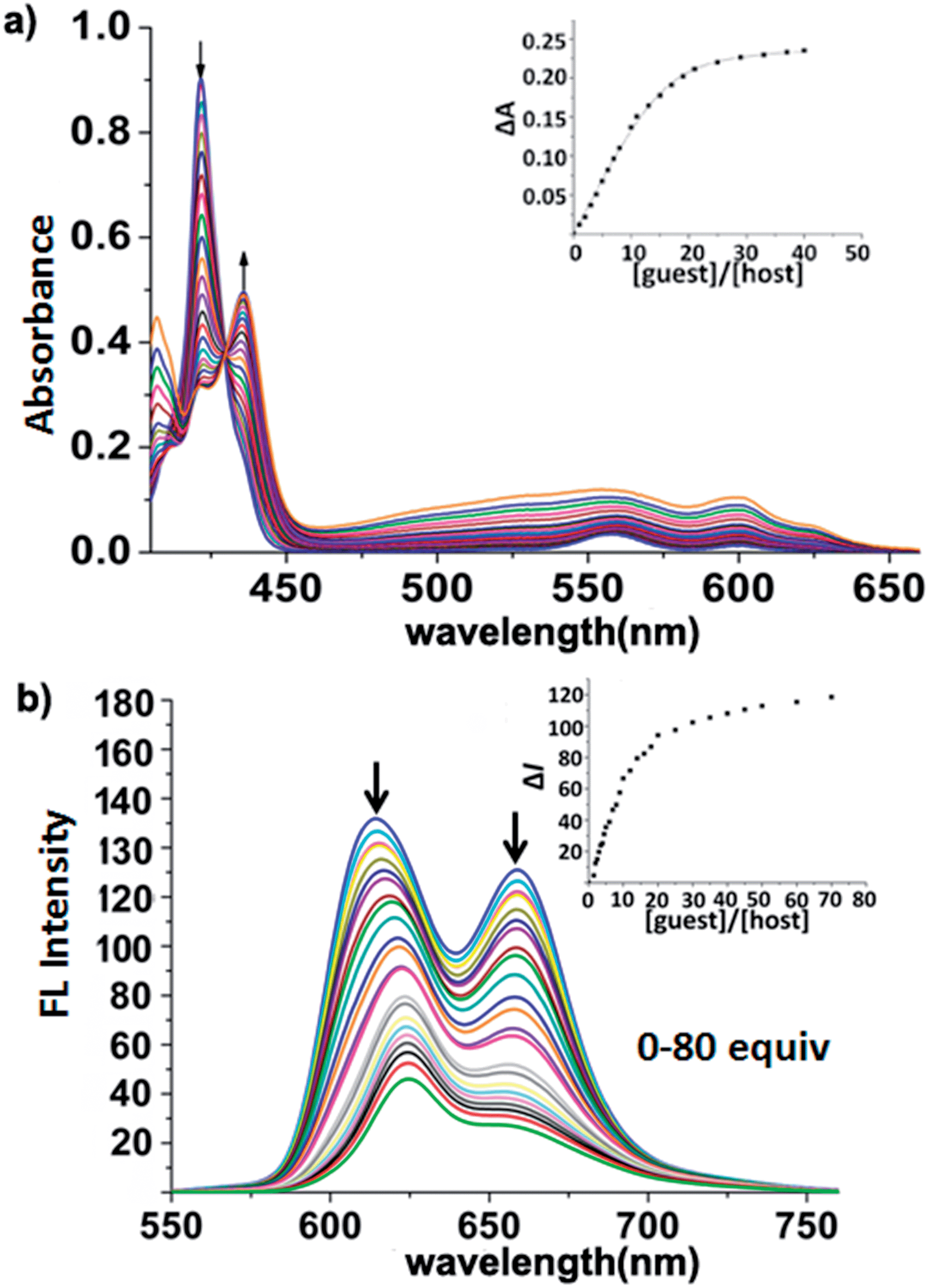 | ||
| Fig. 4 (a) Absorption spectral change of 2 (1 μM) in toluene at 298 K upon titration with C60 (0–40 μM). Inset: plot of ΔA412nm against number of equivalents of C60 added. (b) Fluorescence spectra during the titration of 2 (0.1 μM) with C70 (0–80 equiv.) in toluene at 298 K (λex = 421 nm). Inset: plot of I609nm against number of equivalents of C70 added. | ||
In summary, we successfully synthesized a new zinc porphyrin cage through simple steps, and its flexible skeletons are constructed based on the CuAAC click reaction. We studied the process of interactions between cage 2 and C60 or C70 by 1H NMR titration experiments and TLC analysis. The results demonstrated that cage 2 interacted with C60 quickly and with C70 at a relatively slower rate. The affinities of cage 2 for C60 or C70 are competitive among those of the best-performing fullerene receptors reported so far, but cage 2 is more easily synthesized and modified.
Notes and references
- F. Diederich, J. Effing, U. Jonas, L. Jullien, T. Plesnivy, H. Ringsdorf, C. Thilgen and D. Weinstein, Angew. Chem., Int. Ed., 1992, 31, 1599 CrossRef
.
-
(a) F. Diederich and M. Gomez-Lopez, Chem. Soc. Rev., 1999, 28, 263 RSC
- J. L. Segura and N. Martin, Chem. Soc. Rev., 2000, 29, 13 RSC
- J. P. A. D. J. Williams, A. G. M. Barrett and B. M. Hoffman, J. Chem. Soc., Chem. Commun., 1995, 16, 1703 Search PubMed
-
(a) D. Canevet, M. Gallego, H. Isla, A. de Juan, E. M. Perez and N. Martin, J. Am. Chem. Soc., 2011, 133, 3184 CrossRef CAS PubMed
-
(a) D. Canevet, E. M. Perez and N. Martin, Angew. Chem., Int. Ed., 2011, 50, 9248 CrossRef CAS PubMed
- D. Y. Sun, F. S. Tham, C. A. Reed, L. Chaker and P. D. W. Boyd, J. Am. Chem. Soc., 2002, 124, 6604 CrossRef CAS PubMed
- Y. Kubo, A. Sugasaki, M. Ikeda, K. Sugiyasu, K. Sonoda, A. Ikeda, M. Takeuchi and S. Shinkai, Org. Lett., 2002, 4, 925 CrossRef CAS PubMed
- K. Tashiro, T. Aida, J. Y. Zheng, K. Kinbara, K. Saigo, S. Sakamoto and K. Yamaguchi, J. Am. Chem. Soc., 1999, 121, 9477 CrossRef CAS
- A. R. Mulholland, C. P. Woodward and S. J. Langford, Chem. Commun., 2011, 47, 1494 RSC
- G. Gil-Ramirez, S. D. Karlen, A. Shundo, K. Porfyrakis, Y. Ito, G. A. D. Briggs, J. J. L. Morton and H. L. Anderson, Org. Lett., 2010, 12, 3544 CrossRef CAS PubMed
- J. Song, N. Aratani, H. Shinokubo and A. Osuka, J. Am. Chem. Soc., 2010, 132, 16356 CrossRef CAS PubMed
-
(a) J.-M. Lehn, Chem.–Eur. J., 1999, 5, 2455 CrossRef CAS
- F. Hajjaj, K. Tashiro, H. Nikawa, N. Mizorogi, T. Akasaka, S. Nagase, K. Furukawa, T. Kato and T. Aida, J. Am. Chem. Soc., 2011, 133, 9290 CrossRef CAS PubMed
- C. Zhang, Q. Wang, H. Long and W. Zhang, J. Am. Chem. Soc., 2011, 133, 20995 CrossRef CAS PubMed
- M.-J. Li, C.-H. Huang, C.-C. Lai and S.-H. Chiu, Org. Lett., 2012, 14, 6146 CrossRef CAS PubMed
- J. Zhang, Y. Li, W. Yang, S.-W. Lai, C. Zhou, H. Liu, C.-M. Che and Y. Li, Chem. Commun., 2012, 48, 3602 RSC
- M. J. Frisch, et al., Gaussian 09, Revision D.01, Gaussian, Inc., Wallingford CT, 2009 Search PubMed
- A. D. Becke, J. Chem. Phys., 1993, 98, 5648 CrossRef CAS PubMed
- C. T. Lee, W. T. Yang and R. G. Parr, Phys. Rev. B: Condens. Matter Mater. Phys., 1988, 37, 785 CrossRef CAS
- N. Armaroli, F. Diederich, L. Echegoyen, T. Habicher, L. Flamigni, G. Marconi and J.-F. Nierengarten, New J. Chem., 1999, 23, 77 RSC
- L. Stella, A. L. Capodilupo and M. Bietti, Chem. Commun., 2008, 39, 4744 RSC
- M. Yanagisawa, K. Tashiro, M. Yamasaki and T. Aida, J. Am. Chem. Soc., 2007, 129, 11912 CrossRef CAS PubMed
Footnote |
| † Electronic supplementary information (ESI) available: Synthetic details, UV-vis, and fluorescence spectra. See DOI: 10.1039/c4ra04583a |
| This journal is © The Royal Society of Chemistry 2014 |