DOI:
10.1039/C4RA03207A
(Paper)
RSC Adv., 2014,
4, 49070-49078
A pharmaceutical cocrystal with potential anticancer activity†
Received
9th April 2014
, Accepted 16th September 2014
First published on 18th September 2014
Abstract
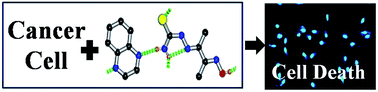
The design of pharmaceutical cocrystals has become a prime thrust of crystal engineering and the pharmaceutical industry in recent times – but the use of pharmaceutical cocrystals as regular drugs is yet to be explored. Quinoxaline acts as a basic skeleton of several potential anticancer drugs. We have successfully cocrystallized quinoxaline with another organic molecule 3-thiosemicarbano-butan-2-one-oxime (TSBO, a virus replication inhibitor) and examined the anticancer activity of the cocrystal. The crystal structure of the cocrystal was determined by single crystal X-diffraction study. According to thermogravimetric study the cocrystal exhibits better thermal stability than quinoxaline. UV-Vis spectroscopic study has shown that in solution state the behavior of the cocrystal and the physical mixture of its components (mixture of quinoxaline and TSBO) are significantly different. The solubility of the cocrystal in distilled water has been found to be 31.9 mg mL−1. The cocrystal exhibits a specific cytotoxic effect on lung cancer cells (A549) at 10−7 M concentration while it shows growth inhibitory effect on normal cells. The detailed mechanistic study of the cytotoxicity of the cocrystal suggests that it follows the mitochondrial mediated cell death pathway through activation of Caspase 9 and Bax. It also shows anticancer activity on breast cancer cells (MCF-7).
1. Introduction
Supramolecular synthesis has evolved as an excellent paradigm for the self-assembly of molecules through non-covalent interactions.1,2 Although these non-covalent interactions are weak in nature, they govern the ultimate solid state structure in most cases. Recently, developing cocrystals of organic molecules by properly tuning the weak interactions has emerged as a thrust area of crystal engineering. A cocrystal can be defined as a supramolecular assembly of two or more different molecular components in the presence or absence of solvent in the solid state.3,4 According to recent reports, cocrystals can be of immense help in designing porous solids for gas storage and separation applications, synthesis of room temperature ferroelectrics, NLO active materials and sensors.5–7 Besides, the physical and chemical properties of drug molecules can be substantially modified through cocrystalazition.8–14 Cocrystallization offers a unique opportunity to modulate physical properties such as solubility along with dissolution rate, stability, bulk density, friability, melting point and hygroscopicity of active pharmaceutical ingredients (APIs) in a controlled manner.15–18
Because of their high intrinsic value in the context of both intellectual property and bioavailability, the design and synthesis of pharmaceutical cocrystals of APIs has attracted much attention from researchers, and as a consequence, a large amount of work and numerous patents on pharmaceutical cocrystals have been reported in recent years.19–23 Researchers have mainly focused on the study of crystal structure, supramolecular structure, solubility, melting point etc.15–18 But, to date, there have been very few studies on the biological activities of cocrystals. Recently, Anand and co-workers cocrystallized quercetin (a plant-derived flavonoid, having anticancer activity) with anti-diabetic agents such as metformin or tolazamide and suggested that the combination drug has different physical properties and biological activity than the individual components.24 Brader and co-workers cocrystallized insulin, a peptide hormone used for the treatment of diabetes, with lipophilically modified, closely related insulin analogue octanoyl-Ne-LysB29-human insulin and found that the activity of the cocrystal is comparable to that of the parent insulin and it can be delivered in a controlled way.25 Solvates (including hydrates) of APIs are well-accepted as drugs in the market, thus there is a great prospect for pharmaceutical cocrystals to be used as regular drugs. Therefore, the use of such cocrystals as pharmaceutical products is a strong challenge to both crystal engineering and the pharmaceutical industry.
Today, cancer is still a threat to human civilization being the second leading cause of death worldwide. The disease transforms normal cells into malignant cells which, in turn, tend to damage the surrounding tissues and organs. Therefore, research on cytotoxic drugs is a challenging area for scientists. Internationally accepted treatments of cancer such as chemotherapy and radiotherapy, though effective on malignant cells, also cause serious side-effects by damaging normal cells.26–29 It is imperative to develop our knowledge about the mechanistic mode of the cytotoxicity of a cancer drug in order to modify it in a controlled manner so that the molecule acts specifically on malignant cells.
Quinoxaline is a simple heterocyclic compound, consisting of a pyrazine ring and a benzene ring. Due to the presence of imino-N atoms, quinoxaline may participate in cocrystallization through X–H⋯N (X = O, N etc.) hydrogen bonding interactions with other molecules containing functional groups with donor hydrogen atoms such as carboxylic acids, amides, diols etc. Quinoxaline acts as a basic skeleton of several potential anticancer drugs.30,31 Owing to their structural diversity, quinoxaline derivatives offer improved potency and lower toxicity.30,31 However, to the best of our knowledge, no report on the anticancer activity of pharmaceutical cocrystals is available in the literature to date. We present here for the first time the specific cytotoxic effect of a quinoxaline-based pharmaceutical cocrystal on lung cancer cells along with its structural property. The present cocrystal has been synthesized by using quinoxaline (API) and 3-thiosemicarbano-butan-2-one-oxime (TSBO) (cocrystallizing agent) by refluxing method. It may be noted that thiosemicarbazone derivatives are potent inhibitors of virus (viz. herpes simplex virus) replication.32 This is a hydrated cocrystal (solvent water molecules participate in cocrystallization) and it is soluble in both water and water–DMSO mixture (9
:
1). We have studied the anticancer behavior of the cocrystal on lung cancer cells. It causes death of lung cancer cells (A549) while showing growth inhibition effect on normal cells in the in vitro condition. Mechanistic explanation of cytotoxicity of the cocrystal towards lung cancer cells (A549) has been illustrated experimentally. The cocrystal augments mitochondrial mediated cell death pathway through activation of Caspase 9 and Bax. Cytotoxicity of the cocrystal is examined for breast cancer cells (MCF-7) also.
2. Experimental
2.1. Materials and reagents
All the reagents quinoxaline, diacetylmonoxime, thiosemicarbazide and all the solvents were purchased from a commercial source (Aldrich) and used without further purification. Elemental analyses (CHN analysis) were carried out using a Perkin-Elmer 240C elemental analyzer. The IR spectra (KBr) were recorded by Perkin Elmer 1330 and L120-000A FT-IR spectrophotometers. 1H NMR spectra of the cocrystal, quinoxaline and TSBO in DMSO-d6 solution were recorded by Bruker Avance 300 NMR spectrometer using tetramethylsilane (TMS) as the internal standard. Thermogravimetric analysis of the samples was performed by Mettler Toledo TGA/SDTA 851e instrument. The UV-Vis spectra of the cocrystal, coformers viz., quinoxaline and 3-thiosemicarbano-butan-2-one-oxime (TSBO) and the physical mixture of the coformers were recorded in solution state using a Shimadzu UV-Vis-NIR scanning spectrophotometer (UV-3101 PC). The powder X-ray diffraction (PXRD) data was collected at room temperature (20 °C) on a Bruker D8 Advance diffractometer operating in reflection mode using Cu-Kα radiation with a wavelength of 1.5418 Å. The generator was set at 40 kV and 40 mA. The PXRD data was collected within 2θ range of 5.0°–40.0° (step size 0.02°) with a scan speed of 1 S per step.
The solubility of the cocrystal in distilled water was examined by Franz diffusion cell consisting of two chambers separated by a 0.50 μm synthetic membrane. The membrane was attached at the bottom face of the upper chamber. For this measurement a supersaturated solution of the cocrystal in distilled water was prepared which was then filtered through a synthetic membrane of 0.50 μm size. Subsequently, the solution obtained was poured into the upper chamber and the bottom chamber was filled with distilled water. During solubility measurement the chamber temperature was maintained at 37 °C. 2 mL of specimen solution was taken out from the bottom chamber and 2 mL of fresh distilled water was poured into it at intervals of 5, 10, 15, 20, 30, 40, 50, 60, 80, 100, 120, 150 and 180 minutes. These solutions were diluted six fold before UV-Vis measurement. The concentrations of these diluted solutions were measured by UV-Vis spectrometer. To determine the concentration of these solutions, the absorbance of the standard solutions of the cocrystal in distilled water was measured by UV-Vis spectrometer and the calibration curve with absorbance vs. concentration was plotted. All these experiments dealing with solubility were repeated five times and the average of the results has been presented.
2.2. Synthesis of the cocrystal
The reaction mixture containing diacetylmonoxime (0.51 g, 0.005 mol) in 25 mL of methanol, and thiosemicarbazide (0.45 g, 0.005 mol) dissolved in 25 mL of boiling water was taken in a clean 100 mL round bottom flask and refluxed for 3 h. To this hot solution, 20 mL of an ethanolic solution of quinoxaline (0.66 g, 0.005 mol) was added and heated for 30 min on a water bath. On cooling, a colorless crystalline compound was precipitated. It was collected by filtration, then washed several times with hot water and small quantities of cold methanol. Afterwards the compound was dried in vacuum then re-crystallized from water–DMSO mixture. The synthesis method is depicted in Scheme 1. Yield: 70%. Anal. Calcd for C13H18N6O2S indicates C: 48.50% (48.44% theo), H: 5.60% (5.59% theo) and N: 26.05% (26.08% theo). IR (KBr, cm−1): 3612, 3380, 3280, 3235, 3144, 1602, 1193. 1H NMR (DMSO-d6, 300 MHz, δ ppm): 7.7386, 8.3402 (s, NH2), 11.5502 (1H, OH), 10.1868 (1H, –NH–N), 8.9474, 8.0844–8.1171 (d, 2H, quinoxaline ring protons), 2.48 [s, (3H,CH3), 2.0665 ppm (s, 3H,CH3)] and 3.3434 (b, water). For quinoxaline: 1H NMR (DMSO-d6, 300 MHz, δ ppm): 8.9438, 7.84701–8.11389 (d, 2H, quinoxaline ring protons) and for TSBO: 1H NMR (DMSO-d6, 300 MHz, δ ppm): 7.7297, 8.3290 (s, NH2), 11.5385 (s, OH), 10.1755 (s, –NH–N), 2.48 [s, (3H,CH3), 2.071 (s, 3H, CH3)]. The results of the 1H NMR study on the cocrystals, quinoxaline and TSBO are depicted in Fig. S1–S3 and Table S1.†
 |
| Scheme 1 Synthesis of the cocrystal of quinoxaline and 3-thiosemicarbano-butan-2-one-oxime (TSBO). | |
2.3. Cell culture
Human lung cancer cell line A549, lung normal fibroblast cell line WI-38 and breast cancer cell line MCF-7 were purchased from the National Centre for Cell Science, Pune, India and cultured in DMEM supplemented with 10% FCS and antibiotics/antimycotic (concentration 100 units) and gentamicin (concentration 50 μg mL−1) solution with Na-pyruvate (1 mM) in a humified incubator at 37 °C in 5% CO2.
2.4. Cell proliferation (MTT) assay
A549 cells were seeded in 96-well plates at a density of 5 × 10³ cells per well in 200 μL of medium and left overnight. After attachment of the cells to the plates, various concentrations (0–20 μg mL−1) of water–DMSO solution of the cocrystals were added and incubated for 24 h. After completion of treatment, 10 μL of MTT was added to each well and incubated for 3 h and again after completion for another 3 h. The formazan complex was dissolved in 100 μL isopropanol/HCl solution after removing the medium to each well. Then, the formazan crystals were dissolved and the optical density (OD) values were read on ELISA at 570 nm.
2.5. Cell cycle distribution by flow cytometry
Cells were seeded in a 6-well culture plate at a density of 5 × 10⁴ cells per well and incubated in DMEM medium containing 10% FCS for 24 and 48 h. After the adherence of cells, the cells were treated with the cocrystal ranging from concentration 0–20 μg mL−1 for 24 h and one well was kept as a control. After completion of treatment, cells were collected by trypsinization. After centrifugation at 1000 rpm for 10 min, the cell pellet was fixed with 70% ethanol. Before the experiment, the cells were centrifuged at 1000 rpm for 10 min and the cells were permeabilized with PBS solution containing 0.1% triton x-100 with RNase (concentration: 40 μg mL−1) for 45 min. Before the experiments were performed, the cells were stained with propidium iodide (PI) solution (concentration 50 μg mL−1) on ice for 30 min. The PI fluorescence was measured through a FL-2 filter (585 nm) and 10
000 events were acquired. Flow cytometry data was analyzed using cell quest software. A histogram of DNA content (X-axis, PI-fluorescence) versus counts (Y-axis) was plotted.
2.6. Detection of apoptosis–annexin V binding assay
A549 cells were incubated with various concentrations of cocrystal for 24 h. The Annexin V-FITC/PI Apoptosis Assay (Biovision, Annexin V-FITC, cat#1001-200) was used to detect apoptotic cells. It may be noted that Annexin V has a strong affinity for phosphatidylserine. Experiments were performed according to the manufacturer's instructions. Apoptosis rates were then assessed by flow cytometry (FACS Verse-BD Biosciences). At least 10
000 events were recorded and represented as dot plots.
2.7. ROS measurement by flow cytometry using H2DCFDA
Intracellular ROS generation was assessed using the cell permeable dye 2′,7′-dichlorodihydrofluoresceindiacetate (H2DCFDA). Intracellular ROS oxidizes non-fluorescent H2DCFDA to the fluorescent 2′,7′-dichlorofluorescein (DCF) which has an emission maximum at 528 nm. Cells were seeded (4 × 10⁵ cells per well) in 6-well plates and allowed to adhere overnight. The cells were then treated with the cocrystal for 24 h. After completion of the treatment, cells were trypsinized and washed with PBS; next cells were resuspended in PBS containing H2DCFDA (at a working concentration of 5 μM mL−1) and incubated for 20 min at 37 °C. Then fluorescence of DCF was detected and quantified by fluorescence-activated cell sorter (FACS) using CellQuest Pro software.
3. Results and discussion
3.1. Molecular and supramolecular structure
The crystal structure of the cocrystal was determined using a single crystal obtained by evaporation of a solution of quinoxaline and Schiff base TSBO by single crystal X-ray diffraction (SC-XRD) study. The PXRD pattern of the bulk sample corroborates the simulated pattern obtained from SC-XRD study (Fig. S4†). Structural analysis reveals that it is a cocrystal of quinoxaline and TSBO. It is a hydrated cocrystal and solvent water molecules participate in the crystallization. The stoichiometric ratio of the three components within the cocrystal is 1
:
1
:
1 (Fig. S5†). Supramolecular structural analysis reveals that each quinoxaline molecule is connected with two adjacent Schiff base TSBO molecules by hydrogen-bonding interactions (O–H⋯N and N–H⋯N) (Table S3†) with formation of infinite 1D supramolecular chains (Fig. 1). These 1D chains are further connected by water mediated hydrogen bonding interactions leading to the formation of a 3D supramolecular structure (Fig. S6†).
 |
| Fig. 1 1D supramolecular chain formation through hydrogen bonding interactions. | |
3.2. Thermal analysis
The thermogravimetric curves of quinoxaline, TSBO and the cocrystal are shown in Fig. S7.† According to the thermogravimetric analysis quinoxaline decomposes between 72–165 °C whereas TSBO decomposes between 200–250 °C. On the other hand, the cocrystal decomposes in three consecutive steps. Within the temperature range of 52–90 °C, ∼6% weight loss has been observed due to removal of one solvent water molecule from the sample. A weight loss of 40% of the sample between 98–150 °C indicates the removal of the quinoxaline moiety. The cocrystal completely decomposes between 200–370 °C. It is well known that the melting point of quinoxaline (CAS number 91-19-0) lies between 29–32 °C whereas the cocrystal under investigation remains stable up to 52 °C. This indicates that the thermal stability of the cocrystal is better than that of quinoxaline.
3.3. UV-Vis study
The UV-Vis spectra of the cocrystal, quinoxaline, TSBO and the physical mixture of quinoxaline and TSBO are presented in Fig. 2. The UV-Vis spectra of the cocrystal and quinoxaline, TSBO and the physical mixture of coformers (quinoxaline and TSBO) were recorded by dissolving them in acetone–water (1
:
1, v/v) mixture. Fig. 2 clearly reveals that the UV-Vis spectrum of the cocrystal is distinctly different from its coformers. More importantly, the spectrum of the cocrystal does not match with that of the physical mixture of quinoxaline and TSBO. Thus the UV-Vis study suggests that in solution state the cocrystal exhibits significantly different optical absorption behavior compared to that of its components which, in turn, indicates that the cocrystal and the physical mixture of the coformers behave differently in the solution state. The band at around 310 nm comprises the π–π* transition of the imine group of TSBO and the n–π* transitions of both the imine and quinoxaline ring (shown by TSBO, quinoxaline and physical mixture of TSBO and quinoxaline) have been blue shifted by ∼15 nm for the cocrystal, possibly due to the fact that hydrogen bonding between the TSBO and the quinoxaline of the cocrystal remains intact in solution. Thus the cocrystal under investigation is not just behaving as a solution of solvated components, but rather it may retain its physical properties in the solution state.
 |
| Fig. 2 UV-Vis spectra of quinoxaline, TSBO, the cocrystal and the physical mixture of quinoxaline and TSBO. | |
3.4. Solubility study
The solubility study was carried out using the peak at 293 nm in the UV-Vis spectra (Fig. S8†) of the samples. The UV-Vis spectra of a series of standard solutions (i.e. 2, 4, 5, 6, 7, 8, 16 mg mL−1) of the cocrystal have been recorded to construct an absorbance vs. concentration calibration curve for the cocrystal. The calibration curve (Fig. 3) is linear in nature. The concentrations of solutions collected from the Franz cell at intervals of 5, 10, 15, 20, 30, 40, 50, 60, 80, 100, 120, 150 and 180 minutes are diluted six fold and the UV-Vis spectra (Fig. S9†) of these samples are recorded. The concentration of these diluted solutions has been determined using the calibration curves. The obtained concentrations are multiplied by six to get the real concentration and the solubility rate curve so obtained is presented in Fig. 4. The solubility of the cocrystal increases between 5 and 120 minutes and afterwards it gradually approaches saturation. The solubility of the cocrystal in distilled water is 31.9 mg mL−1. It is noteworthy that the solubilities of 1,4,7,8,9,10-hexahydro-9-methyl-nitropyrido[3,4-f]quinoxaline-2,3-dione, [(7-bromo-6-vinyl-2,3-dioxo-1,2,3,4 tetrahydroquinoxalin-5-ylmethyl)methylamino]acetic acid hydrochloride, [(7-bromo-6-ethyl-2,3-dioxo-1,2,3,4-tetrahydroquinoxalin-5-ylmethyl)methylamino]acetic acid, [(6-ethyl-7-nitro-2,3-dioxo-1,2,3,4-tetrahydroquinoxalin-5-ylmethyl)methylamino]acetic acid ammonium salt and [(6-ethyl-7-nitro-2,3-dioxo-1,2,3,4-tetrahydroquinoxalin-5-lmethyl)methylamino]acetic acid ethyl ester are 8.6, 170, 140, 420 and 150 μg mL−1 in phosphate buffer at pH 7.4.33 Therefore this cocrystal under investigation exhibits better solubility than these quinoxaline derivatives and may show good bioactivity due to its enhanced solubility.
 |
| Fig. 3 Solubility calibration curve of the cocrystal considering the UV-Vis peak at 293 nm. | |
 |
| Fig. 4 The solubility rate curve of the cocrystal. | |
3.5. Specific cytotoxicity of cocrystal: cell death for lung cancer cells and growth inhibition for normal lung cells
The cytotoxicity of the cocrystal on malignant cells was detected by following the change of cell morphology using the phase contrast microscope. Lung cancer cells (A549) were exposed to a water–DMSO solution of cocrystal at a concentration range of 0–20 μg mL−1 for 24 h. The detachment of cells from the substratum, change of cell morphology and also cell shrinkage were clearly observed after 24 h exposure of cocrystal on A549 cells which directly points to cell death (Fig. 5a). A 5 μg mL−1 solution of the cocrystal was found to be sufficient to cause cell death of A549 cells. For further confirmation and for quantitative evaluation, the MTT assay was performed by using 3-(4,5-dimethylthiazole-2-yl)-2,5-diphenyltetrazolium bromide. MTT results (Fig. S10a†) demonstrated the reduction in cell proliferation with increasing concentration of the cocrystal for 24 h exposure which is due to cell death.
 |
| Fig. 5 The cocrystal shows cytotoxicity on A549 cells. (a) Phase contrast micrograph (at 20× magnification) of A549 cells, after 24 h exposure of the cocrystal according to the respective concentration. Dead cells were marked by arrow heads. Scale bar = 2 μm. (b) Change of nuclear morphology was detected by DAPI staining (at 20× magnification) of A549 cells after 24 h exposure to the cocrystal according to the above dose. Condensed or deformed nucleus was marked by arrow heads. Scale bar = 2 μm. | |
Similarly, the cytotoxicity was also verified on normal lung fibroblast (WI-38). The reduction in cell confluency after 24 h exposure of the cocrystal in comparison to the control indicates the growth inhibition phenomenon (Fig. S11a†). Significant changes in cell morphology, cell shrinkage and detachment of cells from the substratum had not been observed. The growth inhibition phenomenon started at the dose of 10 μg mL−1 for WI-38 cells. Thus, it can be concluded that the cocrystal showed cytotoxicity to cancer cells and a growth inhibitory effect on normal cells. As growth inhibition is clearly observed for normal cells, cell cycle analysis was therefore performed to investigate in which phase the growth arrest took place. The cell cycle distribution pattern (Fig. S11b†) demonstrated that the G0/G1 cells population was increased by 17% compared to that of the control at the dose of 20 μg mL−1 on WI-38 cells. Thus, the G0/G1 growth arrest was noteworthy on normal cells for 24 h exposure of the cocrystal. In the case of malignant cells, the cell cycle distribution pattern (Fig. S11a†) revealed that for A549 cells the sub-G0/G1 cell population increased by 28% compared to that of the control at the dose of 20 μg mL−1 upon treatment for 24 h. The increase in sub-G0/G1 population with the treatment with the cocrystal provides a clear indication of cell death of malignant cells. Annexin V-propidium iodide (PI) staining on A549 cells (Fig. S12b†) also suggests the cocrystal mediated apoptosis of A549 cells and the results indicate that at 15 μg mL−1 and 20 μg mL−1 of treatment consists of 47% and 53% of Annexin V positive A549 cells.
As the cocrystal exhibits a specific cytotoxic effect on malignant cells, we therefore investigated the mechanistic mode of apoptosis by following the change of nuclear morphology. The DAPI nucleus staining (Fig. 5b) showed the deformed or condensed nucleus with treatment with the cocrystal and it was an indication of apoptotic DNA. Inactivation of poly (ADP-ribose) polymerase (PARP) activity has been also reported with DNA fragmentation during apoptosis.34 Induction of cleaved PARP (Fig. S12c†) was investigated after 24 h of exposure to the cocrystal. The generation of reactive oxygen species (ROS) was also confirmed within A549 cells due to 24 h of exposure to the cocrystal by using 2′,7′-dichlorodihydrofluoresceindiacetate (H2DCFDA) in flow cytometry (Fig. S10b†). Excessive ROS generation leads to the activation of the mitochondrial mediated death pathway along with Caspase 9 activation.35 Significant expression of Bax and Caspase 9 (Fig. S12c†) after 24 h of treatment with the cocrystal on A549 cells was also detected and these results indicated the involvement of the mitochondrial mediated death pathway. The antioncogenic activity of the cocrystal is comparable with other quinoxaline derivatives,36 which also indicates that the antioncogenic activity of the cocrystal is linked with presence of the quinoxaline group. It is important to note that the sensitivity of the cocrystal is greater (operates in 10−7 M concentration) than the other derivatives of quinoxaline i.e. low concentration of the cocrystal is effective to show cytotoxicity.
3.6. Cytotoxicity on breast cancer cells
In addition, we have also assessed the anticancer activity of the cocrystal on breast cancer cells (MCF-7) by exposing them to a water–DMSO solution of the cocrystal at a concentration range of 0–20 μg mL−1 for 24 h. The phase contrast micrograph at 20× magnification indicated the cell shrinkage and detachment of MCF-7 cells from the substratum which are characteristics of cell death. Fig. 6 reveals that death of the MCF-7 cells starts at a concentration of 10 μg mL−1. Cell cycle analysis also confirmed that the cocrystal mediated cell death of MCF-7 cells. The cell cycle distribution pattern of MCF-7 cells (Fig. 6b) suggests that the sub G0/G1 cells population increased by 47% in comparison to the control at the dose of 15 μg mL−1 for 24 h of treatment. In the case of cell cycle analysis by flow cytometry, the DNA is stained with propidium iodide and depending on the DNA content in different phases of cell cycle the PI fluorescence also changes. The PI fluorescence intensity (FL-2A) in the case of sub G0/G1 cell population is always less than the fluorescence intensity of 2 N DNA containing cells i.e. from the G0/G1 cell population. Here the increase in sub G0/G1 population with the treatment with the cocrystal gives a clear indication of cell death of malignant cells. We have investigated the change of nuclear morphology by DAPI nucleus staining. The DAPI nucleus staining (Fig. 6c) indicates that the nucleus gets deformed or condensed upon treatment by the cocrystal and it is an indication of apoptotic DNA fragmentation.
 |
| Fig. 6 Cocrystal shows cytotoxicity to MCF-7 cells along with its nuclear condensation. (a) Phase contrast micrograph (at 20× magnification) of MCF-7 cells, after 24 h exposure to the cocrystal according to the respective concentration. Dead cells were marked by arrow heads. (b) Flow cytometry analysis of the cell cycle distribution pattern of MCF-7 cells after 24 h exposure to the cocrystal according to respective concentration. Histogram display represents PI fluorescence along X-axis (FL-2A) vs. events (along Y-axis). Bar graphs represent the % of sub G0/G1 cell population with different concentrations of cocrystal for 24 h exposure. The data represent the mean ± SE of three independent experiments. (c) Change of nuclear morphology was detected by DAPI staining (at 40× magnification) of MCF-7 cells after 24 h exposure to the cocrystal according to the above dose. Condensed or deformed nucleus was marked by arrow heads. Scale bar = 2 μm. | |
3.7. Comparison with other anticancer drugs
By literature review it may be concluded that the anticancer activity of this cocrystal is better than other quinoxaline derivatives.30,31 The cocrystal has been synthesized by a very simple process that does not involve any covalent bond making/breaking step. The reagents/ingredients used for synthesis of the cocrystal are very cheap and easily available. The aqueous solubility of the cocrystal being similar to quinoxaline allows us to assess its anticancer activity.
Most of the quinoxaline derivatives such as 6-trifluoromethyl-3-(1-ethoxycarbonylethyl)-2(1H)-quinoxalinone, 7-trifluoromethyl-3-(1-ethoxycarbonylethyl)-2(1H)-quinoxalinone, 6-fluoro-7-morpholinyl-3-ethyl-2(1H)-quinoxalinone, 7-fluoro-6-morpholinyl-3-ethyl-2(1H)-quinoxalinone and 5-chloro-3-(bromomethyl)-1,2-dihydropyrido-[2,3-g] quinoxalin-2-one, show anticancer activity in the 10−4 to 10−5 M concentration range30,31 while our cocrystal shows anticancer activity at a concentration of 10−7 M. Additionally, the side effects of these previously reported quinoxaline derivatives have not been reported; but our cocrystal shows only a very small amount of growth inhibitor effect on normal cells. There are so many benchmark anticancer drugs on the market but the major concern is their side effects. In this respect, this cocrystal shows very few side effects and the cocrystal shows anticancer activity at nanomolar concentration. Thus this cocrystal may be able to show cytotoxicity in in vivo systems with endurable side effects.
4. Conclusion
In conclusion, we have presented the unprecedented anticancer activity of a pharmaceutical cocrystal (cocrystal of quinoxaline and TSBO) and elucidated the detailed cytotoxic mechanism responsible for its anticancer activity. It has been shown that the cocrystal is thermally more stable than quinoxaline. According to UV-Vis study in solution state the cocrystal and the physical mixture of its components exhibit significantly different behaviors. Interestingly, this cocrystal does not behave just as a solution of solvated components, but rather it does not lose its physical properties in the solution state. Moreover, the cocrystal exhibits an enhancement in solubility compared to some other quinoxaline derivatives. The cocrystal exhibits a potential and specific cytotoxic effect on malignant cells. Exploration of the mechanistic mode of the cocrystal induced apoptosis of A549 cells demonstrates the ROS induced mitochondrial mediated cell death through activation of Bax and Caspase 9 in association with PARP cleavage resulting in chromosomal fragmentation. Our results reveal that this quinoxaline based pharmaceutical cocrystal exhibits potential cytotoxic effect at nanomolar concentration with endurable side effects. The differentiating cell-wounding quality of the cocrystal indicates that therapeutic assessment through in vivo tumor mouse model study may yield very good results.
Acknowledgements
We gratefully acknowledge the financial support of CSIR, New Delhi, Government of India through grant number 60(0106)/13-EMR-II. Thanks to Prof. S. Paul, Department of Life Science and Bio-Technology, Jadavpur University and Dr S. Das, Department of Chemistry, Jadavpur University for scientific suggestions. Thanks to Mr T. Keswani for his kind advice in manuscript preparation. We also acknowledge the active help of Dr K. Kuotsu, Mr R. K. Sahoo and Mr N. Sahoo, Department of Pharmaceutical Technology, Jadavpur University regarding solubility measurement.
References
- J. M. Lehn, Supramolecular Chemistry: Concepts and Perspectives. Wiley-VCH, 1995 Search PubMed.
- G. R. Desiraju, Crystal Engineering: the Design of Organic Solids, Elsevier, 1989 CAS; M. J. Zaworotko, Cryst. Growth Des., 2007, 7(1), 4–9 CAS.
- C. B. Aakeröy and D. J. Salmon, CrystEngComm, 2005, 7, 439–448 RSC; A. S. Bond, CrystEngComm, 2007, 9, 833–834 RSC.
- J. D. Dunitz, CrystEngComm, 2003, 5, 506 RSC.
- J. T. A. Jones, T. Hasell, X. Wu, J. Bacsa, K. E. Jelfs, M. Schmidtmann, S. Y. Chong, D. J. Adams, A. Trewin, F. Schiffman, F. Cora, B. Slater, A. Steiner, G. M. Day and A. I. Cooper, Nature, 2011, 474, 367–371 CrossRef CAS PubMed.
- A. S. Tayi, A. K. Shveyd, A. C.-H. Sue, J. M. Szarko, B. S. Rolczynski, D. Cao, T. J. Kennedy, A. A. Sarjeant, C. L. Stern, W. F. Paxton, W. Wu, S. K. Dey, A. C. Fahrenbach, J. R. Guest, H. Mohseni, L. X. Chen, K. L. Wang, J. F. Stoddart and S. I. Stupp, Nature, 2012, 488, 485–489 CrossRef CAS PubMed.
- K.-S. Huang, D. Britton, M. C. Etter and S. R. Byrn, J. Mater. Chem., 1997, 7, 713–720 RSC.
- J. W. Steed, Cell, 2013, 34, 185–193 CAS.
- D. P. McNamara, S. L. Childs, J. Giordano, A. Iarriccio, J. Cassidy, M. S. Shet, R. Mannion, E. O'Donnell and A. Park, Pharm. Res., 2006, 23, 1888–1897 CrossRef CAS PubMed.
- M. L. Cheney, D. R. Weyna, N. Shan, M. Hanna, L. Wojtas and M. J. Zaworotko, Cryst. Growth Des., 2010, 10, 394–405 CAS.
- M. K. Stanton, S. Tufekcic, C. Morgan and A. Bak, Cryst. Growth Des., 2009, 9, 1344–1352 CAS.
- N. Schultheiss and A. Newman, Cryst. Growth Des., 2009, 9, 2950–2967 CAS.
- K. M. Anderson, M. R. Probert, C. N. Whiteley, A. M. Rowland, A. E. Goeta and J. W. Steed, Cryst. Growth Des., 2009, 9, 1082–1087 CAS.
- A. V. Trask, Mol. Pharmaceutics, 2007, 4, 301–309 CrossRef CAS PubMed.
- A. O. L. Evora, R. A. E. Castro, T. M. R. Maria, M. T. S. Rosado, M. R. Silva, A. M. Beja, J. Canotilho and M. E. S. Eusébio, Cryst. Growth Des., 2011, 11, 4780–4788 CAS.
- S. Ghosh, P. P. Bag and C. M. Reddy, Cryst. Growth Des., 2011, 11, 3489–3503 CAS.
- A. D. Bond, CrystEngComm, 2006, 8, 333–337 RSC.
- S. Chattoraj, L. Shi and C. C. Sun, CrystEngComm, 2010, 12, 2466–2472 RSC.
- S. Karki, T. Friščić, L. Fábián, P. R. Laity, G. M. Day and W. Jones, Adv. Mater., 2009, 21, 3905–3909 CrossRef CAS.
- C. B. Aakeröy, S. Forbes and J. Desper, J. Am. Chem. Soc., 2009, 131, 17048–17049 CrossRef PubMed.
- C. B. Aakeröy, S. Forbes and J. Desper, CrystEngComm, 2012, 14, 2435–2443 RSC.
- D. C. Heather, M. B. Hickey, B. Moulton, J. A. Perman, M. L. Peterson, Ł. Wojtas, O. . Almarsson and M. J. Zaworotko, Cryst. Growth Des., 2012, 12, 4194–4201 Search PubMed.
- G. Springuel, B. Norberg, K. Robeyns, J. Wouters and T. Leyssens, Cryst. Growth Des., 2012, 12, 475–484 CAS.
- A. K. Kruthiventi, I. Javed, S. R. Jaggavarapu, R. Ngalapalli, G. S. Viswanadha and S. K. Anand, US Patent 2012 0258170 A1, 2012.
- M. L. Brader, M. Sukumar, A. H. Pekar, D. S. McClellan, R. E. Chance, D. B. Flora, A. L. Cox, L. Irwin and S. R. Myers, Nat. Biotechnol., 2002, 20, 800–804 CrossRef CAS PubMed.
- A. M. Chen, M. E. Ellison, A. Peresypkin, R. M. Wenslow, N. Variankaval, C. G. Savarin, T. K. Natishan, D. J. Mathre, P. G. Dormer, D. H. Euler, R. G. Ball, Z. Ye, Y. Wang and I. Santos, Chem. Commun., 2007, 419–421 RSC.
- T. Hori, T. Ikehara, S. Takatsuka, T. Fukuoka, M. Tendou, K. Tezuka, N. Dan, H. Nishino and K. Hirakawa, Gan to Kagaku Ryoho, 2009, 36, 2309–2311 Search PubMed.
- S. Hazama, S. Watanabe, M. Ohashi, M. Yagi, M. Suzuki, K. Matsuda, T. Yamamoto, Y. Suga, T. Suga, S. Nakazawa and M. Oka, Anticancer Res., 2009, 2611–2617 CAS.
- A. Zong, H. Cao and F. Wang, Carbohydr. Polym., 2012, 90, 1395–1410 CrossRef CAS PubMed.
- A. Carta, P. Sanna, L. Gherardini, D. Usai and S. Zanetti, Farmaco, 2001, 56, 933–938 CrossRef CAS.
- P. Sanna, A. Carta, M. Loriga, S. Zanetti and L. Sechi, Farmaco, 1999, 54, 161–168 CrossRef CAS.
- C. Shipman Jr, S. H. Smith, J. C. Drach and D. L. Klayman, Antiviral Res., 1986, 6, 197–222 CrossRef CAS.
- S. S. Nikam, J. J. Cordon, D. F. Ortwine, T. H. Hembach, A. C. Blackburn, M. G. Vartanian, C. B. Nelson, R. D. Schwarz, P. A. Boxer and M. F. Rafferty, J. Med. Chem., 1999, 42, 2266–2271 CrossRef CAS PubMed.
- J. D. West, C. Ji and L. J. Marnett, J. Biol. Chem., 2005, 280, 15141–15147 CrossRef CAS PubMed.
- H. U. Simon, A. Haj-Yehia and F. Levi-Schaffer, Apoptosis, 2000, 5, 415–418 CrossRef CAS.
- S. Piras, M. Loriga and G. Paglietti, Farmaco, 2004, 59, 185–194 CrossRef CAS PubMed.
Footnotes |
† Electronic supplementary information (ESI) available: ESI includes extended experimental procedures, figures and tables. CCDC 974652. For ESI and crystallographic data in CIF or other electronic format see DOI: 10.1039/c4ra03207a |
‡ All contributed equally. |
|
This journal is © The Royal Society of Chemistry 2014 |
Click here to see how this site uses Cookies. View our privacy policy here.