Simultaneous determination of dopamine, uric acid and ascorbic acid using a glassy carbon electrode modified with reduced graphene oxide

Huiwen Wang, Fangfang Ren, Caiqin Wang, Beibei Yang, Duan Bin, Ke Zhang and Yukou Du*
College of Chemistry, Chemical Engineering and Materials Science, Soochow University, Suzhou 215123, P. R. China. E-mail: duyk@suda.edu.cn; Fax: +86-512-65880089; Tel: +86-512-65880089

Received 8th April 2014 , Accepted 9th June 2014

First published on 9th June 2014


Abstract

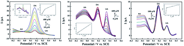
A facile and cost-effective approach has been developed towards electrochemical fabrication of a reduced graphene oxide (RGO) modified glassy carbon electrode (RGO/GCE). Scanning electron microscopy (SEM) images show that RGO is covered completely on the surface of glassy carbon electrodes. The RGO/GCE is used to detect dopamine (DA), uric acid (UA) and ascorbic acid (AA) simultaneously via cyclic voltammetry (CV) and differential pulse voltammetry (DPV) methods. Compared with bare GCE, RGO/GCE exhibits much high electrocatalytic activities toward the oxidation of DA, UA and AA, and three well-defined fully resolved anodic peaks were found in the CV curve at RGO/GCE. The GCEs modified with different amounts of RGO have an obvious influence on the determination of DA, UA and AA. By changing the concentrations of DA, UA and AA in the three substances coexisting system, the linear response ranges for the determination of DA, UA and AA were 0.1–400 μM, 2–600 μM, and 0.7–100 μM with the limit of detection (LOD) (S/N = 3) were estimated to be 0.1 μM, 1 μM and 0.7 μM, respectively. Moreover, it is found that RGO/GCE displays high reproducibility and selectivity for the determination of DA, UA and AA.


1. Introduction

Dopamine (DA) is a crucial neurotransmitter that belongs to the catecholamine family and has been given extensive attention in clinical research because it plays pivotal roles in the function of cardiovascular, metabolism and central nervous system of mammals.1–3 Abnormal levels of DA may result in various neurological diseases, such as Parkinson's disease, Huntington's disease and schizophrenia.4–6 Uric acid (UA) is a primary component in the end product of human metabolism.7,8 The concentration of UA contained in blood or urine is related to the physical condition of a person and disorders of UA are symptoms of several diseases such as gout and hyperuricemia.9–11 Ascorbic acid (AA), a water-soluble vitamin with antioxidant properties, exists in some particular kinds of food plants in abundance.12,13 It plays a crucial role in many biological processes such as free radical scavenging, cancer prevention and immunity improvement14,15 and it is also used for the prevention of scurvy and treatment of common cold and mental illness.16 Hence, AA is commonly used in large scale as an antioxidant in food, animal feed, pharmaceutical formulations.17,18 DA, UA and AA are usually considered as crucial molecules for physiological processes in human metabolism and they are coexisting in our body fluids. Therefore, it is essential to develop selective and sensitive methods for their determination in analytical application and diagnostic research.

So far, there have been various kinds of methods for the DA, UA and AA determination, such as electrochemical techniques,19,20 chemiluminescence,21 colorimetric,22 spectrophotometry23 and chromatography.24 Among these methods, electrochemical detection based on oxidation of these three molecules has proven to be high sensitivity, convenience and rapidity. However these three molecules are oxidized at almost the same potential at traditional electrodes and the oxidation peaks of them overlap each other, which complicates their electrochemical identification. Hence, clearly separating the electrochemical signals of these three compounds is a significant research challenge. To overcome this problem, various chemically modified electrodes have been developed.25,26 Carbon-based nanomaterial, such as carbon nanofiber and carbon nanotube, has been widely utilized for the determination of DA, UA and AA.27,28

Graphene, a two-dimensional monolayer of graphite in a closely packed honeycomb two-dimensional lattice with low cost, is among the most promising materials developed in recent years because of its unique properties such as huge specific surface area, rapid electron transportation, high electrical conductivity and perfect biocompatibility.29–33 It has been employed as modification materials on the surface of glassy carbon electrodes (GCE) and carbon fiber electrodes serving as electrochemical sensor. Its unique electronic properties have enabled its application for the determination of DA, UA and AA. Recently, various strategies have been developed to fabricate reduced graphene oxide (RGO) film modified electrodes.34–36

In previous work, composite materials consist of noble nanoparticles and graphene prepared by chemical methods were used for determination of DA, UA and AA. The graphene/Pt/GCE prepared by Sun et al.37 and the palladium nanoparticle/graphene/chitosan prepared by Wang et al.38 were able to simultaneously detect DA, UA and AA. However, these methods they adopted are complex and time-consuming. In addition, the prepared electrodes are high cost due to involving the noble metals. In this work, we modified graphene oxide (GO) on GCE and adopted electrochemical method for GO reduction obtaining RGO on GCE. The RGO/GCE was prepared by simple electrochemical method distinguishing from other complex chemical methods. Moreover, RGO/GCE was fabricated without any noble metals and complex composites that would be low-cost and have the potential application. The RGO/GCE was endowed large surface area and unique electrochemical activity. Cyclic voltammetry (CV) and differential pulse voltammetry (DPV) were employed to investigate the electrochemical behaviors of DA, UA and AA at the prepared RGO. It is also obvious that the RGO/GCE showed perfect selectivity and excellent electrochemical activity towards the oxidation of the three molecules.

2. Experimental

2.1 Reagents

Graphite powder (Sinopharm Chemicals Reagent Co., Ltd., China) was used as received. Dopamine, uric acid and ascorbic acid were purchased from Acros Organics Co., Ltd., China. Sodium nitrate, sulfuric acid, potassium permanganate, hydrogen peroxide (30%), disodium hydrogen phosphate, sodium dihydrogen phosphate (Shanghai shiji Chemicals Reagent Co., Ltd., China), were analytical grade. Double-distilled water was used throughout the experiments.

2.2 Apparatus

The scanning electrode microscopy (S-4700, Hitachi High Technologies Corporation, Japan) was used to characterize the morphology of as-prepared electrodes. All the electrochemical experiments were carried out in a conventional three-electrode system using a CHI660D electrochemical workstation (Shanghai Chenhua Instrument Co., Ltd., China). The traditional three-electrode system was used for electrochemical experiments in this paper. GCE with a surface area of 0.07 cm2 was used as the working electrode. A platinum wire and a saturated calomel electrode (SCE) were used as the counter electrode and the reference electrode, respectively. All the experiments were performed at room temperature.

2.3 Electrode preparation

Graphene oxide (GO) was prepared by the Hummer's method,39,40 and then 6 mg GO product was added into 10 mL of double-distilled water under ultrasonic homogenizer for 90 min. Impurities in GO solution were wiped off by centrifuging. The mass concentration of the aqueous GO dispersion was estimated to be 0.3 mg mL−1. 10 μL of the obtained GO dispersion was dropped onto the surface of the highly polished GCE and dried in air then repeated it for 0–8 times and the modified GCE with different amount GO was obtained. The GO/GCE was used as the working electrode and the electrochemical reduction of GO was carried out at a constant voltage as −0.9 V vs. SCE for 1000 s in a 0.1 M Na–PBS solution (pH = 4.12). The it curve for GO reduction is shown as Fig. 1. The current has a high initial value and decreases rapidly in the first 200 s. The result demonstrates that the surface oxygen group of GO could be reduced at this constant voltage quickly. At last the reduced graphene oxide was modified on GCE.
image file: c4ra03148b-f1.tif
Fig. 1 it curve of GO reduction modified GCE at −0.9 V.

3. Results and discussion

3.1 Characterization of RGO

The surface morphology of GCE and RGO/GCE were characterized by SEM, as shown in Fig. 2a and c. It can be seen that RGO covers on the surface of GCE without any breakages. Many wrinkles are observed on the surface of the electrode, indicating the increase in surface area of the RGO/GCE. Fig. 2b and d shows the cyclic voltammograms (CVs) for 2.5 mM Fe(CN)63−/4− recorded at GCE and RGO/GCE. Compared the two electrodes, a pair of redox peaks is observed at RGO/GCE clearly. It exhibits well-defined peaks with a smaller peak potential separation (ΔE) of 75 mV, and the peak currents at RGO/GCE are also much higher than those at GCE. It is indicated that the RGO/GCE has excellent conductivity and electrochemical activity. The enhanced electrochemical properties at the RGO might be due to the high electric conductivity of graphene and the plenty of graphitic edges on the electrode surface.
image file: c4ra03148b-f2.tif
Fig. 2 SEM images of GCE (a) and RGO/GCE (modified with 0.018 mg GO) (c); cyclic voltammograms of GCE (b) and RGO/GCE (d) in 2.5 mM Fe(CN)63−/4− + 0.1 M KCl solution. Scan rate: 50 mV s−1.

3.2 Electrochemical behaviors of single DA, UA and AA

The typical CVs of 0.5 mM DA, 1 mM UA, and 10 mM AA at GCE in a 0.1 M PBS (pH = 7.0) solution are shown in Fig. 3a, respectively. In Fig. 3a, for DA, the cathodic and anodic peaks appear at 0.02 V and 0.42 V, respectively, and the peak potential separation (ΔE) is about 0.4 V. For AA and UA, the electrochemical reactions are irreversible and the signals show broader oxidation peaks with the peak potentials of 0.45 V (UA) and 0.56 V (AA). These indicate the sluggish electron transfer kinetics at bare GCE, which may be related to the weak conductive and the electrode fouling caused by the deposition of DA, UA and AA and their oxidation products on the electrode surface. From Fig. 3a, the oxidation peaks of the three molecules overlap with each other and the oxidation peak potentials are so near that DA, UA, and AA cannot be detected simultaneously at bare GCE. In comparison with the bare GCE, seen from Fig. 3b, it is found that the three oxidation peaks of DA, UA, and AA at RGO/GCE separate from each other obviously. The entire current peaks of these three molecules are also higher than those in Fig. 3a. The cathodic and anodic peaks of DA appear at 0.10 V and 0.26 V, respectively. The peak potential separation (ΔE) is about 0.16 V, suggesting that the reversibility of DA at the RGO is remarkably improved. For UA, a sharp oxidation peak at 0.35 V and a small reduction peak at 0.27 V are obtained. In the case of AA, it displays a substantial negative shift of the oxidation peak potential to 0.02 V which is about 0.54 V more negative than that at GCE. The above results demonstrate that the RGO/GCE has higher electrochemical surface area and electron transfer for the oxidation of DA, UA and AA. Such an improvement could be attributed to the special structure of RGO and the huge surface area of RGO/GCE.
image file: c4ra03148b-f3.tif
Fig. 3 Cyclic voltammograms of 0.5 mM DA, 1 mM UA, and 10 mM AA recorded at the GCE (a) and RGO/GCE (b). Supporting electrolyte: 0.1 M PBS (pH = 7.0). Scan rate: 100 mV s−1.

3.3 Cyclic voltammetric simultaneous determination of ternary mixtures of DA, UA, and AA

It is very important to get the optimal amount of GO which modified on GCE with the best determination results of DA, UA and AA. A series of RGO/GCEs were prepared by using the different volumes of GO solution. Fig. 4A shows the preparation process of the RGO/GCEs dropped with 10 μL, 20 μL, 30 μL, 40 μL, 50 μL, 60 μL, 70 μL, 80 μL and 90 μL 0.3 mg mL−1 GO solution. The thickness of GO sheets increases with the amount of GO. After electrochemical reduction, the brown GO sheets are reduced to black RGO sheets, and the modified GCEs with different amount RGO have been obtained. Fig. 4B shows the CVs of GCE and RGO/GCEs with different amount RGO in a 0.1 M PBS (pH = 7.0) solution containing 0.5 mM DA, 1 mM UA and 10 mM AA. Only a broadened peak at 0.75 V is observed in the curve a. The peak is the overlapped signal of DA, UA and AA. Therefore, the oxidation peak potentials of DA, UA and AA are indistinguishable, and it is impossible to determine the individual concentration of these compounds from the overlapped oxidation peak at the GCE. Compare with bare GCE, the peaks in curve b separate at the RGO/GCE modified with 0.003 mg GO. The peaks of DA and UA are enhanced and well-defined, but the peak of AA is very weak. In curve c, the peaks at the RGO/GCE modified with 0.009 mg GO are enhanced significantly. Among all the curves, the RGO/GCE modified with 0.018 mg GO shows the highest peak currents and well-defined peaks at approximately 0.02 V, 0.30 V and 0.45 V, corresponding to the oxidation of AA, DA, and UA, respectively. The calculated peak potential separations (ΔE) are 0.28 V for AA–DA, 0.15 V for DA–UA, and 0.43 V for UA–AA, suggesting the selective determination of one component in the presence of other two species or simultaneous determination of these three species is feasible at this RGO/GCE. The peak currents of three molecules at the RGO/GCE modified with 0.027 mg GO decline somewhat. The reason may be few layers RGO have lower electrical resistivity than multilayer RGO,41 but the limited surface area of them cannot provide enough electrochemical active area for DA, UA and AA oxidation. Meanwhile GCE modified with excessive RGO sheets has the increased electrical resistivity and decreased conductivity, which results in the decline of peak currents. From the results, the RGO/GCE modified with 0.018 mg GO were chosen for the simultaneous determination of DA, UA and AA.
image file: c4ra03148b-f4.tif
Fig. 4 (A) The preparation process of the RGO/GCEs modified with different amount of GO. (B) Cyclic voltammograms of GCE (a) and RGO/GCEs modified with 0.003 mg (b), 0.009 mg (c), 0.018 mg (d) and 0.027 mg (e) GO. Supporting electrolyte: 0.1 M PBS (pH = 7.0) solution containing 0.5 mM DA, 1 mM UA and 10 mM AA. Scan rate: 100 mV s−1.

3.4 Effect of scan rate

The kinetics of electrode reaction was investigated by evaluating the effect of scan rate on the oxidation peak current and peak potential. As shown in Fig. 5a–c, the scan rate of cyclic voltammetry exhibits a profound effect on the oxidation peak current of 0.5 mM DA, 1 mM UA and 10 mM AA 0.1 M PBS (pH = 7.0) solution. When the scan rate increases in the range of 20–200 mV s−1, small positive shifts in the three molecules oxidation peaks are observed, suggesting that the adsorption of DA, UA, and AA does not occur on RGO/GCE in 0.1 M PBS (pH = 7.0) solution. And for them, the redox peak currents gradually increase as the scan rate increases, while a linear relationship is established between the anodic peak current and the square root of scan rate. The linear equations are expressed as follows: DA: Ipa (μA) = 8.795υ1/2 (mV s−1) − 19.017, R2 = 0.993; UA: Ipa (μA) = 18.923υ1/2 (mV s−1) −65.517, R2 = 0.989; AA: Ipa (μA) = 1.7451υ1/2 (mV s−1) + 0.7113, R2 = 0.994; the above results indicate that the oxidation of DA, UA, and AA on RGO/GCE is a diffusion-controlled process.
image file: c4ra03148b-f5.tif
Fig. 5 Cyclic voltammograms of RGO/GCE in 0.1 M PBS (pH = 7.0) solution containing 0.5 mM DA (a), 1 mM UA (b) and 10 mM AA (c) at different scan rates of 20, 40, 80, 120, 160 and 200 mV s−1. Inset: plot of oxidation peak current versus the square root of scan rate.

3.5 Simultaneous determination of DA, UA, and AA

Based on the above results, the difference in the oxidation peak potentials for DA, UA and AA at RGO/GCE is large enough for separation and simultaneous determination of three molecules in a mixture. Differential pulse voltammetry (DPV), a widely used analytical technique for the enhancement of specificity and sensitivity, was carried out in their mixture when the concentration of one species changed, while the other two species remained constant. As shown in Fig. 6a–c, three anodic peaks corresponding to the oxidation of DA, UA, and AA are observed at 0.17 V, 0.33 V and −0.05 V, respectively. It can be seen that the RGO possesses improved and comparable performance for the simultaneous determination of DA, UA, and AA obviously. The oxidation peak currents of these three molecules increase linearly with their concentrations, indicating the stable and efficient electrocatalytic activity of the RGO. For DA detection (Fig. 6a), 50 μM UA and 100 μM AA are mixed in the PBS (pH = 7) solution and the corresponding linear regression equation is defined as Ipa (μA) = 0.3304CDA (μM) + 0.8003, R = 0.991 (CDA = 0.1–100 μM) and Ipa (μA) = 0.0828CDA (μM) + 21.545, R = 0.997 (CDA = 100–400 μM). For UA detection (Fig. 6b), 5 μM DA and 100 μM AA are mixed in the PBS (pH = 7) solution and the corresponding linear regression equation is defined as Ipa (μA) = 0.0153CUA (μM) + 0.325, R = 0.995 (CUA = 2–600 μM). For AA detection (Fig. 6c), 5 μM DA and 50 μM UA are mixed in the PBS (pH = 7) solution and the corresponding linear regression equation is defined as Ipa (μA) = 0.4342CAA (μM) + 1.9675, R = 0.998 (CAA = 0.7–5 μM) and Ipa (μA) = 0.0983CAA (μM) + 4.1756, R = 0.997 (CAA = 5–100 μM). The detection limits (S/N = 3) for the determination of DA, UA, and AA are evaluated as 0.1 μM, 1 μM and 0.7 μM, respectively. Table 1 shows the linear range and detection limit for DA, UA and AA at RGO/GCE compared with some sensors of other research paper. In general, RGO/GCE has a wide linear range and a low detection limit for detecting the three molecules.
image file: c4ra03148b-f6.tif
Fig. 6 (a) DPVs of DA at RGO/GCE in the presence of 50 μM UA and 100 μM AA concentrations: 10, 20, 30, 50, 70, 100, 200, 300, and 400 μM. Insets: DPVs of DA at RGO/GCE in the presence of 50 μM UA and 100 μM AA concentrations: 0, 0.1, 0.3, 0.5, 0.7, 1, 2, 3, 5, 7 and 10 μM; plots of oxidation currents versus the concentration of DA; (b) DPVs of UA at RGO/GCE in the presence of 5 μM DA and 100 μM AA concentrations: 0, 1, 2, 10, 20, 30, 50, 70, 100, 200, 300, 400, 500 and 600 μM. Insets: plots of oxidation currents versus the concentration of UA; (c) DPVs of AA at RGO/GCE in the presence of 5 μM DA and 50 μM UA concentrations: 0, 0.7, 1, 3, 10, 20, 50 and 100 μM. Insets: plots of oxidation currents versus the concentration of AA. Supporting electrolyte: 0.1 M PBS (pH = 7.0).
Table 1 The analytical performances of different modified electrodes for the simultaneous determination of DA, UA and AA
Electrodes Detection limit (μ mol L−1) Linear range (μ mol L−1) Ref.
DA UA AA DA UA AA
a Poly-Evans Blue modified glassy carbon electrode.b Multi walled carbon nanotube modified carbon-ceramic electrode.c Ordered mesoporous carbon/Nafion composite film modified glassy carbon electrode.d Polyaniline/graphene oxide modified carbon-ceramic electrode.e 2-Amino-1,3,4,thiadiazole modified indium tin oxide.
Poly (Evans Blue)/GCEa 0.25 2.0 0.3 1–10 30–110 5–105 42
MWCNT/CCEb 0.31 0.42 7.71 0.5–100.0 0.55–90.0 15.0–800.0 43
OMC/Nafion/GCEc 0.5 4.0 20 1–90 5–80 40–800 44
PANI-GO/GCEd 0.5 0.2 20 2–18 2–18 25–200 27
p-ATD/ITOe 0.33 0.19 2.01 5–50 10–100 30–300 45
RGO/GCE 0.1 1 0.7 0.1–400 2–600 0.7–100 This work


3.6 Reproducibility, stability of RGO/GCE and real sample analysis

To assess the reproducibility of the single electrode, RGO/GCE was used to (n = 6) determine 0.4 mM DA, 0.6 mM UA and 0.1 mM AA repeatedly in the mixture solution. It is found that the relative standard deviations (RSD) for DA, UA and AA are 1.42%, 2.16% and 2.64% respectively, indicating the excellent reproducibility of the prepared composite electrode. Moreover, the stability of RGO/GCE was also an important factor. The modified electrode was stored for 7 days at room temperature before the experiment. The peak current intensity of DA, UA and AA decayed by 2.5%, 2.7% and 3.6%, respectively, which demonstrates a storage stability of RGO/GCE.

The RGO/GCE for the real samples determination was performed by measuring rat serum and urine using the standard addition technique. All samples were diluted with 0.1 M PBS (pH = 7.0) and transferred to the electrochemical cell before the measurements. In order to ascertain the correctness of the results, certain amounts of DA, UA and AA were added into the diluted samples and were then detected. The results were summarized in Table 2. One can see that that RGO/GCE exhibited exact recovery results, which indicated that RGO/GCE has the potential application for determining DA, UA and AA in real samples.

Table 2 Determination of DA, AA and UA in real samplesa
Sample UA DA AA
Original (μM) Add (μM) Found (μM) Recovery (%) Original (μM) Add (μM) Found (μM) Recovery (%) Original (μM) Add (μM) Found (μM) Recovery (%)
a Three replicate measurements were made on each sample.
Serum 3.1 10.0 13.5 103.1 5.0 5.1 102.0 10.0 10.6 106.0
Urine 6.2 10.0 16.6 102.2 5.0 5.2 104.0 10.0 9.8 98.0


4. Conclusions

In this work, the RGO modified GCE has been fabricated for detecting DA, UA and AA simultaneously. RGO with many wrinkles covers on the surface of GCE completely without any breakages. By the electrochemical characterization, RGO can enhance electron transfer activity because of its great electric conductivity and the ultrahigh surface area. Compared with the bare GCE, the clear and well-separated electro-oxidation peaks of DA, UA, and AA at RGO/GCE are observed. In addition, the modified electrode exhibits high selectivity and good reproducibility towards the determination of DA, UA and AA with low detection limit and wide detection range. It is proved that GCEs modified with different amounts of RGO have obvious influence on DA, UA and AA detection result. The fabrication of RGO/GCE is simple, cost-effective, and user-friendly and it is expected to have good application prospects.

Acknowledgements

This work was supported by the National Natural Science Foundation of China (Grant nos 51373111, 51073114 and 20933007), Suzhou Nano-project (ZXG2012022), the Opening Project of Xinjiang Key Laboratory of Electronic Information Materials and Devices (XJYS0901-2010-01), the Academic Award for Young Graduate Scholar of Soochow University, the Priority Academic Program Development of Jiangsu Higher Education Institutions (PAPD).

Notes and references

  1. M. A. Kurian, P. Gissen, M. Smith, S. J. Heales and P. T. Clayton, Lancet Neurol., 2011, 10, 721 CrossRef CAS.
  2. W. Schultz, Annu. Rev. Neurosci., 2007, 30, 259 CrossRef CAS PubMed.
  3. P. N. Tobler, C. D. Fiorillo and W. Schultz, Science, 2005, 307, 1642 CrossRef CAS PubMed.
  4. X. H. Cao, L. X. Zhang, W. P. Cai and Y. Q. Li, Electrochem. Commun., 2010, 12, 540 CrossRef CAS PubMed.
  5. S. C. Fernandes, I. C. Vieira, R. A. Peralta and A. Neves, Electrochim. Acta, 2010, 55, 7152 CrossRef CAS PubMed.
  6. T. Thomas, R. J. Mascarenhas, C. Nethravathi, M. Rajamathi and B. E. K. Swamy, J. Electroanal. Chem., 2011, 659, 113 CrossRef CAS PubMed.
  7. D. Lakshmi, M. J. Whitcombe, F. Davis, P. S. Sharma and B. B. Prasad, Electroanalysis, 2011, 23, 305 CrossRef CAS.
  8. H. Manjunatha, D. H. Nagaraju, G. S. Suresh and T. V. Venkateshab, Electroanalysis, 2009, 21, 2198 CrossRef CAS.
  9. J. P. Dong, Y. Y. Hu, S. M. Zhu, J. Q. Xu and Y. J. Xu, Anal. Bioanal. Chem., 2011, 396, 1755 CrossRef PubMed.
  10. P. L. Riches, A. F. Wright and S. H. Ralston, Hum. Mol. Genet., 2009, 18, 177 CrossRef PubMed.
  11. C. Wang, R. Yuan, Y. Q. Chai, Y. Z. Zhang, F. X. Hu and M. H. Zhang, Biosens. Bioelectron., 2011, 30, 315 CrossRef CAS PubMed.
  12. M. W. Davey, M. Van Montagu, D. Inze, M. Sanmartin, A. Kanellis and N. Smirnoff, J. Sci. Food Agric., 2000, 80, 825 CrossRef CAS.
  13. U. Yogeswaran and S. Chen, Electrochim. Acta, 2007, 52, 5985 CrossRef CAS PubMed.
  14. S. Englard and S. Seifter, Annu. Rev. Nutr., 1986, 6, 365 CrossRef CAS PubMed.
  15. P. Kalimuthu and S. A. John, Bioelectrochemistry, 2009, 77, 13 CrossRef CAS PubMed.
  16. P. Kalimuthu and S. A. John, Talanta, 2010, 80, 1686 CrossRef CAS PubMed.
  17. C. Andre, I. Castanheira, J. M. Cruz, P. Paseiro and A. Sanches, Trends Food Sci. Technol., 2010, 21, 229 CrossRef CAS PubMed.
  18. A. M. Yu and H. Y. Chen, Anal. Chim. Acta, 1997, 344, 181 CrossRef CAS.
  19. J. Du, R. R. Yue, F. F. Ren, Z. Q. Yao, F. X. Jiang, P. Yang and Y. K. Du, Biosens. Bioelectron., 2014, 53, 220 CrossRef CAS PubMed.
  20. R. Suresh, K. Giribabu, R. Manigandan, A. Stephen and V. Narayanan, RSC Adv., 2014, 4, 17146 RSC.
  21. H. Chen, R. B. Li, L. Lin, G. S. Guo and J. M. Lin, Talanta, 2010, 81, 1688 CrossRef CAS PubMed.
  22. Y. H. Lin, C. E. Chen, C. Y. Wang, F. Pu, J. S. Ren and X. G. Qu, Chem. Commun., 2011, 47, 1181 RSC.
  23. A. Abbaspour, A. Khajehzadeh and A. Ghaffarinejad, Analyst, 2009, 134, 1692 RSC.
  24. V. Carrera, E. Sabater, E. Vilanova and M. A. Sogorb, J. Chromatogr. B, 2007, 847, 88 CrossRef CAS PubMed.
  25. Z. Xue, Y. Feng, H. Guo, C. Hu, A. Mahmoud idris Mohmed, J. Li and X. Lu, RSC Adv., 2014, 4, 5849 RSC.
  26. E. M. Zahran, M. I. Prodromidis, D. Bhattacharyya and L. G. Bachas, Anal. Bioanal. Chem., 2012, 404, 1637 CrossRef CAS PubMed.
  27. P. Manivel, M. Dhakshnamoorthy, A. Balamurugan, N. Ponpandian, D. Mangalaraj and C. Viswanathan, RSC Adv., 2013, 3, 14428 RSC.
  28. M. Noroozifar, M. Khorasani-Motlagh, R. Akbari and M. B. Parizi, Biosens. Bioelectron., 2011, 28, 56 CrossRef CAS PubMed.
  29. S. Eissa, L. L'Hocine, M. Siaj and M. Zourob, Analyst, 2013, 138, 4378 RSC.
  30. F. F. Ren, H. W. Wang, C. Y. Zhai, M. S. Zhu, R. R. Yue, Y. K. Du, P. Yang, J. K. Xu and W. S. Lu, ACS Appl. Mater. Interfaces, 2014, 6, 3607 CAS.
  31. H. W. Wang, J. Du, Z. Q. Yao, R. R. Yue, C. Y. Zhai, F. X. Jiang, Y. K. Du, C. Y. Wang and P. Yang, Colloids Surf., A, 2013, 436, 57 CrossRef CAS PubMed.
  32. R. R. Yue, F. Ren, C. Wang, J. K. Xu and Y. K. Du, RSC Adv., 2014, 4, 500 RSC.
  33. X. X. Liu, H. Zhu and X. R. Yang, RSC Adv., 2014, 4, 3706 RSC.
  34. T. Kuila, S. Bose, P. Khanra, A. K. Mishra, N. H. Kim and J. H. Lee, Biosens. Bioelectron., 2011, 26, 4637 CrossRef CAS PubMed.
  35. C. B. Liu, K. Wang, S. L. Luo, Y. H. Tang and L. Y. Chen, Small, 2011, 7, 1203 CrossRef CAS PubMed.
  36. Z. Q. Yao, R. R. Yue, C. Y. Zhai, F. X. Jiang, H. W. Wang, Y. K. Du, C. Y. Wang and P. Yang, Int. J. Hydrogen Energy, 2013, 38, 6368 CrossRef CAS PubMed.
  37. C. L. Sun, H. H. Lee, J. M. Yang and C. C. Wu, Biosens. Bioelectron., 2011, 26, 3450 CrossRef CAS PubMed.
  38. X. Wang, M. Wu, W. R. Tang, Y. Zhu, L. W. Wang, Q. J. Wang, P. G. He and Y. Z. Fang, J. Electroanal. Chem., 2013, 695, 10 CrossRef CAS PubMed.
  39. M. S. Zhu, P. L. Chen and M. H. Liu, ACS Nano, 2011, 5, 4529 CrossRef CAS PubMed.
  40. W. S. Hummers and R. E. Offeman, J. Am. Chem. Soc., 1958, 80, 1339 CrossRef CAS.
  41. P. Nirmalraj, T. Lutz, S. Kumar, G. Duesberg and J. Boland, Nano Lett., 2011, 11, 16 CrossRef CAS PubMed.
  42. L. Q. Lin, J. H. Chen, H. Yao, Y. Z. Chen, Y. J. Zheng and X. H. Lin, Bioelectrochemistry, 2008, 73, 11 CrossRef CAS PubMed.
  43. B. Habibi and M. H. Pournaghi-Azar, Electrochim. Acta, 2012, 55, 5492 CrossRef PubMed.
  44. D. Zheng, J. S. Ye, L. Zhou, Y. Zhang and C. Z. Yu, J. Electroanal. Chem., 2009, 625, 82 CrossRef CAS PubMed.
  45. P. Kalimuthu and S. A. John, Talanta, 2010, 80, 1686 CrossRef CAS PubMed.

This journal is © The Royal Society of Chemistry 2014
Click here to see how this site uses Cookies. View our privacy policy here.