DOI:
10.1039/C4RA03122A
(Paper)
RSC Adv., 2014,
4, 28683-28690
Effects of template removal on both morphology of mesoporous silica-coated gold nanorod and its biomedical application
Received
8th April 2014
, Accepted 6th June 2014
First published on 6th June 2014
Abstract
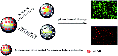
Mesoporous silica-coated Au nanorods (Aurod@SiO2) have recently attracted considerable interest in nanomedicine, and the template removal procedure is crucial for the preparation of such hybrid nanostructure. Herein, two kinds of typical extraction solvents, i.e., NH4NO3–CH3OH and HCl–CH3OH, were separately used to extract the surfactant cetyltrimethylammonium bromide (CTAB) from the obtained Aurod@SiO2. The results show that CTAB molecules could be completely removed from the pores of Aurod@SiO2 without damaging the internal Au nanorod by NH4NO3–CH3OH, while Aurod@SiO2 treated with HCl–CH3OH suffered from both poor extraction efficiency and the shape transformation of Au nanorod due to selective etching in the presence of oxygen. In addition, the consequent drug loading experiment shows that Aurod@SiO2 extracted by NH4NO3–CH3OH possesses a larger drug loading capacity with a loading efficiency of 82.5%. Furthermore, the in vitro photo-thermal therapy experiment shows that Aurod@SiO2 extracted by NH4NO3–CH3OH is more efficient in killing the YCC-2 gastric cancer cells as compared with that extracted by HCl–CH3OH.
Introduction
Recently, the development of hybrid nanomaterials with multiple functionalities, such as diagnostic imaging, drug delivery and therapy, has been attracting more attention in the biomedical fields. Compared with pure nanoparticles (NPs), the hybrid nanomaterial can cleverly integrate various properties into unique nanoparticle formulation; moreover, it has potential improvement on the previously established therapeutic and diagnostic regimes.1 Among various hybrid nanomaterials, mesoporous silica-coated Au nanorods (Aurod@SiO2) have specially stirred up great interest. In this hybrid structure, Au nanorods exhibit intriguing nonlinear optical properties owing to their localized surface plasmon resonances that can be synthetically tuned across a broad spectral range, covering the visible and near-infrared regions by tailoring their aspect ratios (the ratio between the length and diameter). Such splendid plasmon-related property makes Au nanorods usable in both imaging and photothermal conversion-based therapy.2–5 Moreover, mesoporous silica shell (MSS) has also gained much attention due to its unique features, such as large surface area and pore volume, high chemical and thermal stability, excellent biocompatibility, and versatile surface chemistry for further functionalization.6,7 Therefore, it can work as “nanocarrier” for the delivery of drugs or other cargos to cells and also be used as the ideal platform for constructing multifunctional materials.
To date, the main procedure to fabricate Aurod@SiO2 always involves the use of cetyltrimethylammonium bromide (CTAB), which is first used as stabilizer in the synthesis of Au nanorods, and subsequently as the soft template for the formation of mesoporous silica shell.8 In the post-synthesis treatments, CTAB should be efficiently removed to get a more open and hollow pore channel so as to increase its drug loading capacity. In addition, the residual CTAB is potentially toxic for human system when using Aurod@SiO2 as drug carrier. Therefore, how to effectively remove CTAB template from the obtained Aurod@SiO2 is an important issue before its real application as a theranostic platform. To date, numerous methods, including high-temperature thermal–chemical calcinations processes,9 dialysis,10 and solvent extraction,11 have been proposed to remove the template from silica based mesostructured materials. The calcination processes, owing to their aggressive thermal and/or chemical environments, often damage the intrinsic physicochemical characteristics of the underlying substrate, thus limiting their application in the removal of CTAB from the obtained Aurod@SiO2. The “dialysis” process is time-consuming, and the treatment often requires a large amount of water containing acetic acid, which is neither environmentally friendly nor cost-effective for the real production. Therefore, to the best of our knowledge, solvent extraction is the most effective way to remove CTAB from the silica based mesostructured hybrid NPs.
However, compared with pure silica based mesostructured materials, the effect of solvent extraction on the property of hybrid silica based mesostructured NPs has rarely been reported. As we know, Au nanorods have been reported to easily reshape or to suffer from size reduction upon the various conditions such as the treatment with cyanide,12 oxygen,13 ferric chloride,14 Cu2+ and ascorbic acid in a hot aqueous solution.15 In order to guarantee its potential efficacy as a theranostic platform, it is crucial for us to determine a kind of extraction solvent, which can not only extract all the CTAB molecules from Aurod@SiO2, but also maintain the original shape of both mesoporous silica NPs and internal Au nanorod. Generally, the high efficiency of template removal requires that the suitable solvent not only has good accessibility to highly cross-linked regions but also can break the interactions between CTAB and the siliceous mesostructured framework. Therefore, in most cases, the methanol solvent-containing cations such as H+, NH4+ is most recommended, in which CTAB is expected to be removed through both its dissolution in methanol and the ion exchange between H+ or NH4+ and CTAB.
For the reasons mentioned above, herein we comparatively studied the effect of two different extraction solvents (NH4NO3–CH3OH and HCl–CH3OH) on the efficiency of CTAB removal from the obtained Aurod@SiO2. Their effects on both morphology and plasmon-related properties of the obtained Aurod@SiO2 were mainly investigated. Then, the drug loading experiment was carried out to assess the effect of two extraction solvents on the drug loading capacity. Finally, the in vitro photo-thermal therapy experiment was implemented to evaluate the photo-thermal therapy (PTT) effect of Aurod@SiO2 after solvent extraction. These results reported here will lay a solid foundation for the Aurod@SiO2 based photo-thermal therapy and drug delivery.
Experimental section
Materials
Gold(III) chloride (HAuCl4·3H2O), cetyltrimethylammonium bromide (CTAB), silver nitrate (AgNO3), sodium borohydride (NaBH4), sodium hydroxide (NaOH), methanol (CH3OH), L-ascorbic acid (AA), and tetraethyloxysilane (TEOS) were purchased from Sinopharm Chemical Reagent Limited Corporation and used as received. 3-Aminopropyltriethoxysilane (APTES), polyacrylic acid (PAA, MW = 1800), and DOX in the form of the hydrochloride salt were purchased from Aladdin. α-mPEG-ω-amine (MW = 10
000) was purchased from Shanghai Yanyi Biotechnology Corporation. Fetal bovine serum (FBS), Dulbecco's modified Eagle's medium, penicillin/streptomycin were purchased from Thermo Fisher Scientific. Calcein-AM and propidium iodide (PI) were obtained from Invitrogen. All glassware and Teflon-coated magnetic stirring bars were thoroughly cleaned with aqua regia, followed by rinsing with purified water. Note that water was purified with a Millipore system.
Synthesis of Au nanorods
Au nanorods were fabricated by a seed-mediated, Ag(I)-assisted growth procedure according to a previous report.8 The seed solution was prepared as follows: briefly, 0.25 mL of an aqueous solution of HAuCl4 solution (0.01 M) was added to 7.5 mL of CTAB solution (0.10 M) in a test tube. The solutions were gently mixed by inversion, and they appeared bright brown-yellow in color. Then, 0.6 mL of an aqueous ice-cold NaBH4 solution (0.01 M) was added all at once, followed by rapid inversion mixing for 2 min. The resultant solution developed a pale brown-yellow color. After that, the test tube was kept in a water bath maintained at 25 °C to obtain stable seed solution. The growth solution was prepared by mixing 285 mL of CTAB (0.1 M), 12 mL of HAuCl4 (0.01 M) and 1.8 mL of AgNO3 (0.01 M) in a 500 mL reagent bottle. Another 100 μL of HCl (37%, wt) was added into the above solution. After this step, 1.92 mL of L-ascorbic acid (0.01 M) was added to the resultant solution with gentle stirring, which changed the color of the growth solution from dark yellow to colourless. Finally, 0.6 mL of seed solution was added to the resulting solution at 27–30 °C, and the color of the solution gradually changed within 10–20 min. The temperature of the growth medium was maintained constant at 27–30 °C during the whole procedure.
Synthesis of Aurod@SiO2
The post-mesoporous silica coating of Au nanorods was achieved by a modified Stöber method. Firstly, 200 mL of as-synthesized Au nanorods were washed twice with Milli-Q water to remove excess CTAB and re-dispersed in 100 mL of Milli-Q water. Subsequently, 10 mL of NaOH solution (0.1 M) was added to adjust the pH value to 11, and then 0.9 mL of 20% TEOS in methanol solution was added to the resultant solution at the rate of 300 μL every 30 min. The mixture was allowed to react at 27 °C for at least 2 days. The resulting Aurod@SiO2 was washed two times with water, and then three times with methanol, and finally dispersed into 100 mL of methanol.
The removal of CTAB from the as-synthesized Aurod@SiO2 by HCl–CH3OH (denoted as “route A”) or NH4NO3–CH3OH (denoted as “route B”) extraction
The removal of CTAB from the as-synthesized Aurod@SiO2 was carried out by two different solvent extraction methods. Typically, the removal of CTAB by HCl–CH3OH extraction was performed in 0.25 M HCl–CH3OH solution at 60 °C. Firstly, 2 mg of as-synthesized Aurod@SiO2 was added to 5 mL of 0.25 M HCl–CH3OH solution. Then, the resultant mixture was heated to reflux at 60 °C with stirring. In comparison, the removal of CTAB by NH4NO3–CH3OH extraction is similar to the above the procedure, except that HCl–CH3OH was replaced by 5 mL of methanol solution containing 30 mg of NH4NO3. For the sake of comparison, the extracted Aurod@SiO2 was extracted and washed by methanol at a selected time for both UV-vis and TEM characterization.
Evaluation of drug loading capacity of Aurod@SiO2 treated with route A or route B
DOX was chosen as a model drug to assess the effect of different extraction solvents on drug loading capacity of the extracted Aurod@SiO2. Briefly, 1 mg of the extracted Aurod@SiO2 was mixed with 1 mL of PBS buffer containing 100 μg of DOX and stirred at room temperature for 24 h. Then, the supernatant was collected and the residual DOX content was measured by UV-vis measurement at a wavelength of 480 nm. The loading efficiency (LE) is obtained using the following equation:
Preparation of PEGylated Aurod@SiO2
To improve biocompatibility of Aurod@SiO2, their surface was further functionalized with α-mPEG-ω-amine group prior to the in vitro photo-thermal therapy experiment. Firstly, 5 mg of Aurod@SiO2 treated by route A or route B was dispersed in 5 mL of methanol, and then 100 μL of APTES was rapidly added to the above solution. After 24 h of reaction under room temperature, the APTES functionalized Aurod@SiO2 (denoted as Aurod@SiO2@NH2) was obtained by centrifugation at 15
000 rpm for 10 min to remove supernatant, washing with ethanol for four times, and then dried under vacuum. After that, polyacrylic acid (PAA, MW = 1800) was then covalently attached to the surface of Aurod@SiO2@NH2 using EDC coupling chemistry with the aim to obtain carboxylate-functionalized Aurod@SiO2 (denoted as Aurod@SiO2@COOH). Briefly, 0.03 g of PAA dissolved in 2 mL of MES buffer (pH = 6.0) was first activated with the help of 1 mg of EDC. Then, 5 mg of the obtained Aurod@SiO2@NH2 was added into the above solution, and the reaction lasted for 30 min. Next, the resultant solution was purified by centrifugation and subsequently washed with PBS buffer for at least 3 times. For further PEGylation, 100 μL of EDC (10 mg mL−1) and 100 μL of NHS (10 mg mL−1) were separately added to 2 mL of aqueous solution containing 5 mg of Aurod@SiO2@COOH. After activation for 30 min, 1 mL of mPEG10000–NH2 solution (100 mg mL−1) was added to the above mixture, and the solution was stirred overnight. Finally, the PEGylated Aurod@SiO2 (denoted as Aurod@SiO2@PEG) was collected and washed with distilled water by centrifugation at 15
000 rpm, and the resultant particles were dispersed in distilled water.
In vitro photo-thermal therapy (PTT) of Aurod@SiO2 treated with route A or route B
The in vitro photo-thermal therapy experiment was carried out to investigate the cancer killing efficiency of the Aurod@SiO2 treated with two extraction solvents. Firstly, gastric cancer cells YCC-2 were seeded in a 24-well flat culture plate. To allow cell attachment, the cells were incubated overnight, followed by incubation with 50 μL of 1 mg mL−1 PEGylated Aurod@SiO2 in normal DMEM cell medium containing 10% fetal bovine serum (FBS) and 1% penicillin–streptomycin at 37 °C in a humidified atmosphere containing 5% CO2. After 24 h incubation, free Aurod@SiO2 was removed from the culture medium by rinsing three times with PBS. Cells were then irradiated with an 808 nm optical fiber-coupled diode near-infrared (NIR) laser at power density of 3 W cm−2 for 1, 2, 4 min. After irradiation, the cell viability was assessed by calcein-AM/propidium iodide double staining as per a previously reported method.16 Cells cultured without the addition of the resultant Aurod@SiO2 at the same time intervals were used as the negative control. Fluorescence microscopic images of stained cells were then taken using an Olympus fluorescent microscope.
Characterization
The morphology and size of both Au nanorods and Aurod@SiO2 were acquired by transmission electron microscopy (TEM). Before the measurement, samples were prepared by drying a drop of dispersion of Au nanorods or Aurod@SiO2 suspension on a carbon-coated copper grid at room temperature and were analyzed using a JEOL JEM 2100 F electron microscope operating at 200 kV. The UV-vis spectrum was recorded on a Shimadzu spectrometer spectrophotometer between 450 and 1000 nm wavelength. The samples were measured in a 1 cm quartz cuvette using the corresponding pure solvent as a reference. FT-IR Spectrum was acquired by a Nicolet 5700 Fourier transform infrared spectrometer using KBr pellets. Zeta potential measurement was conducted in Zetasizer NanoS from Malvern Instruments. Before the measurement, the particles were directly dispersed in deionized water.
Results and discussion
Aurod@SiO2 was synthesized by combining the seed-mediated growth and modified Stöber method according to the reported method with some modifications,8 in which CTAB was separately employed as both stabilizing agent for the synthesis of Au nanorods and the soft template for the formation of mesoporous silica shell. Fig. 1a shows the representative TEM image of Au nanorods, and it can be seen that the average length and width of Au nanorods are about 30 and 8.5 nm, respectively. After silica coating, the obtained Aurod@SiO2 shows the typical core–shell structure made of a Au nanorod core and uniform silica shell with the average thickness of 11 nm (as shown in Fig. 1b); moreover, disordered mesopores can be observed in the silica shell. The corresponding UV-vis absorbance spectra further confirms the successful silica coating of Au nanorods. Fig. 2 shows the typical UV-vis absorbance spectra of Au nanorods before and after silica coating. It can be seen that the as-synthesized Au nanorod has a longitudinal surface plasmon resonance (LSPR) peak at 752 nm, and after silica shell coating, it only exhibits a red shift of 10 nm. This result is consistent with a previous report, in which the red shift of LSPR peak was attributed to an increase in the local refractive index of the surrounding medium for Au nanorod after replacing CTAB ligands with silica shell.23 Taking all these above results together, it can be concluded that the typical Aurod@SiO2 with core–shell structure is obtained.
 |
| Fig. 1 TEM images of Au nanorod before (a) and after (b) silica coating. | |
 |
| Fig. 2 UV-vis absorbance spectra of Au nanorod and Aurod@SiO2. | |
As mentioned above, herein we mainly studied the effect of two kinds of extraction solvents (route A and route B) on both morphology and plasmon-related property of Aurod@SiO2. Fig. 3a firstly shows the real-time UV-vis absorbance spectra of the extracted Aurod@SiO2 at various stages in route A. In the initial stage of HCl–CH3OH extraction, Aurod@SiO2 exhibits two adsorption peaks located at 522 and 752 nm, corresponding to the transverse surface plasmon resonance (TSPR) and LSPR, respectively. As the solvent extraction procedure progresses, the LSPR peak gradually moves to lower wavelength and the corresponding absorbance decreases. When the extraction time is extended to 24 h, the LSPR peak nearly disappears from the absorption spectra and becomes a shoulder of the TSPR peak. Because the LSPR peak is directly determined by the aspect ratio of the Au nanorod, its blue-shift indicates the decrease of the aspect ratio of Au nanorod core and a preferential shortening along the axial direction of the Au nanorod, which is further confirmed by the corresponding TEM images. As shown in Fig. 4a, the initial Au nanorod in Aurod@SiO2 exhibits an average length of 30 ± 3.5 nm and a width of 8.6 ± 0.7 nm, corresponding to an aspect ratio of 3.50. After 3 h extraction, its average length and width are reduced to 25.0 ± 3.2 and 8.2 ± 0.4 nm, respectively, corresponding to an aspect ratio of 3.03, as presented in Fig. 4b. When extending the extraction time to 8 h, there is a remarkable shortening in the length of Au nanorods but little narrowing in width, and the aspect ratio of Au nanorod at this stage decreases from 3.03 to 2.82. At the same time, more spherical Au NPs are observed in Fig. 4c. Further extension of the extraction time to 24 h results in the complete shape transformation from the rod-like structure to sphere. As shown in Fig. 4d, it can be seen that, initially, the rod-like outline of the interior Au nanorod almost completely disappeared and became spherical Au particles with the diameter of 22.6 nm. These above results indicate that when HCl–CH3OH mixture is used as the extraction solvent, the etching of Au nanorods easily occurs during the removal of CTAB, and the shape transformation preferentially occurs along the axial direction of the Au nanorods, which could be attributed to less surface passivation or higher reactivity at the tips of Au nanorods.17,18
 |
| Fig. 3 UV-vis absorbance spectra of Aurod@SiO2 treated by HCl–CH3OH extraction (a) at different time intervals (0, 1, 3, 6, 8, 10, 12, 24 h), and (b) Aurod@SiO2 treated by NH4NO3–CH3OH extraction at different time intervals (0, 3, 8, 24 h). | |
 |
| Fig. 4 TEM images of Aurod@SiO2 treated by HCl–CH3OH extraction at different time intervals: (a) 0 h; (b) 3 h; (c) 8 h; (d) 24 h. | |
As a control, when using NH4NO3–CH3OH mixture as extraction solvent in route B, the results are completely different from the above study, in which no significant shape transformation of Aurod@SiO2 can be observed within 24 h. Fig. 3b shows the typical UV-vis absorbance spectra of Aurod@SiO2 before and after NH4NO3–CH3OH extraction. It can be seen that there is only a red shift of 13 nm even we extend the extraction time to 24 h. Moreover, the LSPR peak does not show any obvious peak broadening, suggesting that surfactant removal by NH4NO3–CH3OH does not induce any obvious shape transformation, which is further confirmed by the corresponding TEM image. As shown in Fig. 5, it can be seen that no significant shape transformation of Aurod@SiO2 is observed after 24 h extraction in comparison with the original morphology of non-extracted Aurod@SiO2 and most cores still maintain the rod-like structure.
 |
| Fig. 5 TEM image of Aurod@SiO2 treated by NH4NO3–CH3OH extraction for 24 h. | |
In order to explain the above phenomena, the plausible mechanism is presented as follows: according to Stucky's previous studies,13 one of the main reasons for the shape transformation of Au nanorods is presumably associated with oxidation etching caused by environmentally benign oxygen. Moreover, the oxidation rate is controlled by the temperature and acid concentration. According to the Nernst equation of the reduction of O2 (see eqn (1)),
|
 | (1) |
the electrode potential of O
2/H
2O is proportional to the concentration of H
+. In the above experiment, when using NH
4NO
3–CH
3OH mixture as extraction solvent, the corresponding pH value of this system is 5.20,
i.e., the concentration of H
+ is about 10
−5.2 M. In contrast, the concentration of H
+ in HCl–CH
3OH is 0.25 M. Therefore, the oxidative capability of O
2 for HCl–CH
3OH is much higher than that for NH
4NO
3–CH
3OH, indicating that Au nanorods are easily reshaped in the former case. Moreover, another study has also suggested that Cl
− from the HCl–CH
3OH could effectively reduce the electron potential of the gold species,
19 which accelerates the selective etching of elemental gold. In addition, as is well known, most of the Au nanorods obtained in the presence of Ag(
I) are in the form of a single crystal grown along (001) direction. The side surfaces are surrounded by (110) and (100) facets, and the tips are enclosed by (111), (110) and (001) facets.
20,21 Due to the presence of CTAB molecules, (110) and (100) facets become more stable than (111) planes;
22,23 therefore, the tips with (111) facets might be preferentially etched in route A. On the other hand, high surface curvature would lead to less passivation of the CTAB molecules at the tip, which also enables the etching reagent (HCl–CH
3OH) to more easily approach the tip for the reaction.
In order to further evaluate the effect of two extraction solvents on the extraction efficiency of CTAB, FT-IR analysis was conducted to measure the amount of residue CTAB in Aurod@SiO2 after treatment with the above two extraction solvents. The curve a in Fig. 6A shows the FT-IR adsorption spectrum of Aurod@SiO2 before HCl–CH3OH extraction. Two bands at ∼2930 and ∼2850 cm−1 are due to the asymmetrical and symmetrical vibrations of CH2 units from CTAB molecules, respectively. Therefore, the removal of CTAB could be confirmed by a comparison of the peak absorbance at∼2930 or ∼2850 cm−1 during the extraction procedure. As shown in Fig. 6A, after HCl–CH3OH extraction for 1 h, the intensity of these CH2-stretching vibration bands slightly weakens. With the prolongation of extraction time from 1 to 24 h, no obvious decrease is seen for the intensity of 2930 and 2850 cm−1 bands, indicating that there is still some residual CTAB inside the mesopore of siliceous framework. However, when using NH4NO3–CH3OH mixture as extraction solvent, as shown in Fig. 6B, it can be seen that the intensity of those two CH2-stretching vibration bands significantly decrease as the extraction time reaches 8 h, which indicates that most of CTAB has been successfully extracted. When the extraction time is extended to 24 h, both CH2-stretching vibration bands almost disappear, suggesting that CTAB has been completely removed from Aurod@SiO2. As mentioned above, the purpose of introduction of NH4NO3 or HCl in methanol is to break the electrostatic force between the oligomeric silicate anions and cationic headgroups (CTA+) of the CTAB. According to the previous study,24 the NH4+ ion can directly spread to the internal mesopore of siliceous framework and exchange with the CTA+ due to a weak interaction between the NH4+ ions and SiO− groups at the surface of the mesopore, followed by the consequent desorption of ammonia from the extracted samples, which is probably generated by heating NH4+ ions in the Aurod@SiO2. Thus, such behavior allows more NH4+ ions to further exchange with CTA+ as long as there are CTAB molecules left inside the mesopore of Aurod@SiO2. As a control, ion exchange rate between H+ and CTA+ has been reported to be comparatively slower, and therefore decreasing the extraction efficiency of CTAB. In addition, the structure transformation of Aurod@SiO2 during the solvent extraction may be also presumably responsible for the lower extraction efficiency of CTAB removal considering the fact that the reshaped Au nanorod in the inner core could block the channel for the free spread of proton, which further makes the ion exchange between proton and CTA+ even more difficult.
 |
| Fig. 6 FT-IR adsorption spectra of Aurod@SiO2 before and after different solvent extractions at different time intervals. (A) by HCl–CH3OH solvent (a: before the extraction; b: 1 h; c: 3 h; d: 6 h; e: 8 h; f: 10 h; g: 12 h; h: 24 h); (B) by NH4NO3–CH3OH solvent (a: before the extraction; b: 3 h; c: 8 h; d: 24 h). | |
As previously stated in the introduction section, one of the potential applications of Aurod@SiO2 is to be used as drug carrier. Therefore, we further investigate the effect of two extraction solvents on the drug loading capacity of the resulting Aurod@SiO2. Fig. 7 shows the UV-vis adsorption spectra of the original DOX solution (a), the residual DOX solution after being absorbed by Aurod@SiO2 treated with HCl–CH3OH (b) or with NH4NO3–CH3OH (c), respectively. It can be seen that the adsorption intensity of DOX decreased greatly after interaction with Aurod@SiO2, indicating that DOX molecules can be loaded into Aurod@SiO2. Further comparison between Aurod@SiO2 treated with different extraction solvents shows that Aurod@SiO2 treated with NH4NO3–CH3OH possesses a larger drug loading capacity than that treated with HCl–CH3OH. According to the decrement of absorbance of DOX, the loading efficiency of Aurod@SiO2 treated with NH4NO3–CH3OH and HCl–CH3OH reach 82.5% and 71.1%, respectively. Such loading efficiency difference may be explained as follows: firstly, NH4NO3–CH3OH extraction route possesses higher extraction efficiency; therefore, leads to a more open internal channel after the removal of CTAB, which in turn results in an enhanced drug loading capacity. Secondly, the long-time extraction with HCl–CH3OH would undoubtedly cause the non-negligible pore contraction, which also leads to a poorer drug loading capacity compared with samples treated with NH4NO3–CH3OH. Thus, it can be expected that Aurod@SiO2 treated with NH4NO3–CH3OH would possess much larger drug loading capacity than that treated with HCl–methanol.
 |
| Fig. 7 UV-vis adsorption spectra of the DOX solution before being loaded into Aurod@SiO2 (a) and after being loaded into the Aurod@SiO2 treated with HCl–CH3OH (b) or with NH4NO3–CH3OH (c). | |
To further improve biocompatibility of Aurod@SiO2 to be used for photo-thermal therapy, their surfaces were also functionalized with α-mPEG-ω-amine group. Fig. 8 shows the FT-IR adsorption spectra of Aurod@SiO2 (a) and PEGylated Aurod@SiO2 (b), respectively. It can be seen that after the linkage of PEG group to the surface of Aurod@SiO2, the Aurod@SiO2@PEG shows the typical C–H mode at 2850–2930 cm−1 and 1462 cm−1, which should be associated with C–H bonds in the PEG block. In addition, a peak at 1650 cm−1 can be assigned to the amide carbonyl groups, indicating the successful linkage of mPEG10000–NH2 to the surface of Aurod@SiO2@COOH via the amidation reaction. In order to further track the evolvement of linked groups on the surface of Aurod@SiO2, the zeta potential characterization was also conducted. As shown in Fig. 9, the zeta potential of Aurod@SiO2 is −15.6 mV before APTES functionalization, showing the existence of a lot of silanol groups. Moreover, amine-functionalized Aurod@SiO2 presents a positive zeta potential of +15.6 mV. After grafting with PAA, the potential of Aurod@SiO2@COOH decreases to −32.0 mV, indicating the existence of a great amount of carboxyl groups on the surface of PAA modified Aurod@SiO2. As an evidence for the successful conjugation of mPEG10000–NH2, the zeta potential of Aurod@SiO2@PEG increases from −32.0 to −6.0 mV, suggesting that most carbonyl groups from the surface of Aurod@SiO2@COOH have reacted with mPEG10000–NH2. Taking all the above results together, it can be concluded that PEGylated Aurod@SiO2 is obtained.
 |
| Fig. 8 FT-IR spectra of Aurod@SiO2 after NH4NO3–CH3OH extraction (a) and PEGylated Aurod@SiO2 (b). | |
 |
| Fig. 9 Zeta potential of Aurod@SiO2, Aurod@SiO2@NH2, Aurod@SiO2@COOH and Aurod@SiO2@PEG. | |
Finally, we studied the enhanced in vitro photo-thermal therapy efficiency of Aurod@SiO2 after treatment with two extraction solvents by calcein-AM/propidium iodide double staining assay. As is well known, calcein AM, a live cell stain, is a nonfluorescent dye that can permeate the cell membrane and be further hydrolyzed by intracellular esterases to a green fluorescent calcein dye in live cells. Propidium iodide, a dead cell stain, is membrane nonpermeable and generally excluded from viable cells; moreover, it can bind to the DNA of dead cells by intrabase intercalation, and thus generate red fluorescence.25 Fig. 10a shows the fluorescence images of negative control after irradiation at selected time intervals (1, 2, 4 min). Note that all the YCC-2 cells remain healthy with green calcein fluorescence even when we extend the irradiation time to 4 min, suggesting that 808-nm laser irradiation alone is not harmful for cells. However, remarkably enhanced cancer cell-killing efficiency can be observed for the YCC-2 cells cultured with PEGylated Aurod@SiO2 treated with NH4NO3–CH3OH (Fig. 10c) when the irradiation time is 2 min. As a control, the same light irradiation induces no cellular damage for cells cultured with PEGylated Aurod@SiO2 treated with HCl–CH3OH (Fig. 10b) even when we extend the irradiation time to 4 min. Such cancer-killing efficiency difference can be explained as follows: Aurod@SiO2 treated with NH4NO3–CH3OH maintains the original structure of Au nanorod, whereas the original structure of Au nanorod in Aurod@SiO2 is completely damaged when treated with HCl–CH3OH. Therefore, photo-thermal conversion efficiency of the Aurod@SiO2 treated with NH4NO3–CH3OH is greater than that that of the Aurod@SiO2 treated with HCl–CH3OH, which in turn leads to an enhanced cancer cells killing efficiency. Our results collectively demonstrate that the selection of extraction solvent for the removal of CTAB from Aurod@SiO2 could have great impact on its photothermal conversion property, which further influences its efficacy as a theranostic platform.
 |
| Fig. 10 Fluorescence images of calcein-AM/propidium iodide co-stained YCC-2 cells after the same PTT treatment. (a) control experiment, without the addition of any Aurod@SiO2; (b) YCC-2 cells incubated with Aurod@SiO2 treated with HCl–CH3OH extraction; (c) YCC-2 cells incubated with Aurod@SiO2 treated with NH4NO3–CH3OH extraction. | |
Conclusion
In summary, this paper mainly studied the effect of template removal on both the morphology of Aurod@SiO2 and its biomedical applications such as drug loading and in vitro photo-thermal therapy. We have found that when using NH4NO3–CH3OH as the extraction solvent, it only takes 8 h to remove most of CTAB from the pores of Aurod@SiO2 without damaging the morphology of the internal Au nanorod. Moreover, the LSPR of Au nanorod does not show any obvious peak broadening after the extraction. However, Aurod@SiO2 treated with HCl–CH3OH suffers from both poor extraction efficiency and shape transformation of the internal Au nanorod, which is presumably associated with oxidation etching during the extraction. After 24 h extraction in this case, the LSPR peak of Au nanorods nearly disappears from the absorption spectra and becomes a shoulder of the TSPR peak. The consequent drug loading experiment shows that Aurod@SiO2 treated with NH4NO3–CH3OH has a larger drug loading capacity than that treated with HCl–CH3OH with a loading efficiency of 82.5%. Furthermore, the in vitro photo-thermal therapy experiment shows that Aurod@SiO2 treated with NH4NO3–CH3OH is much more efficient in killing the YCC-2 gastric cancer cells as compared with that extracted by HCl–CH3OH. All these above results have laid a solid foundation for the application of Aurod@SiO2 in both photo-thermal therapy and drug delivery.
Acknowledgements
This work was supported by the State key Basic Research Program of the PRC (2014CB744501, 2010CB933903), and the NSF of China (61271056).
Notes and references
- G. Kickelbick, in Hybrid Materials: Synthesis, Characterisation and Applications, Wiley-VCH, Weinheim,Germany, 2007 Search PubMed.
- Z. Wang, S. Zong, J. Yang, J. Li and Y. Cui, Biosens. Bioelectron., 2011, 26, 2883 CrossRef CAS PubMed.
- J. Qian, L. Jiang, F. Cai, D. Wang and S. He, Biomaterials, 2011, 32, 1601 CrossRef CAS PubMed.
- D. K. Yi, I. C. Sun, J. H. Ryu, H. Koo, C. W. Park, I. C. Youn, K. Choi, I. C. Kwon, K. Kim and C. H. Ahn, Bioconjugate Chem., 2010, 21, 2173 CrossRef CAS PubMed.
- T. B. Huff, L. Tong, Y. Zhao, M. N. Hansen, J.-X. Cheng and A. Wei, Nanomedicine, 2007, 2, 125 CrossRef CAS PubMed.
- W. X. Mai and H. Meng, Integr. Biol., 2013, 5, 19 RSC.
- Y. Chen, H. Chen and J. Shi, Adv. Mater., 2013, 25, 3144 CrossRef CAS PubMed.
- I. Gorelikov and N. Matsuura, Nano Lett., 2008, 8, 369 CrossRef CAS PubMed.
- M. T. Keene, R. D. Gougeon, R. Denoyel, R. K. Harris, J. Rouquerol and P. L. Llewellyn, J. Mater. Chem., 1999, 9, 2843 RSC.
- C. Urata, Y. Aoyama, A. Tonegawa, Y. Yamauchi and K. Kuroda, Chem. Commun., 2009, 5094 RSC.
- H. Ji, Y. Fan, W. Jin, C. Chen and N. Xu, J. Non-Cryst. Solids, 2008, 354, 2010 CrossRef CAS PubMed.
- N. R. Jana, L. Gearheart, S. O. Obare and C. J. Murphy, Langmuir, 2002, 18, 922 CrossRef CAS.
- C.-K. Tsung, X. Kou, Q. Shi, J. Zhang, M. H. Yeung, J. Wang and G. D. Stucky, J. Am. Chem. Soc., 2006, 128, 5352 CrossRef CAS PubMed.
- R. Zou, X. Guo, J. Yang, D. Li, F. Peng, L. Zhang, H. Wang and H. Yu, CrystEngComm, 2009, 11, 2797–2803 RSC.
- T. Sreeprasad, A. Samal and T. Pradeep, Langmuir, 2007, 23, 9463 CrossRef CAS PubMed.
- B. Van de Broek, N. Devoogdt, A. D'Hollander, H.-L. Gijs, K. Jans, L. Lagae, S. Muyldermans, G. Maes and G. Borghs, ACS Nano, 2011, 5, 4319 CrossRef CAS PubMed.
- S. Link, M. Mohamed and M. El-Sayed, J. Phys. Chem. B, 1999, 103, 3073 CrossRef CAS.
- K. Caswell, J. N. Wilson, U. H. Bunz and C. J. Murphy, J. Am. Chem. Soc., 2003, 125, 13914 CrossRef CAS PubMed.
- J. A. Dean, Lange's Handbook of Chemistry, McGraw-Hill, 15th edn, 1999 Search PubMed.
- Y. Xiang, X. Wu, D. Liu, L. Feng, K. Zhang, W. Chu, W. Zhou and S. Xie, J. Phys. Chem. C, 2008, 112, 3203 CAS.
- X. Kou, S. Zhang, C. K. Tsung, Z. Yang, M. H. Yeung, G. D. Stucky, L. Sun, J. Wang and C. Yan, Chem.–Eur. J., 2007, 13, 2929 CrossRef CAS PubMed.
- R. T. Tom, A. Samal, T. Sreeprasad and T. Pradeep, Langmuir, 2007, 23, 1320 CrossRef CAS PubMed.
- C. J. Johnson, E. Dujardin, S. A. Davis, C. J. Murphy and S. Mann, J. Mater. Chem., 2002, 12, 1765 RSC.
- N. Lang and A. Tuel, Chem. Mater., 2004, 16, 1961 CrossRef CAS.
- D. K. Kirui, D. A. Rey and C. A. Batt, Nanotechnology, 2010, 21, 105105 CrossRef PubMed.
|
This journal is © The Royal Society of Chemistry 2014 |
Click here to see how this site uses Cookies. View our privacy policy here.