Taiki Iharaa,
Hajime Wagatab,
Toshihiro Kogurec,
Ken-ichi Katsumataa,
Kiyoshi Okadaa and
Nobuhiro Matsushita*a
aMaterials and Structures Laboratory, Tokyo Institute of Technology, 4259 Nagatsuta, Midori-ku, Yokohama 226-8503, Japan. E-mail: matsushita.n.ab@m.titech.ac.jp; Fax: +81-45-924-5358; Tel: +81-45-924-5310
bDepartment of Environmental Science and Technology, Shinshu University, 4-17-1 Wakasato, Nagano 380-8553, Japan
cDepartment of Earth & Planetary Science, Graduate School of Science, The University of Tokyo, 7-3-1 Hongo, Bunkyo-ku, Tokyo 113-0033, Japan
First published on 28th May 2014
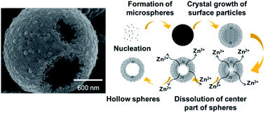
ZnO hollow microspheres were prepared by a solvothermal method, without using a template. The morphology and structure of the microspheres were investigated by scanning and transmission electron microscopies. A series of controlled experiments was carried out to better understand the formation mechanism. The sample prepared at 150 °C for 1 h consisted of ZnO microspheres 1–2 μm in diameter. Microspheres were composed of crystallites of tens of nm in diameter. The surfaces of the microspheres prepared at 150 °C for 3 h were surrounded by hexagonal pyramid-like crystals, of a few hundred nm in diameter. The sample prepared by reaction at 150 °C for 6 h consisted of hollow spheres, even in the absence of a template. The microsphere surface consisted of ZnO c planes. ZnO hollow microspheres prepared at 150 °C for 12 h exhibited intense narrow ultraviolet emission.
) the c(−)-plane, which are terminated with Zn and O atomic planes, respectively. The chemical stability and catalytic activity of ZnO depend on its polarity.8–10 The photocatalytic activity of ZnO strongly depends on the specific crystal planes involved. The (0001) and (000
) faces exhibit higher photocatalytic activity than the perpendicularly-orientated nonpolar (10
0) and (11
0) faces. Thus, ZnO morphologies predominantly exposing c planes are likely to exhibit good photocatalytic behavior. Matsumoto et al. reported ZnO solid microspheres covered with c(+)-planes.11
The preparation of ZnO hollow spheres has received much attention, because of their low density, high surface area and good surface permeability.12,13 ZnO hollow spheres have potential in gas sensing, drug delivery, microcapsule reactors and photoelectric devices.14–16 They also exhibit large light-harvesting efficiencies, so are also applicable in photocatalysts and dye sensitized solar cells. ZnO hollow spheres with surface-exposed c planes are likely to exhibit better performance than ZnO solid microspheres. However, preparing well-crystallized ZnO hollow microspheres with controlled surface morphologies and crystal orientations remains a challenge.17,18
Thermal evaporation19–21 and template hydrothermal processing22,23 are commonly used to prepare ZnO hollow microspheres. The thermal evaporation process generally involves the vaporization of Zn powder, solidification of liquid Zn droplets, oxidation of spherical Zn particle surfaces, and sublimation of Zn within particles. Its disadvantages include the requirement for high temperatures (typically >500 °C) and vacuum conditions. The hydrothermal synthesis of ZnO hollow spheres is usually achieved by either hard22 or soft templating.23 Such templates complicate the synthesis process, and often require subsequent removal steps. The development of facile, low temperature and template-free methods for preparing ZnO hollow spheres is desirable. Zhu et al. reported the template-free preparation of ZnO hollow spheres, in aqueous solution at low temperature.24 Li et al. reported the template-free solvothermal preparation of ZnO hollow spheres, which was assisted by polyoxometalate.25 However, these reports did not clarify the crystal orientation in the prepared hollow spheres. There have been few reports of ZnO spherical hollow powders with surfaces covered with c planes.26,27 Hu et al. prepared ZnO hollow microspheres consisting of ZnO nanorods with surface-exposed c planes, using a water–glycerol solvent.26 Qiu et al. prepared hierarchical Zn1−xCoxO nanodiscs into hollow spheres with surface-exposed c planes, using a water–ethylene glycol solvent. However, further investigation is required to reveal the formation mechanism.27
In the current study, we report the template-free solvothermal preparation of ZnO hollow microspheres, using ethylene glycol (EG) and hexamethylenetetramine (HMT) as the solvent and pH buffer, respectively. The synthetic process is simple, and does not require specialized equipment or templates. Highly crystalline microspheres are obtained at a solvothermal temperature of 150 °C. The morphology of the ZnO particles can be tuned from solid to hollow spheres, by varying the reaction time. The ZnO hollow spheres are covered with c planes. A formation mechanism is proposed, based on the Zn2+ solubility and microsphere inner structure.
![]() | ||
| Fig. 1 SEM images of ZnO particles prepared at 150 °C for (a) 1, (c) 3, (e) 6 and (g) 12 h, and their higher magnification views in (b), (d), (f) and (h), respectively. | ||
Fig. 2 shows the particle size distributions of spheres prepared at 150 °C for different reaction times. Data were obtained from 500 individual spheres observed in SEM images. The mean particle size (standard deviation) for samples prepared for 1, 3 and 12 h were 2.3 (0.5), 3.1 (0.6) and 2.8 (0.6) μm, respectively. The particle size increased with reaction time from 1 to 3 h, because of crystal growth. The particle size of the sample prepared for 12 h was slightly lower, because of dissolution of the nanoparticles.
The size, shape and surface morphology of the solid and hollow microspheres were characterized by TEM. The TEM images of the products prepared for 3, 6 and 12 h dispersed on carbon films are shown in Fig. 3. Contrast between the sphere edges and cores was not observed for the sample reacted for 3 h, indicating that the microspheres were solid (Fig. 3a). Contrast between edges and the core was apparent in some spheres reacted for 6 h (Fig. 3b). When the reaction time was 12 h, the shells appeared porous, and contrast between shells and cores was apparent for all spheres, indicating hollow structures (Fig. 3c). Fig. 4 shows SEM and corresponding cross-sectional TEM images of solid and hollow spheres prepared at 150 °C for 3 h (Fig. 4a–c) and 12 h (Fig. 4d and e). Solid spheres appeared dense, and a radial contrast was observed inside the sphere (Fig. 4b). The region around the center of the sphere appeared porous, while that within ∼500 nm of the surface appeared dense. Different crystal sizes were observed at the surface and core. A selected-area electron diffraction (SAED) pattern of an individual sphere exhibited a ring-like pattern, as shown inset in Fig. 3b. This indicated that the center was composed of polycrystalline ZnO, rather than being amorphous. A high-resolution TEM (HRTEM) image of a sphere surface (Fig. 4c) showed clear and continuous lattice fringes, with spacing of 0.52 nm. This corresponds to the 0001 plane, and indicates that the surfaces of the spheres were covered with c planes. Fig. 4e shows that the sample prepared for 12 h consisted of hollow microspheres, whose shells were composed of needle-like ZnO crystals.
![]() | ||
| Fig. 3 TEM images of top views of ZnO microspheres prepared at 150 °C for (a) 3, (b) 6 and (c) 12 h. | ||
The Zn concentration in the supernatants of reacted solutions was measured, to investigate the formation mechanism of the ZnO hollow microspheres. Fig. 5 shows the variation of supernatant Zn concentration, measured by ICP-AES after each reaction duration at 150 °C. The Zn concentration at 3 h was lower than that at the beginning of the reaction, because Zn2+ was consumed during the formation of ZnO spheres. The concentration increased after 3 h, and became saturated by 9 h. This increase can be attributed to the dissolution of particles at the center of individual spheres.
![]() | ||
| Fig. 5 ICP-AES analysis of the variation in supernatant Zn concentration after various reaction times. | ||
The effect of reaction temperature on the formation of ZnO hollow microspheres was also investigated. For the sample prepared at 100 °C for 12 h, solid microspheres were clearly obtained, as shown in Fig. 6. Their surfaces consisted of hexagonal crystals of hundreds of nm in diameter, and were similar to the surfaces of the sample prepared at 150 °C for 3 h (Fig. 1d). An SEM image of a broken sphere revealed a solid hierarchal structure consisting of pyramid-like crystals (Fig. 6b). Microspheres prepared at 200 °C were composed of larger particles (Fig. 6c), and an SEM image of one of these broken sphere revealed a hollow structure (Fig. 6d). The shell was much thicker than that of the sample prepared at 150 °C. ICP-AES was also used to measure the variation of supernatant Zn concentrations for reactions at 100 and 200 °C (Fig. S2†). No precipitate was obtained after reaction for 1 h at 100 °C, and the Zn concentration was comparable to the initial Zn concentration (∼6000 ppm). For reaction at 100 °C, the supernatant Zn concentration gradually decreased with increasing reaction time, because of the formation of ZnO. An increase in Zn concentration was not observed until a reaction time of 12 h. The supernatant Zn concentration exhibited an unstable change upon reaction at 200 °C. For reaction after 1 h, the Zn concentration was much lower than the initial concentration (∼6000 ppm), and the concentration fluctuated with increasing reaction time. These results indicated that 150 °C was the optimal temperature for preparing ZnO hollow microspheres. This was because dissolution did not occur at lower reaction temperatures (100 °C), and the reaction was too fast to readily control at higher temperatures (200 °C). Fig. 7 shows SEM images and corresponding cross-sectional TEM images of ZnO microspheres prepared at 200 °C for 1 and 4 h. A cross-sectional TEM image of the microspheres prepared at 200 °C reveals a clear difference between the sphere surfaces and centers. The cross-sectional TEM image of the sample prepared for 1 h in Fig. 7b is similar to that of the sample prepared at 150 °C for 3 h (Fig. 3b). The center of the sphere appears porous, and the surface appears to be dense to a depth of ∼200 nm. A high magnification TEM image of the sphere surface shown inset in Fig. 7b indicates the surface consists of pyramid-like crystals. The HR-TEM image in Fig. 7c shows that surface particles were single crystals with 0.52 nm lattice fringes which correspond to the [0001] ZnO growth direction. This indicated that the surface of the ZnO spheres was covered with c planes. Fig. 7d shows that the centers of spheres prepared by reaction for 4 h had started to become hollow. A difference in the sphere's structure was clearly observed from the center to surface, as the structure changed from granular, needle-like to pyramid-like crystals (Fig. 7e). The surface of this sphere was surrounded by pyramid-like crystals 300–600 nm in diameter. Some surface crystals had side facets appearing as brilliant-cut diamonds, which are shown inset in Fig. 7e. The well-resolved lattice fringes also indicate that the surface was highly crystalline with flat planes, as shown in Fig. 7f.
![]() | ||
| Fig. 6 SEM images of ZnO microspheres prepared at (a) 100 and (c) 200 °C for 12 h, and their corresponding high magnification images in (b) and (d), respectively. | ||
Fig. 8a shows a proposed mechanism for formation of the ZnO hollow microspheres, based on the SEM, TEM, XRD and ICP-AES results. The following reactions are proposed to have occurred in the first step of the formation of solid ZnO microspheres:
| C6H12N4 + 6H2O → 6HCHO + 4NH3, | (1) |
| NH3 + H2O → NH4+ + OH−, | (2) |
| Zn2+ + 2OH− → Zn(OH)2, | (3) |
| Zn(OH)2 → ZnO + H2O. | (4) |
The role of HMT (C6H12N4) in the growth of the solid microspheres can be regarded as a pH buffer, because it slowly releases OH− during thermal decomposition (eqn (1) and (2)).28 OH− reacts with Zn2+ to form Zn(OH)2 nanoclusters in solution (eqn (3)), which subsequently transform into an initial ZnO crystal (eqn (4)). The initial crystals agglomerate after nucleation, to form ZnO solid microspheres and thus minimize their surface energy. Extended reaction time results in particles on or near the surface growing into hexagonal crystals. After the degree of supersaturation decreased, the following reaction occurred:
| ZnO + NH3 + 2H+ → Zn(NH3)2+ + H2O. | (5) |
Particles at sphere centers possessing low crystallinity preferentially dissolved according to eqn (5), and hollow spheres are formed through Ostwald ripening.29
We investigated the optical properties of the ZnO hollow microspheres by room-temperature PL measurements. Fig. 9 shows the PL spectrum of the ZnO hollow microspheres, which exhibited strong UV emission at ∼392 nm. While weak emission was observed at ∼420 nm, emission at visible wavelengths was negligible.26,30,31 The UV emission originated from the recombination of free excitons in the ZnO near band edge,32 so that the intensity of the room-temperature UV emission indicated the ZnO quality. Visible emission is associated with interstitial Zn ions,33 oxygen vacancies,34 cationic impurities35 and damaged crystals.36 Visible emission indicates the existence of deep centers within the ZnO. Therefore, the strong band-edge emission accompanied by insignificant visible emission suggests that the ZnO hollow spheres were of high crystal quality. The effect of non-radiative recombination centers was not considered in the current study, so it should not be concluded that the ZnO hollow spheres had a lower defect concentration than previous studies.26,30,31 Further investigation of the total defect concentration of the ZnO hollow spheres is required.
Footnote |
| † Electronic supplementary information (ESI) available. See DOI: 10.1039/c4ra02877e |
| This journal is © The Royal Society of Chemistry 2014 |