Recent advances in H2PO4− fluorescent sensors
Dawei Zhang
ab,
James Robert Cochrane
b,
Alexandre Martinez
*b and
Guohua Gao
*a
aShanghai Key Laboratory of Green Chemistry and Chemical Processes, Department of Chemistry, East China Normal University, 3663 North Zhongshan Road, Shanghai, 200062, P. R. China. E-mail: ghgao@chem.ecnu.edu.cn; Fax: +86 (21)62233323; Tel: +86 (21)62233323
bLaboratoire de Chimie, CNRS, École Normale Supérieure de Lyon, 46, Allée d'Italie, F-69364, Lyon, France. E-mail: alexandre.martinez@ens-lyon.fr
First published on 10th June 2014
Abstract
Dihydrogen phosphate (H2PO4−) plays an essential role in a number of chemical and biological processes. The sensitive and selective detection of H2PO4− is of great interest to many scientific fields, ranging from supramolecular chemistry to life sciences. For the detection of H2PO4−, fluorescent methods have plenty of distinct advantages, for example they are simplistic and allow low levels of determination. Therefore, this review will focus on the current progress in the development of H2PO4− fluorescent sensors based on organic scaffolds, for sensing in both organic and aqueous solutions. Three main types of fluorescent probes will be categorized in this review: (i) intensity-based “turn-off” fluorescent sensors; (ii) intensity-based “turn-on” fluorescent sensors; and (iii) ratiometric fluorescent sensors that involve a ratio of two emission outputs. This review should provide a comprehensive description of this research area to date and be instructive for the design and synthesis of new fluorescent sensors for H2PO4−. In addition, the principles and mechanisms employed in the design of H2PO4− sensors will be thoroughly described.
 Dawei Zhang | Dawei Zhang obtained his BS degree in chemistry from Northeastern University (NEU) in 2011, China. Subsequently, he started a co-sponsored PhD project between the École Normale Supérieure de Lyon (ENS-Lyon) in France and East China Normal University (ECNU) in China. He spent the first two years (2011–2013) in ECNU under the supervision of Prof. Gao. In September 2013, he moved to ENS-Lyon for three years supervised by Dr A. Martinez. His research interests are focused on anion recognition and fluorescent sensing as well as supramolecular catalysis. |
 James Robert Cochrane | James Robert Cochrane completed his PhD at the University of Sydney in 2011.Working with Katrina A. Jolliffe he studied the synthesis of cyclic peptide natural products. He then moved to the University of Melbourne, where he undertook a two year postdoctoral research fellowship with Craig A. Hutton. Here he worked on a number of projects including the synthesis of cyclic peptide hemicryptophanes for molecular recognition. After migrating to France, James is currently working as a postdoc in The Supramolecules, Materials and Stereochemistry team of the École Normale Supérieure de Lyon. |
 Alexandre Martinez | Alexandre Martinez received his Ph.D. in chemistry in 2004 from the University of Toulouse France, for studies asymmetric oxidation under the supervision of Dr B. Meunier. He did a postdoctoral work in 2004–2005 at the University of Geneva under the supervision of Prof. J. Lacour, working on supramolecular chemistry. He joined The Ecole Normale Superieure de Lyon in 2006 as associated professor, with Dr J.-P. Dutasta. His research activities include stereochemistry, catalysis, and supramolecular chemistry, in particular he focusses on hemicryptophane hosts. |
 Guohua Gao | Guohua Gao, Ph.D., Professor, received his Ph.D. in 1993 from Lanzhou Institute of Chemical Physics, Chinese Academy of Sciences. He subsequently joined the Research Institute of Beijing Yanshan Petrochemical Corporation, SINOPEC. From May 1997 to December 2003, he worked respectively at National University of Singapore, Hokkaido University and University of Ottawa. Since August 2003, he has been a member of the Shanghai key laboratory of green chemistry and chemical processes, East China Normal University, China. His research interests are focused on imidazolium chemistry, with the special emphases on the catalysis by imidazolium based ionic liquids and the recognition by imidazolium based receptors. |
1. Introduction
Inorganic phosphate species are biologically relevant anions that have essential roles in genetic information storage, gene regulation, energy transduction, signalling processing and muscle contraction.1,2 Phosphate is also a key constituent of two important biopolymers, DNA and RNA as well as many chemotherapeutic and antiviral drugs.3,4 On the other hand, the over-use of inorganic phosphate in agriculture can lead to excessive algal growth, followed by decomposition and depletion of dissolved oxygen, and ultimately, the eutrophication of aquatic ecosystems.5 Dihydrogen phosphate (H2PO4−) is the predominant equilibrium species of inorganic phosphate at physiological pH. Therefore, H2PO4− is an important target anion, and methods for its detection have received increasing attention of late.6–10The development of sensors for the recognition and detection of anions is of great importance in the field of modern supramolecular chemistry.11–15 In this field fluorescent sensors bear inherent advantages, these include their high sensitivity, simple manipulation and facile visualization. All of these are essential properties for bio-imaging and thus the design of fluorescence-based probes for the detection of H2PO4− is an important area of research.16–26 Artificial fluorescent anion sensors belong to one of the following three design approaches (Scheme 1A): (a) the “binding site-signalling unit”, the interaction of the binding site(s) with anions, causes the change of the electronic properties of the signalling unit. Fluorescent mechanisms that commonly involved in this approach include photo-induced electron transfer (PET), the rigidity effect, fluorescence resonance energy transfer (FRET), excimer/exciplex formation/extinction, photo-induced charge transfer (PCT), and less frequently, excited-state proton transfer (ESPT). (b) The “displacement” protocol, in which the introduction of anions to the coordinated metal complex revives the non-coordinated spectroscopic properties of the indicator. (c) The reaction-based strategy which occurs between the target anions and the “chemodosisensor”. It is worth mentioning that until now, all the H2PO4− fluorescent sensors have fallen into the first two categories, and there has been no reported chemodosisensor for H2PO4− sensing.
 | ||
| Scheme 1 (A) The approaches for designing fluorescent sensors: (a) “binding site-signalling unit” approach; (b) displacement approach; (c) reaction-based chemodosisensor. (B) The possible fluorescent behaviours after anion binding: (i) “turn off” fluorescence; (ii) “turn on” fluorescence; (iii) ratiometric fluorescence. (C) The possible fluorescent mechanisms involved in the corresponding fluorescent behaviours. | ||
The fluorescent phenomena observed after H2PO4− binding also follows three main patterns of behaviour (Scheme 1B): fluorescence quenching, enhancement at the original wavelength, and ratiometric sensing that involves a comparison of intensities at two different emission outputs. Generally the fluorescent behaviour observed depends on the initial design of the sensor (Scheme 1C). (i) For sensors adopting the “binding site-signalling unit” approach, the mechanisms of PET and rigidity effect may quench or enhance the fluorescence of the probe after anion binding, while the FRET, excimer/exciplex formation/extinction, PCT, and ESPT can cause the bathochromic/hypochromatic-shift of the emission resulting in a ratiometric sensing behaviour. (ii) For sensors utilizing the “displacement” protocol, the fluorescent change of the coordinated complex induced by H2PO4− is usually consistent with the initial non-coordinated indicator.
In the last decade, there have been reviews dealing with the subject of phosphate detection,8,9 bio-phosphate recognition,6,7,10 and pyrophosphate fluorescent sensing,27 however to the best of our knowledge, there is no comprehensive review which thoroughly, systematically and timely describes fluorescent sensing of H2PO4−. As H2PO4− is in equilibrium with two other basic anions HPO42− and PO43− at physiological pH,8 the selective sensing of H2PO4− is especially important as well as challenging. For this reason in this review, we only highlight the detection of H2PO4−. In terms of the fluorescent behaviour observed upon H2PO4− binding, we have classified the fluorescent probes based on their modes of action. Fluorescent probes exhibiting “turn-off” detection are covered in Section 2, “turn-on” in Section 3 and ratiometric sensing in Section 4. We subdivide the content of each section into the sensing mechanisms involved. This classification should promote a better understanding of the anion-induced fluorescent behaviour and will be instructive for the design of more selective sensors with the desired fluorescent properties. It should be made clear that in some cases, the mechanism of fluorescence is inferred by the authors and are not demonstrated absolutely. In some instances, two or more possible mechanisms might simultaneously exist in the sensing process, and in these situation, we place the sensor into the category where it can be best explained. It should also be highlighted that the selective recognition of H2PO4− over other common anions is one of the most important issues to be addressed, thus the selectivity of each sensor has been evaluated.
2. Intensity-based “turn-off” fluorescent sensors for H2PO4−
In this section, fluorescent sensors that provide “turn-off” fluorescence detection of H2PO4− will be described. Fluorescence quenching by H2PO4− is a commonly encountered phenomenon which provides a facile approach for monitoring this important anion. PET is one of the most extensively adopted mechanisms for fluorescence quenching (Section 2.1 and Scheme 2). Upon the binding of H2PO4−, the PET process of the sensor is initiated or promoted (Scheme 2a), causing a corresponding decrease in the fluorescence of the sensor. The other methods used in fluorescence detection, such as “displacement” and ligand-to-metal charge transfer are discussed in Sections 2.2 and 2.3. For convenient comparison, the spectroscopic and analytical parameters of each “turn off” fluorescent sensor for H2PO4− have been summarized in Table 1.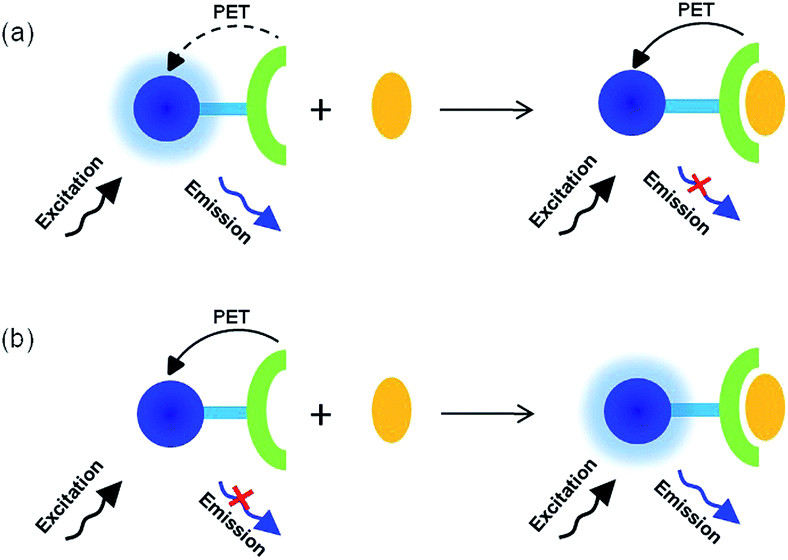 | ||
| Scheme 2 Diagrams for (a) the initiation or promotion of PET and (b) the inhibition of PET after anion binding. | ||
| Sensor | Solvent | Fluorophore | λem (nm) | Fluorescent mechanism | H–G stoichiometry | Ka determined from fluorescence | Ref. |
|---|---|---|---|---|---|---|---|
| 1 | CH3CN | Anthracene | 415 | PET | 1![[thin space (1/6-em)]](https://www.rsc.org/images/entities/char_2009.gif) :1 :1 |
1.3 × 10⁶ M−1 | 28 |
| 2 | CH3CN–DMSO 9:1 (v/v) |
Anthracene | 415 | PET | 1:1 |
>1.3 × 10⁶ M−1 | 29 |
| 3 | CH3CN | Anthracene | 426 | PET | 1:1 |
1.6 × 10⁵ M−1 | 30 |
| 4 | CH3CN | Anthracene | 420 | PET | 1:1, 1:2 |
5.6 × 10³ M−1 | 32 |
| 5a | CH3CN–DMSO–H2O 98:1:1 (v/v/v) |
Benzthiazole | 452 | PET | 1:1 |
7.9 × 10³ M−1 | 33 |
| 6 | CH3CN | Isoquinolyl | 395 | PET | 1:1 |
2.5 × 10⁶ M−1 | 34 |
| 7 | CH3CN | Quinolyl | 350 | PET | 1:1 |
2.8 × 10⁶ M−1 | 34 |
| 8 | H2O | Naphthalene | 478 | Displacement | 1:1 |
1.8 × 10⁶ M−1 | 35 |
| 9 | CH3OH–HEPES buffer 1:1 (v/v) |
2,2′-Dihydroxyazobenzene | 610 | Displacement | 1:1 |
1.6 × 10⁴ M−1 | 36 |
| 10 | CH3OH | — | 330 | Ligand-to-metal charge transfer | 1:2 |
1.0 × 10⁵ M−2 | 37 |
2.1 Initiation or promotion of PET
The fluorescent sensor (1) reported by Kim et al.28 (Fig. 1) bearing two imidazolium groups at the 1, 8-position of anthracene showed significant fluorescence quenching in CH3CN upon addition of H2PO4−. This was due to the formation of (C–H)+⋯X− hydrogen bonds. The binding constant of 1 with H2PO4− was found to be relatively large (1.3 × 10⁶ M−1), however strong competition by F− for H2PO4− binding was observed. To circumvent this, their group subsequently designed the rigid fluorescent sensor 2. Based on the scaffold of 1 two anthracene units were directly connected by the two imidazolium moieties forming a cyclic receptor.29 Significant fluorescence quenching of sensor 2 occurred after addition of H2PO4− in CH3CN–DMSO (9:1, v/v) due to the PET process. More importantly, competitive binding studies demonstrated that there is no interference of the binding of H2PO4− even when in the presence of 1.5 equiv. of F−. The binding constant of sensor 2 with H2PO4− was also found to be larger than that of sensor 1 (>1.3 × 10⁶ M−1).
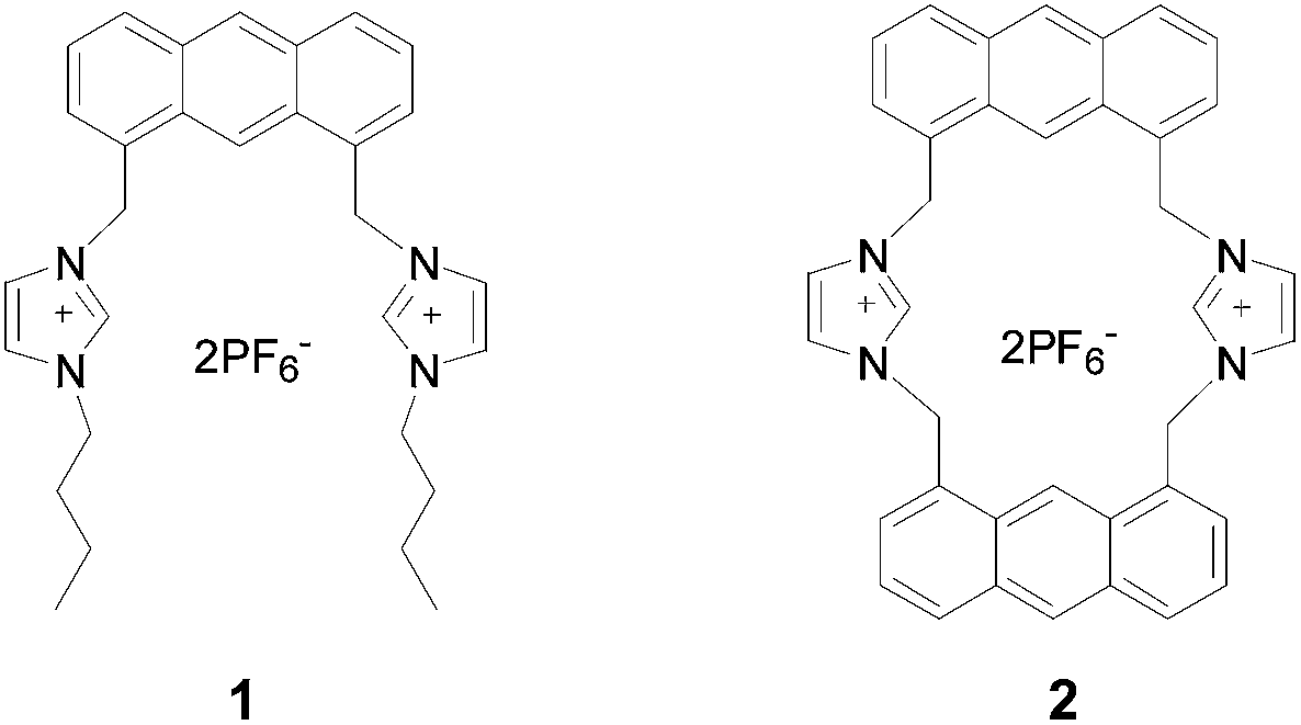 | ||
| Fig. 1 Structures of sensors 1 and 2. | ||
Bearing the concept of the PET process between imidazolium binding sites and the excited state of anthracene in mind, Jadhav et al.30 developed an anthracene-imidazolium-based macrocyclic sensor (3) (Fig. 2). In this sensor the two pendant imidazolium arms are connected by attachment to a molecule of cholestane.31 The sensor 3 exhibited 95% fluorescence quenching after addition of 10 equiv. of H2PO4− in CH3CN with a binding constant of 1.6 × 10⁵ M−1, while only 20–40% decrease in intensity was induced by other common anions (F−, Cl−, Br−, I−, AcO−, HSO4−). Competitive experiments demonstrated that the presence of excess anions (50 equiv.) did not cause any significant changes in the emission of 3 with H2PO4− (5 equiv.).
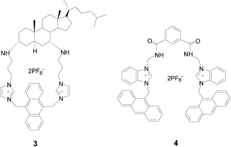 | ||
| Fig. 2 Structures of sensors 3 and 4. | ||
Ghosh and co-workers have32 designed and synthesised a flexible anthracene linked benzimidazolium-based receptor (4) (Fig. 2) which could be used as a “turn-off” fluorescent sensor for H2PO4− over other common anions. The fluorescence was quenched by 72% after addition of 2 equiv. of H2PO4− in CH3CN, while no other changes such as excimer emission were observed. The binding constant was found to be 5.6 × 10³ M−1. A Stern–Volmer plot of sensor 4 with H2PO4− indicated both static and dynamic quenching effects. Since further addition of H2PO4− induced an increase in emission of sensor 4, the authors suggested a two-step process of complexation: 1:1 binding occurred initially, and then changed to a 1:2 (host–guest) stoichiometry when in the presence of excess H2PO4−.
A benzthiazole-based fluorescent sensor (5a) (Fig. 3) was developed for the detection of H2PO4− by Lee and co-workers.33 In order to evaluate its anion recognition performance, receptor 5b was synthesised. The results showed that among all the tested common anions, only H2PO4− resulted in the significant fluorescence quenching of sensor 5a in CH3CN–DMSO–H2O (98:1:1, v/v/v) with a binding constant of 7.9 × 10³ M−1, and no significant anion-binding interactions occurred with sensor 5b. The high selectivity of sensor 5a was attributed to the specific combination of both hydrogen bond-donating and -accepting moieties within the rigid cleft.
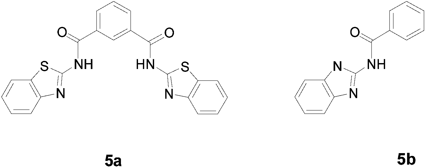 | ||
| Fig. 3 Structures of sensors 5a and 5b. | ||
Recently, Kondo et al.34 reported selective detection of H2PO4− in CH3CN utilizing the fluorescence quenching effect. The synthesised tetraamide-based sensors (6 and 7) contain isoquinolyl and quinolyl moieties respectively (Fig. 4). Especially, sensor 7 bearing 2-quinolyl groups showed selective and nearly complete quenching by H2PO4−, whereas it showed small or no changes towards other anions. The high selectivity of sensors 6 and 7 for H2PO4− (Ka = 2.5 × 10⁶ M−1 and 2.8 × 10⁶ M−1 respectively) can be attributed to the additional hydrogen bonds formed between H2PO4− and the nitrogen atom of the isoquinolyl and quinolyl moieties.
 | ||
| Fig. 4 Structures of sensors 6 and 7. | ||
2.2 “Displacement”: releasing the non-fluorescent indicator
A pyrimidine-naphthalene anchored Schiff base (8) was synthesised by Kumar et al.35 which was used as a “turn-on” fluorescent sensor for Al3+ in aqueous solution due to the inhibition of C![[double bond, length as m-dash]](https://www.rsc.org/images/entities/char_e001.gif) N isomerization after Al3+ complexation (Fig. 5). The formed 8–Al3+ complex could also achieve the “turn-off” sensing of H2PO4− with a detection limit of 2.27 × 10⁻⁷ M via protonation of aldimine-nitrogen of sensor 8 by H2PO4−. This releases the non-fluorescent 8 from the formed Al3+ complex. Unfortunately, HSO4− which has a lower pKa value (1.99 vs. 3.88) also gave rise to the similar fluorescence quenching behaviour, while other common anions showed no fluorescent response.
N isomerization after Al3+ complexation (Fig. 5). The formed 8–Al3+ complex could also achieve the “turn-off” sensing of H2PO4− with a detection limit of 2.27 × 10⁻⁷ M via protonation of aldimine-nitrogen of sensor 8 by H2PO4−. This releases the non-fluorescent 8 from the formed Al3+ complex. Unfortunately, HSO4− which has a lower pKa value (1.99 vs. 3.88) also gave rise to the similar fluorescence quenching behaviour, while other common anions showed no fluorescent response.
 | ||
| Fig. 5 Sensing mechanism of sensor 8 towards H2PO4−. | ||
The mononuclear 2,2′-dihydroxyazobenzene (DHAB)–Zn2+ complex (Fig. 6) was developed as a cost-effective “on-off” H2PO4− sensor based on the “displacement” protocol in CH3OH–HEPES buffer (1:1, v/v).36 Obvious fluorescence quenching of the Zn2+–9 complex, after addition of H2PO4− was observed due to H2PO4−-induced decomposition of the DHAB–Zn(II) complex releasing the non-fluorescent DHAB. An opposite increase in fluorescence was induced by CN−, while minimal or no changes were observed for other anions. The Zn2+–9 complex could also be used as a colorimetric receptor for H2PO4− due to the marked solution colour changes.
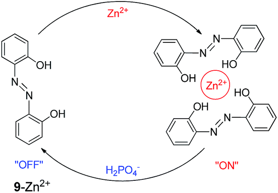 | ||
| Fig. 6 Sensing mechanism of sensor 9 towards H2PO4−. | ||
2.3 Other mechanisms
Chen et al.37 prepared a fluorescent tetranuclear pentacoordinated Zn(II) complex (10) based on a cresolic oxygen bridging ligand (L) shown in Fig. 7. The fluorescent properties of 10 were attributed to the chelation of L to the Zn2+ centers, which enhanced the rigidity of L. After addition of H2PO4− to a CH3OH solution of sensor 10 remarkable fluorescence quenching was observed with a binding constant of 1.0 × 10⁵ M−2 (1:2 binding mode). This may be a result partly from the electron repelling effect of H2PO4− and partly from the decrease of ligand rigidity because of H2PO4− binding. Both effects can prohibit the ligand-to-metal charge transfer reducing the fluorescence intensity. Other common anions only caused moderate fluorescence quenching or even enhancement of the fluorescence of sensor 10.
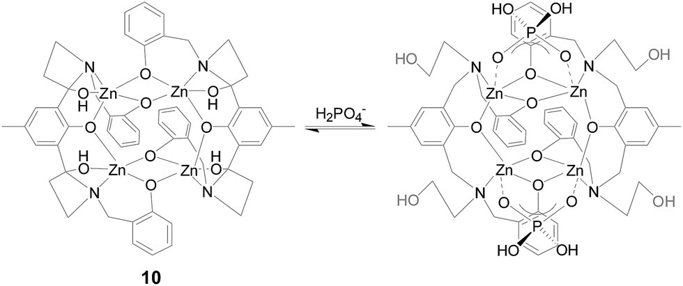 | ||
| Fig. 7 Proposed binding mode of sensor 10 with H2PO4−. | ||
3. Intensity-based “turn-on” fluorescent sensors for H2PO4−
In this section, the advances in “turn-on” H2PO4− fluorescent probes will be described. Compared with the “turn-off” type, these are preferable as they circumvent some of the drawbacks of fluorescence quenching, for example, the limited sensitivity and restricted practical applications. Fluorescence enhancement can occur by a number of pathways such as the prohibition of PET (Section 3.1 and Scheme 2b), the increase of rigidity of the sensors (Section 3.2) and “displacement” which releases the fluorescent indicator after addition of H2PO4− (Section 3.3). The spectroscopic and analytical parameters of each “turn on” fluorescent sensor for H2PO4− have been summarized in Table 2.| Sensor | Solvent | Fluorophore | λem (nm) | Fluorescent mechanism | H–G stoichiometry | Ka determined from fluorescence | Ref. |
|---|---|---|---|---|---|---|---|
| 11a | CH3CN | Binaphthol | 365 | PET | 1:1 |
1.2 × 10⁴ M−1 | 38 |
| 12 | CH3CN | Anthracene | 420 | PET | 1:1 |
— | 39 |
| 13 | CH2Cl2–CH3OH 9:1 (v/v) |
Anthracene | 421 | PET | 1:1 |
2.8 × 10³ M−1 | 40 |
| 14 | CH3CN | Anthracene | 432 | PET, rigidity effect | 1:2 |
5.5 × 10⁹ M−2 | 41 |
| 15 | DMSO | Schiff-base | 333 | PET, rigidity effect | 1:1 |
— | 42 |
| 16 | CH3CN | Quinoline | 393 | PET, rigidity effect | 1:1 |
3.3 × 10⁴ M−1 | 43 |
| 17 | CH3CN | Ru-complex | 608 | Rigidity effect | 1:2 |
7.6 × 10⁹ M−2 | 44 |
| 18 | DMSO | Binaphthol | 396 | Rigidity effect | 1:1 |
5.0 × 10³ M−1 | 45 |
| 19 | CH3CN–MOPS buffer 3:1, (v/v) |
Naphthalimide | 540 | Displacement | — | — | 46 |
| 20 | CH3CN | Acridine | 422 | Additional hydrogen bond | 1:2 |
>108 M−2 | 47 |
| 21 | DMSO–HEPES buffer 1:9 (v/v) |
Schiff-base | 525 | Intramolecular hydrogen bond | 1:1 |
2.0 × 10³ M−1 | 48 |
3.1 Inhibition of PET
A benzimidazolium-based macrocyclic fluorescent sensor (11a) (Fig. 8) was reported by Ghosh et al.38 Significant fluorescence enhancement of sensor 11a after binding H2PO4− was observed in CH3CN which was assumed to be related to the deactivation of the PET process occurring between the macrocyclic binding domain and the excited state of the BINOL fluorophore. Furthermore, other anions, except fumarate, interacted weakly with sensor 11a. The selectivity and binding affinity of sensor 11a towards H2PO4− was greater than the acyclic sensor 11b (Ka = 1.2 × 10⁴ M−1 and 7.5 × 10³ M−1 respectively). | ||
| Fig. 8 Structures of sensors 11a and 11b. | ||
By inhibiting the PET process from an anthracene fluorophore to pyridinium moieties, Gong et al.39 developed a H2PO4− fluorescent sensor (12) (Fig. 9). The sensor 12 displayed an excellent H2PO4− selectivity over the tested common anions in both CHCl3 and CH3CN by significant enhancement of monomer emission of anthracene. Interestingly, it was found that during the titration in CHCl3, two-mode sensing of H2PO4− was exhibited: addition of less than equimolar H2PO4− induced the enhancement of excimer emission of the anthracene moiety alone, while continuous addition until 3 equiv. of H2PO4− made significant enhancement of only monomer emission overwhelming the excimer emission. However, in the polar solvent CH3CN, only the enhancement of monomer emission was observed during the whole titration. The possible binding process of sensor 12 with H2PO4− in CHCl3 was proposed in Fig. 9.
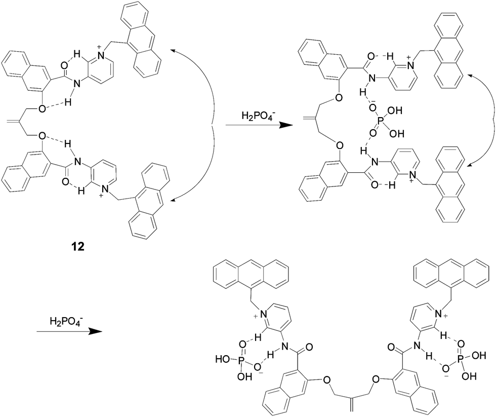 | ||
| Fig. 9 Possible binding mode of sensor 12 with H2PO4− in CHCl3. | ||
Recently, Cao et al.40 synthesised an anthracene-based fluorescent sensor (13) bearing two 1,2,3-triazolium groups (Fig. 10). The sensor 13 showed efficient fluorescence enhancement after addition of H2PO4− in CH2Cl2–CH3OH (9:1, v/v) with a binding constant of 2.8 × 10³ M−1, while no obvious change was observed for other common anions. The presence of 100 equiv. of other anions did not cause any significant change for the emission of sensor 13 with 50 equiv. of H2PO4−. The unique fluorescence enhancement of 13 induced by H2PO4− can be attributed to the formation of the (C–H)+⋯O interaction which diminished acceptor properties of the triazolium ions inhibiting the PET process from anthracene to the charged triazoliums.
 | ||
| Fig. 10 The structure of sensors 13. | ||
A neutral fluorescent sensor (14) (Fig. 11) based on a calix[4]arene tetraamide derivative and anthracene was synthesised by Chen and co-workers.41 The fluorescence of sensor 14 was efficiently enhanced about 130% [(I − I0)/I0] upon addition of 5 equiv. of H2PO4− in CH3CN. More importantly, it exhibited a high selectivity for H2PO4− over a wide range of common anions. The binding constant between 14 and H2PO4− was 5.5 × 10⁹ M−2 with a 1:2 stoichiometry. The effective emission enhancement of sensor 14 towards H2PO4− is probably due to the inhibition of PET or the increased rigidity.
 | ||
| Fig. 11 Structures of sensors 14–16. | ||
A simple H2PO4− fluorescent sensor (15) bearing phenol and thiourea binding sites was developed by Shao and co-workers.42 The presence of H2PO4− induced the “off-on” fluorescence of 15 in DMSO. This is presumably due to both the inhibition of PET and the binding-induced increased rigidity of the sensor. A distinct colour change was also observed, which could be attributed to the deprotonation of the phenol. The binding constant of 15 with H2PO4− was determined to be 5.6 × 10⁴ M−1. Unfortunately, F− and AcO− exhibited significant interference for the detection of H2PO4− using sensor 15.
Goswami et al.43 developed a Cu2+-based fluorescent sensor (16) for H2PO4− in CH3CN. In the absence of H2PO4−, the Cu2+-complex was in a fluorescence-quenching state. However, when in the presence of H2PO4−, the emission intensity of the complex increased dramatically, while much smaller increases in intensity were observed for other anions. The binding constant between 16 and H2PO4− was measured to be 3.3 × 10⁴ M−1. The metal complex might form a suitable cavity for the selective inclusion of H2PO4−, as a consequence, the rigidity of the formed complex increased after H2PO4− complexation. In addition, the anion binding may also suppress the extent of electron transfer between the quinolines and Cu2+ resulting in the fluorescence enhancement.
3.2 Increase of rigidity effect
As mentioned for sensors 14–16, apart from the inhibition of PET, the rigidity effect has been employed to explain the fluorescence enhancement upon anion binding. Indeed the increased rigidity of the formed binding complex could make the non-radiative decay from the excited state less probable and consequently, the emission intensity increases.17Ruthenium(II) complex (17) (Fig. 12) has been developed as a selective fluorescent sensor for H2PO4− in CH3CN.44 Almost 3-fold fluorescence enhancement of sensor 17 was observed after addition of 10 equiv. of H2PO4−. The enhanced emission of 17 in the presence of H2PO4− was caused by the formation of a hydrogen bond between H2PO4− and imidazolyl NH which increased its rigidity and planarity. The binding constant determined by fluorescence was as large as 7.6 × 10⁹ M−2 (1:2 binding mode). In contrast, the same amount of F− and AcO− resulted in significant fluorescence quenching due to the deprotonation of NH, while the changes induced by other common anions were negligible. It is worth mentioning that sensor 17 can also be used as a colorimetric receptor for Fe2+ in CH3CN–HEPES (1:71, v/v) and is thus a bifunctional sensor.
 | ||
| Fig. 12 Structures of sensors 17–18. | ||
Huang et al.45 designed and synthesised an easily prepared H2PO4− fluorescent sensor (18) containing urea group based on binaphthyl. The sensor 18 displayed switch-on fluorescence and the largest binding constant towards H2PO4− in DMSO. Upon complexation with H2PO4−, sensor 18 was rigidified, causing the vibrational and rotational relaxation modes of non-radiative decay inhibited, leading to the increase of emission. Unfortunately, F− and AcO− can also give rise to the fluorescence enhancement with similar binding constants (∼103 M−1).
3.3 “Displacement”: releasing the fluorescent indicator
A naphthalimide derivative (19) (Fig. 13) was developed as a fluorescent sensor by Chen and co-workers.46 The sensor 19 displayed obvious fluorescence quenching in the presence of Cu2+ in CH3CN–MOPS buffer (3:1 v/v) due to the inherent paramagnetic nature of Cu2+. The formed metal complex exhibited a reversible fluorescence enhancement after addition of H2PO4−, while the disturbances of other common anions were subtle. The retrievable “off-on” fluorescent behaviour of sensor 19 can be attributed to the release of the fluorescent 19 from the formed Cu2+–19 complex after addition of H2PO4−.
 | ||
| Fig. 13 Sensing mechanism of sensor 19 towards H2PO4−. | ||
3.4 Other mechanisms
An acridine derivative, bearing two imidazolium groups, as a selective fluorescent sensor (20) (Fig. 14) for H2PO4− has been developed by Kim and co-workers.47 Significant increase of the fluorescence of 20 was observed after addition of H2PO4− in CH3CN, while fluorescence quenching behaviour was observed for other anions. The binding constant of 20 with H2PO4− was measured to be larger than 108 M−2 with a 1:2 stoichiometry. Compared with their previously reported H2PO4− “turn-off” fluorescent sensor (1 in Fig. 1), the only difference is the central nitrogen atom on the acridine fluorophore. Thus the authors inferred that the additional hydrogen bond formed between the nitrogen on the acridine moiety and H2PO4− afforded the anion-induced fluorescence enhancement.
 | ||
| Fig. 14 Structures of sensors 20 and 21. | ||
Recently, a new fluorescent sensor (21) for H2PO4− based on a Schiff-base was reported by Sen and co-workers.48 After excitation in the visible region wavelength (480 nm), the sensor 21 could detect H2PO4− optically by the unique fluorescence enhancement at 525 nm in DMSO–HEPES buffer (1:9, v/v) with a detection limit of 3.5 × 10⁻⁶ M. The binding constant was determined to be 2.0 × 10³ M−1. The response was due to the formation of the intermolecular hydrogen bonds between H2PO4− and sensor breaking the intramolecular hydrogen bonding network between the three phenol residues. In addition, the presence of other common anions did not affect the detection of H2PO4−. Bio-imaging studies indicated that sensor 21 is an efficient staining agent and could be used for monitoring intracellular H2PO4−.
4. Ratiometric fluorescent sensors for H2PO4−
Realizing ratiometric fluorescent sensing of H2PO4−, which involves the ratio of fluorescence intensities at two different wavelengths before and after H2PO4− binding, is a current research focus. Compared with the “turn-off” or “turn-on” fluorescent sensors, ratiometric fluorescent probes present several advantages, for example they permit signal rationing and thus increase the dynamic range and provide built-in correction for environmental effects. In addition, the distinct fluorescent colour change after anion complexation will be practically useful for both visual sensing and convenient bio-imaging of anions. A typical mechanism affording this phenomenon is the formation or extinction of an intramolecular (Section 4.1, Scheme 3a and Table 3) or intermolecular (Section 4.2, Scheme 3b and Table 4) excimer. This will give rise to a change of the intensity ratio between monomer and excimer emissions of the fluorophore based upon the amount of the added anions. ESPT and other principles will also be described in Section 4.3 and 4.4 (Table 5). | ||
| Scheme 3 Diagrams for (a) the intramolecular excimer formation and (b) the intermolecular excimer formation after anion binding. | ||
| Sensor | Solvent | Fluorophore | λmonomer emission (nm) | λexcimer emission (nm) | H – G stoichiometry | Ka determined from fluorescence | Ref. |
|---|---|---|---|---|---|---|---|
| 22 | CH3CN | Naphthalene | 350 | 456 | 1:1 |
9.1 × 10³ M−1 | 49 |
| 23 | CH3CN | Anthracene | 419 | 525 | 1:1, 1:2 |
5.4 × 10³ M−1 | 50 |
| 24 | CH3CN–DMSO 99–1 (v/v) | Anthracene | 418 | 500 | 1:1 |
3.1 × 10⁴ M−1 | 51 |
| 25 | CHCl3–CH3CN 98:2 (v/v) |
Anthracene | 413 | 513 | 1:1, 1:2 |
1.2 × 10⁴ M−1 | 52 |
| 26 | CH3CN | Pyrene | 403 | 482 | 1:1 |
2.2 × 10⁴ M−1 | 53 |
| 27a | CH3CN | Anthracene | 412 | 507 | 1:2 |
2.5 × 10⁴ M−1, 2.2 × 10⁴ M−1 | 54 |
| 28 | CH3CN | Anthracene | 420 | 485 | 1:1 |
2.2 × 10⁵ M−1 | 55 |
| 29 | CH3CN | Anthracene | 418 | 500 | 1:1, 1:3 |
— | 57 |
| 30 | THF | Pyrene | 377 | 477 | 1:1 |
1.9 × 10⁵ M−1 | 58 |
| 31 | THF | Pyrene | 375 | 477 | 1:2 |
— | 59 |
| 32 | CH3CN | Pyrene | 380 | 483 | 1:1 |
— | 60 |
| 33 | CH3CN–CH2Cl2–H2O 1000:1:5 (v/v/v) |
Pyrene | 396 | 485 | 1:1 |
1.0 × 10⁵ M−1 | 61 |
| Sensor | Solvent | Fluorophore | λmonomer emission (nm) | λexcimer emission (nm) | H – G stoichiometry | Ka determined from fluorescence | Ref. |
|---|---|---|---|---|---|---|---|
| 34 | CHCl3–DMSO 99.9:0.1 (v/v) |
Naphthalene | 338 | 420 | 1:1 |
— | 63 |
| 35 | HEPES buffer | Anthracene | 432 | 490 | 1:1 |
>4.6 × 10⁵ M−1 | 64 |
| 36 | CH3CN | Anthracene | 429 | 500 | 1:1 |
3.0 × 10⁶ M−1 | 65 |
| 37 | CH3CN | Pyrene | 400 | 493 | 1:1 |
2.4 × 10⁶ M−1 | 66 |
| 38a | CH3CN | Acridine | 430 | 480 | 1:1 |
5.1 × 10⁴ M−1 | 67 |
| 39 | CH3CN | Acridine | 430 | 556 | 1:1 |
— | 68 |
| 40 | CH3CN | Acridine | 430 | 544 | 1:1 |
— | 68 |
| 41 | CH3CN | Acridine | 430 | 503 | 1:1 |
— | 68 |
| 42 | CH3CN | Acridine | 430 | 466 | 1:1 |
— | 68 |
| 43 | CH3CN | Acridine | 430 | 501 | 1:1 |
2.9 × 10⁶ M−1 | 69 |
| 44a | CH3CN | Anthracene | 428 | 500 | 1:1 |
2.6 × 10⁵ M−1 | 69 |
| Sensor | Solvent | Fluorophore | Original λem (nm) | Induced λem (nm) | H–G stoichiometry | Ka determined from fluorescence | Ref. |
|---|---|---|---|---|---|---|---|
| 45 | DMSO–1,4-dioxane 1:1 (v/v) |
Coumarin | 430 | 540 | 1:1 |
2.0 × 10⁶ M−1 | 70 |
| 46 | CH3CN–H2O 9:1 (v/v) |
Naphthalene | 343 | 412 | 1:1 |
1.0 × 10⁵ M−1 | 74 |
| 47 | CHCl3 | Acridine | 420 | 510 | — | — | 75 |
| 48 | CH3CN–DMSO 99.99:0.01 (v/v) |
Azaindole | 365 | 480 | 1:1 |
2.0 × 10⁴ M−1 | 76 |
| 49 | CH3CN–DMSO 99.99:0.01 (v/v) |
Azaindole | 365 | 480 | 1:1 |
1.1 × 10⁴ M−1 | 77 |
| 50 | CH3CH2OH–THF 3:1 (v/v) |
Hexaphenylbenzene | 438 | 366 | — | — | 78 |
4.1 Intramolecular excimer formation/extinction
A series of tweezer-like fluorescent sensors (22–27) (Fig. 15 and 16) have been developed by Ghosh and co-workers.49–54 These sensors utilized an intramolecular excimer based on either naphthalene, anthracene or pyrene fluorophores. By incorporating benzimidazolium or amide moieties as anion binding sites, these sensors could achieve ratiometric sensing of H2PO4− either in organic or aqueous media with moderate binding constants (∼104 M−1) but high sensing selectivity.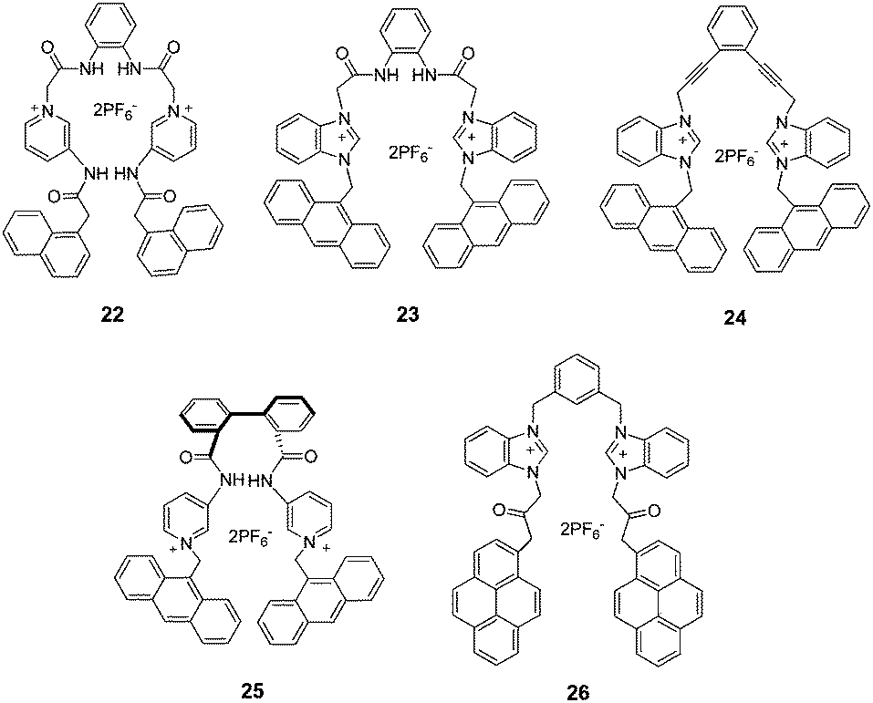 | ||
| Fig. 15 Structures of sensors 22–26. | ||
 | ||
| Fig. 16 Structures of sensors 27a and 27b. | ||
The ortho-phenylenediamine based sensor 22 (ref. 49) could selectively bind H2PO4− in CH3CN by exhibiting excimer emission at 456 nm due to the π–π stacking between two pendant naphthalene fluorophores. The sensor 23,50 based on the same ortho-phenylenediamine unit, could recognize H2PO4− in CH3CN displaying a significant decrease of monomer emission of anthracene, along with a weak increase of excimer emission at 525 nm. A ratiometric change in emission after addition of H2PO4− was also achieved by the enediyne scaffold based sensor 24 (ref. 51) in CH3CN containing 1% DMSO. Compared with 23, in spite of the absence of the amide moieties as hydrogen bonding sites in sensor 24, the binding constant of sensor 24 with H2PO4− was larger than that of 23 (5.4 × 10³ vs. 3.1 × 10⁴ M−1) possibly because the linear nature of the triple bonds in 24 make the sensor more rigid. Pyridinium amide-based sensor 25 (ref. 52) built on a biphenyl scaffold showed selective complexation of H2PO4− with the enhancement of both monomer and excimer emission at 413 and 513 nm respectively in CHCl3 containing 2% CH3CN. By utilizing pyrene as the signalling unit, the benzimidazolium-based sensor 26 (ref. 53) was synthesised and used as an efficiently ratiometric fluorescent sensor for H2PO4− in CH3CN due to its capacity of forming an excimer with pyrene.
An anthracene-labeled 1,2,3-triazole-linked bispyridinium sensor (27a)54 (Fig. 16) was also developed as a ratiometric fluorescent sensor for H2PO4− over other common anions in CH3CN. Job's plot analysis demonstrated a 1:2 stoichiometry between host and guest, and the binding constants were 2.5 × 10⁴ M−1 and 2.2 × 10⁴ M−1 for the successive complexation of the guest molecules. Additionally, the sensor 27a could form a stable gel after addition of H2PO4− in CHCl3–CH3CN (9:1, v/v), which also provided a convenient approach for visually sensing H2PO4−. Interestingly, the control sensor 27b without the triazole links failed to form the gel with H2PO4−, highlighting the crucial role played by the triazole links on gel formation and H2PO4− recognition.
Xu et al.55 also developed a structurally similar ratiometric fluorescent sensor (28) (Fig. 17) for H2PO4− in CH3CN based on imidazolium and anthracene groups. The sensor 28 displayed a unique excimer peak at 485 nm only in the presence of H2PO4− with a binding constant of 2.2 × 10⁵ M−1. The addition of 10 equiv. of other anions did not cause any obvious change to the emission of 28 with H2PO4− (2 equiv.). The rigid phenanthroline moiety in sensor 28 allowed specific hydrogen bonds with H2PO4− accounting for the remarkable selectivity observed.
 | ||
| Fig. 17 The structure of sensor 28. | ||
Most H2PO4− ratiometric fluorescent sensors involving the formation of an intramolecular excimer contain two appended rigid fluorophores. However some sensors bearing multiple fluorophore substituents have been reported. It was believed that tripodal shaped receptors with anion binding groups attached on the three pendant arms would be able to complex anions effectively, due to their preferable pre-organization and conformational flexibility.56 Thus Ghosh et al.57 synthesised a new benzimidazolium-based tripodal fluorescent sensor (29) (Fig. 18) bearing three pendant anthracene rings. The sensor 29 exhibited a high selectivity towards H2PO4− among all the common anions in CH3CN via significant quenching of monomer emission (418 nm) along with the formation of a weak excimer at 500 nm.
 | ||
| Fig. 18 Structures of sensors 29–31. | ||
A two-arm pyridine amide sensor (30) (Fig. 18) bearing four pyrenes as signal units was synthesised by Liao and co-workers.58 This sensor, which provided a pseudo-tetrahedral cleft and multiple hydrogen bonds, formed a 1:1 complex with H2PO4− in THF leading to a significant decrease of the emission intensity ratio between the pyrene monomer (377 nm) and the excimer (477 nm). Further experiments demonstrated that the excimer emission of 30 was formed between two pyrene rings in different arms. Subsequently, a similar two-arm compound (31) based on ferrocene was developed as a ratiometric fluorescent sensor for H2PO4− by the same group.59 The introduction of ferrocene offered another approach, i.e. cyclic voltammetry, for investigation of anion binding. Interestingly, in this case, the two-arm ferrocene hexamide sensor 31 formed a complex with H2PO4− with 1:2 stoichiometry: with each arm bound to one H2PO4− molecule independently. A synclinal conformation was adopted by sensor 31 in THF which was further stabilized by complexation with H2PO4−.
The above-documented H2PO4− ratiometric sensors mostly relied on hydrogen bonding interactions. However, taking advantage of the ion-pairing electrostatic interaction between a metal ion and an anion, would also be useful as this would permit simultaneous binding of cationic and anionic species. It would also allow for a potential co-operative effect to be investigated, in which the anion/cation binding ability of the sensor could be significantly enhanced when in the presence of a bound cation/anion.
A fluorescent sensor (32) (Fig. 19a) bearing pyreneamides as anion binding sites and a signalling unit based on calix[4]crown-5 has been designed and synthesised by Choi and co-workers.60 The fluorescent excimer emission of 32 was first significantly increased after addition of K+ in CH3CN. Subsequently, it was observed that the excimer emission between the two pyrenes of 32 K+ declined obviously upon subsequent addition of H2PO4−, while no change for other common anions occurred. The fact that the 32 K+ complex could achieve ratiometric sensing of H2PO4− while 32 cannot demonstrated that the binding of K+ for 32 favoured the following association with H2PO4− as illustrated in Fig. 19a, i.e. the ion-pairing electrostatic interaction between K+ and H2PO4− played an important role in the sensing behaviour. However, it cannot be neglected that to saturate sensor 32 a large excess of H2PO4− was required (10000 fold) and the K+ coordinated complex might be decomposed at such high concentrations of H2PO4−.
 | ||
| Fig. 19 Structures of sensors 32 and 33, and their binding modes with metal ions and H2PO4−. | ||
A Similar fluorescent sensor 33 (Fig. 19b) based on a pyrene-linked triazole-modified homooxacalix[3]arene was synthesised by Ni and co-workers.61 The sensor showed ratiometric sensing of Zn2+ by enhancing the monomer emission while weakening the excimer in CH3CN–CH2Cl2–H2O (1000:1:5, v/v/v). Interestingly, the subsequent addition of H2PO4− to the solution of 33 Zn2+ resulted in a reversed ratiometric fluorescent change with a detection limit of 1.52 × 10⁻⁷ M. The binding constant was determined to be 1.0 × 10⁵ M−1. The observation that only small fluorescence intensity changes of monomer and excimer emissions of 33 were induced by H2PO4− indicated that Zn2+ played a key role in sensing of H2PO4−. Furthermore, there is no interference for the detection of 40 equiv. of H2PO4− when in the presence of 40 equiv. of other anions.
4.2 Intermolecular excimer formation/extinction
Apart from intramolecular π–π stacking between two appended fluorophores, the intermolecular excimer which occurs between molecules is also a widely used strategy for designing ratiometric sensors. The change of the intensity ratio between the monomer and excimer emissions of the sensor when in the presence of a certain equivalent of anions in various concentrations can confirm whether the formation of excimer is intramolecular or intermolecular. If the values remain constant at different concentrations, the anion-induced excimer occurs intramolecularly. However if there is a change in the ratio, the excimer is being formed intermolecularly.62A naphthalene-based 2,5-diketopiperazine (34) (Fig. 20) was developed as a ratiometric fluorescent sensor for H2PO4− in CHCl3 containing 0.1% DMSO.63 The monomer emission (338 nm) of sensor 34 was dramatically quenched when in the presence of H2PO4− along with the appearance of a weak excimer peak at 420 nm. The binding constant was measured to be 4.7 × 10² M−1 with UV method. The H2PO4−-induced excimer was assigned to be an intermolecular interaction, since the bifunctional hydrogen bonding donor sites in H2PO4− are able to bring two naphthalene residues into close proximity by forming hydrogen bonds with the oxygen atoms between two neighboring diketopiperazines. Unfortunately, aliphatic dicarboxylic acids, such as malonic, succinic, glutaric and adipic acids, could also give rise to the similar fluorescent behaviour, showing the poor selectivity of the sensor.
 | ||
| Fig. 20 Structures of sensors 34–36. | ||
As metal–ligand receptors have a strong binding affinity for anions in competitive media, Huang et al.64 synthesised an anthracene derivative (35) containing Zn2+ sites as a ratiometric fluorescent sensor for H2PO4−. This sensor showed a selective fluorescence enhancement of the excimer emission at 490 nm as well as decrease of monomer emission at 432 nm with H2PO4− in 0.01 M HEPES buffer, pH = 7.4. The binding constant was measured to be larger than 4.6 × 10⁵ M−1 by fluorescence. The formation of the anthracene dimer between two molecules induced by H2PO4− was attributed to binding of H2PO4− within the four zinc binding sites as well as due to a favorable π–π interaction between two flat hydrophobic anthracene rings. Other common anions except SO42− only cause a minor fluctuation in monomer emission. In the case of SO42−, a weak excimer emission could be induced when 10 equiv. were added.
A similar anthracene derivative (36) bearing amidopyridinium groups as anion binding sites was developed as a fluorescent sensor for H2PO4−.65 A concentration-dependent experiment showed that a new excimer peak was observed at high concentration of 36, demonstrating that the sensor has a tendency to aggregate in solution. The anion sensing results revealed that among all the tested anions, only H2PO4− could induce the strong excimer emission of 36 in CH3CN with a binding constant of 3.0 × 10⁶ M−1, which can be attributed to H2PO4−-templated assembly of sensor molecules forming the anthracene excimer. The detection limit was determined to be 3.62 × 10⁻⁷ M.
Taking advantage of the strategy of anion-induced intermolecular π–π stacking, recently, in our group we also synthesised a series of acyclic and macrocyclic H2PO4− ratiometric fluorescent sensors bearing benzimidazolium and urea groups as binding sites built on pyrene, acridine or anthracene fluorophore.66–69
Initially, the synthesis of a flexible pyrene-based fluorescent sensor 37 (Fig. 21) bearing benzimidazolium and urea groups was achieved.66 This sensor was able to distinguish H2PO4− from other anions in CH3CN by displaying ratiometric fluorescent sensing behaviour. Even though there are two pyrene rings per sensor molecule, the pyrene excimer was formed through an intermolecular pattern as illustrated in Fig. 21. This was demonstrated by the change of the intensity ratio of IE/IM which varied with the concentration of sensor 37 when in the presence of 2 equiv. of H2PO4−. The detection limit of H2PO4− was achieved as 5.02 × 10⁻⁷ M. A synergistic effect between benzimidazolium and urea moieties can account for the high binding constant obtained (Ka = 2.4 × 10⁶ M−1).
 | ||
| Fig. 21 The binding mode of sensor 37 with H2PO4−. | ||
To investigate the synergistic binding effect more accurately, we further designed an acridine derivative (38a) containing benzimidazolium and urea moieties (Fig. 22).67 For comparison, sensors 38b and 38c which only possess one type of binding site were prepared. Sensing results showed that 38a could be used as a ratiometric fluorescent sensor for H2PO4− in CH3CN by exhibiting a significant decrease of monomer emission at 430 nm and increase of the excimer at 480 nm with a binding constant of 5.1 × 10⁴ M−1. In contrast, the ratiometric sensing behaviours of sensors 38b and 38c toward H2PO4− were rather poor. A 2:2 binding complex was proposed between 38a and H2PO4−, which was directly detected by HRMS analysis. In addition, the sensor 38a was also able to detect HSO4− in CH3CN according to obvious fluorescence quenching (Ka = 1.7 × 10⁶ M−1), while almost no response towards other anions was observed.
 | ||
| Fig. 22 Structures of sensors 38a–c. | ||
Macrocyclic anion receptors tend to display higher binding constants than their acyclic counterparts due to their well-preorganized topology. Therefore, we synthesised a series of acridine derived benzimidazolium macrocyclic sensors (39–42) (Fig. 23).68 X-ray crystal structures revealed that these sensors had a tendency to aggregate forming acridine dimers. Anion binding studies showed that all the four sensors displayed “turn-on” as well as a bathochromic-shift in fluorescence emission only in the presence of H2PO4− in CH3CN. Furthermore, different cavity size (39, 40 vs. 41) or rigidity (40 vs. 42) of the sensors exhibited different bathochromic-shifts (from 36 to 126 nm) giving rise to various fluorescent colour changes. The tunable bathochromic-shifted emissions of these sensors induced by H2PO4− were attributed to anion-directed assembly of sensors forming the acridine excimers to a different extent.
 | ||
| Fig. 23 Structures of sensors 39–42 and their binding modes with H2PO4−. | ||
In order to further improve the binding affinities of the macrocyclic sensors 39–42 with H2PO4−, a urea moiety was then introduced. The aim being that the urea binding site would synergistically bind H2PO4− along with benzimidazolium moiety.69 Results showed that compared with sensors 39–42, the sensor 43 (Fig. 24) had a better ratiometric sensing performance toward H2PO4− in CH3CN, such as a higher binding constant (2.9 × 10⁶ M−1) and lower detection limit (1.0 × 10⁻⁶ M). In addition, no significant variation in the intensity ratio (I501/I430) was found for the detection of 2 equiv. of H2PO4− when in the presence of 20 equiv. of other anions. Furthermore, interesting results were also achieved from a structurally similar anthracene cyclophane (44a) which exhibited an improved anion binding performance toward H2PO4− compared to the sensor 44b, which incorporated only benzimidazolium groups (Ka = 2.6 × 10⁵ M−1 and 1.6 × 10⁵ M−1 respectively).
 | ||
| Fig. 24 Structures of sensors 43, 44a and 44b. | ||
4.3 Intermolecular excited-state proton transfer
Excited-state proton transfer (ESPT) is an efficient approach utilized in the design of the ratiometric fluorescent sensors. The ESPT process generally incorporates a fast excited-state proton transfer from a proton donor (usually hydroxyl or amino group) to an acceptor group (often either oxygen or nitrogen) mediated by a hydrogen bond. In addition, a large apparent Stokes shift can be observed, which makes it very suitable for designing ratiometric fluorescent sensors. A specific illustration involving both PCT and ESPT can be seen in Scheme 4, apart from the emission channel from CT excited state, the proton transfer from the fluorophore to the anion can open the second emission channel, thus resulting in a ratiometric sensing behaviour.70 | ||
| Scheme 4 The process of intermolecular excited state proton transfer between the receptor and anion. | ||
With the strategy shown in Scheme 4, in 2001, Choi et al. reported the first ESPT-based ratiometric fluorescent sensor (45) (Fig. 25) built on the macrocyclic amide containing a coumarin fluorophore.70 In DMSO–1,4-dioxane (1:1, v/v), the sensor 45 showed preferential binding towards tetrahedral anions, especially for H2PO4− with a binding constant of 2.0 × 10⁶ M−1. In addition to a weak increase of the initial CT band at 430 nm after addition of H2PO4−, a remarkable new emission band at 540 nm was observed. This was argued to be induced by proton transfer from the coumarin excited state to H2PO4−. The intensity of the second emission band was related with the basicity of the anions tested. It should be noted that after this case, further examples using this mechanism have been reported for the detection of basic anions such as F− and AcO−.71–73
 | ||
| Fig. 25 The structure of sensor 45. | ||
4.4 Other mechanisms
The receptor 46 (Fig. 26) bearing theophyllinium as the key binding motif and a naphthalene moiety as the sensing unit was synthesised by Mahapatra and co-workers.74 This sensor could selectively recognize H2PO4− in CH3CN–H2O (9:1, v/v) by exhibiting a significant decrease of emission at 343 nm and increase of a broad emission at 412 nm with a binding constant of 1.0 × 10⁵ M−1. The appearance of the new fluorescence band at longer wavelength (412 nm) might be attributed to the ‘conformational restriction’ after anion binding or the inhibition of PET via charge transfer between the excited state of naphthalene and theophyllium binding sites. For other common anions, the emission of 46 was decreased to a small extent.
 | ||
| Fig. 26 Structures of sensors 46–49. | ||
Martí-Centelles et al.75 synthesised a macrocyclic fluorescent sensor (47) based on acridine fluorophore. Upon addition of various acids in CHCl3, there is an increase in emission at 510 nm while the original emission at 420 nm disappeared only in the case of H3PO4. The fluorescent recognition of H3PO4 via a bathochromic-shift was attributed to the supramolecular interactions between H2PO4− and the strongly fluorescent acridinium ion generated by the acid protonation of 47. This assumption was supported by the fact that no change in fluorescence was observed when 47 was titrated with H2PO4− or TFA alone, but the fluorescence significantly increased when simultaneously adding both TFA and H2PO4−.
Recently, Ghosh et al. developed two azaindole-1,2,3-triazole based ratiometric fluorescent sensors 48 (ref. 76) and 49 (ref. 77) for H2PO4− in CH3CN containing 0.01% DMSO. Both of the two sensors selectively exhibited a decrease in emission at 365 nm and a weak increase at 480 nm upon H2PO4− binding. The binding constants of 48 and 49 with H2PO4− were determined to be 2.0 × 10⁴ M−1 and 1.1 × 10⁴ M−1 respectively. The fluorescence quenching at 365 nm was argued to be due to a reduction in the difference between the lowest energy singlet excited state (nπ*) and the ground singlet state after H2PO4− complexation. The appearance of the peak at 480 nm was attributed to the increase in conjugation between azaindoles and triazoles. In addition, the sensor 49 could be used as an efficient “turn-on” fluorescent sensor for Cl− in the same solvent. Furthermore, for practical applications, the sensor 48 showed no selectivity towards ATP, ADP and AMP in CH3CN containing 0.01% DMSO–H2O (4:1, v/v), while 49 could selectively recognize ATP over ADP and AMP and was used for the bio-imaging of ATP in Hela cells.
Bhalla et al.78 designed and synthesised a hexaphenylbenzene-based derivative (50) (Fig. 27). This sensor showed fluorescence enhancement at 438 nm in the presence of Zn2+ in ethanol–THF (3:1, v/v) due to the suppression of PET from the imino nitrogens to the hexaphenylbenzene scaffold. Additionally, the Zn2+ ensemble of compound 50 exhibited a selective fluorescent response towards H2PO4−: the emission band at 438 nm was quenched and a new hypochromatic-shifted band at 366 nm appeared. The fluorescent phenomenon was attributed to the weakening of the existing 50–Zn2+ bonds due to the interaction of H2PO4− with Zn2+. The detection limit was 10−8 M. In the case of other anions, no significant change in emission was observed except with AMP which induced the enhancement of emission along with a slight hypochromatic-shift from 438 to 431 nm.
 | ||
| Fig. 27 The structure of sensor 50. | ||
5. Concluding remarks
In this review, H2PO4− fluorescent sensors based on synthetic organic molecules have been discussed. These sensors fluorescently detect H2PO4− utilizing either “turn-off”, “turn-on” or ratiometric sensing behaviour. Promotion/inhibition of PET, binding-induced rigidity effect and intra/inter-molecular excimer formation/extinction are the most commonly employed principles involved in the process of H2PO4− recognition. Ratiometric fluorescent sensors utilizing the intra/inter-molecular excimer formed between fluorophores have been developed as the most successful strategy and are a current focus for future H2PO4− sensing.Careful design of the binding site and structure has allowed the development of sensors that can selectively distinguish H2PO4−. This can be achieved even in the presence of basic anions, such as F− and AcO−, and structurally similar tetrahedral anions such as HSO4−. However an even greater challenge, which still remains, is to selectively sense H2PO4− among inorganic/bio-phosphates such as P2O74−, ATP and ADP. In addition, most sensors utilize hydrogen bonding and so are only effective in organic solvents. Even though there are some metal complex based sensors that realize the recognition in water, the development of H2PO4− selective sensors for detection in aqueous media, is a major challenge for chemists. Furthermore, almost all of the reported sensors are only able to achieve qualitative detection of H2PO4− rather than quantitative determination. In spite of these deficiencies, science is advancing step by step, and so it can be expected that in the near future a relatively simple optical method for the selective, quantitative detection of the biologically important anion H2PO4− in aqueous solution will be available. This will be an important advance, as it may lead to a greater understanding of processes occurring within living cells and organs.
Acknowledgements
The authors are grateful to financial support from the National Natural Science Foundation of China (Grant no. 21072061) and Shanghai Leading Academic Discipline Project (Grant no. B409).Notes and references
- W. Saenger, Principles of Nucleic Acid Structure, Springer, New York, 1998 Search PubMed.
- R. L. P. Adams, J. T. Knower and D. P. Leader, The Biochemistry of Nucleic Acids, Chapman and Hall, New York, 10th edn, 1986 Search PubMed.
- P. A. Furman, J. A. Fyfe, M. H. St Clair, K. Weinhold, J. L. Rideout, G. A. Freeman, S. N. Lehrman, D. P. Bolognesi, S. Broder and H. Mitsuya, Proc. Natl. Acad. Sci. U. S. A., 1986, 83, 8333 CrossRef CAS.
- A. Ojida, Y. Mito-oka, K. Sada and I. Hamachi, J. Am. Chem. Soc., 2004, 126, 2454 CrossRef CAS PubMed.
- P. A. Gale, Chem. Commun., 2005, 30, 3761 RSC.
- A. E. Hargrove, S. Nieto, T. Zhang, J. L. Sessler and E. V. Anslyn, Chem. Rev., 2011, 111, 6603 CrossRef CAS PubMed.
- C. Bazzicalupi, A. Bencini and V. Lippolis, Chem. Soc. Rev., 2010, 39, 3709 RSC.
- C. Warwick, A. Guerreiro and A. Soares, Biosens. Bioelectron., 2013, 41, 1 CrossRef CAS PubMed.
- T. Lawal and S. B. Adeloju, Talanta, 2013, 114, 191 CrossRef PubMed.
- C. Spangler, M. Schaeferling and O. S. Wolfbeis, Microchim. Acta, 2008, 161, 1 CrossRef CAS.
- M. Wenzel, J. R. Hiscock and P. A. Gale, Chem. Soc. Rev., 2012, 41, 480 RSC.
- (a) A. Caballero, F. Zapata and P. D. Beer, Coord. Chem. Rev., 2013, 257, 2434 CrossRef CAS PubMed; (b) H. T. Ngo, X. Liu and K. A. Jolliffe, Chem. Soc. Rev., 2012, 41, 4928 RSC.
- L. Fabbrizzi and A. Poggi, Chem. Soc. Rev., 2013, 42, 1681 RSC.
- (a) S. Kubik, Chem. Soc. Rev., 2010, 39, 3648 RSC; (b) S. J. Butler and D. Parker, Chem. Soc. Rev., 2013, 42, 1652 RSC.
- B. M. Rambo, H. Y. Gong, M. Oh and J. L. Sessler, Acc. Chem. Res., 2012, 45, 1390 CrossRef CAS PubMed.
- T. Gunnlaugsson, M. Glynn, G. M. Tocci, P. E. Kruger and F. M. Pfeffer, Coord. Chem. Rev., 2006, 250, 3094 CrossRef CAS PubMed.
- R. Martínez-Máñez and F. Sancenón, Chem. Rev., 2003, 103, 4419 CrossRef PubMed.
- J. S. Kim and D. T. Quang, Chem. Rev., 2007, 107, 3780 CrossRef CAS PubMed.
- R. M. Duke, E. B. Veale, F. M. Pfeffer, P. E. Kruger and T. Gunnlaugsson, Chem. Soc. Rev., 2010, 39, 3936 RSC.
- X. Chen, X. Tian, I. Shin and J. Yoon, Chem. Soc. Rev., 2011, 40, 4783 RSC.
- M. E. Moragues, R. Martínez-Máñez and F. Sancenón, Chem. Soc. Rev., 2011, 40, 2593 RSC.
- J. Fan, M. Hu, P. Zhan and X. Peng, Chem. Soc. Rev., 2013, 42, 29 RSC.
- Z. Xu, X. Chen, H. N. Kim and J. Yoon, Chem. Soc. Rev., 2010, 39, 127 RSC.
- M. J. Culzoni, A. M. de la Peña, A. Machuca, H. C. Goicoechea and R. Babiano, Anal. Methods, 2013, 5, 30 RSC.
- P. de Silva, T. S. Moody and G. D. Wright, Analyst, 2009, 134, 2385 RSC.
- X. Qian, Y. Xiao, Y. Xu, X. Guo, J. Qian and W. Zhu, Chem. Commun., 2010, 46, 6418 RSC.
- S. K. Kim, D. H. Lee, J. I. Hong and J. Yoon, Acc. Chem. Res., 2008, 42, 23 CrossRef PubMed.
- S. K. Kim, N. J. Singh, S. J. Kim, H. G. Kim, J. K. Kim, J. W. Lee, K. S. Kim and J. Yoon, Org. Lett., 2003, 5, 2083 CrossRef CAS PubMed.
- J. Yoon, S. K. Kim, N. J. Singh, J. W. Lee, Y. J. Yang, K. Chellappan and K. S. Kim, J. Org. Chem., 2004, 69, 581 CrossRef CAS PubMed.
- J. R. Jadhav, C. H. Bae and H. S. Kim, Tetrahedron Lett., 2011, 52, 1623 CrossRef CAS PubMed.
- P. R. Brotherhood and A. P. Davis, Chem. Soc. Rev., 2010, 39, 3633 RSC.
- K. Ghosh and D. Kar, Beilstein J. Org. Chem., 2011, 7, 254 CrossRef CAS PubMed.
- G. W. Lee, N. Singh, H. J. Jung and D. O. Jang, Tetrahedron Lett., 2009, 50, 807 CrossRef CAS PubMed.
- S. Kondo and R. Takai, Org. Lett., 2013, 15, 538 CrossRef CAS PubMed.
- A. Kumar, V. Kumar and K. K. Upadhyay, Analyst, 2013, 138, 1891 RSC.
- J. Wang and C. S. Ha, Analyst, 2010, 135, 1214 RSC.
- Z. Chen, X. Wang, J. Chen, X. Yang, Y. Li and Z. Guo, New J. Chem., 2007, 31, 357 RSC.
- K. Ghosh and I. Saha, Org. Biomol. Chem., 2012, 10, 9383 CAS.
- W. Gong, S. Bao, F. R. Wang, J. W. Ye, G. L. Ning and K. Hiratani, Tetrahedron Lett., 2011, 52, 630 CrossRef CAS PubMed.
- Q. Y. Cao, Z. C. Wang, M. Li and J. H. Liu, Tetrahedron Lett., 2013, 54, 3933 CrossRef CAS PubMed.
- Q. Y. Chen and C. F. Chen, Eur. J. Org. Chem., 2005, 2468 CrossRef CAS.
- J. Shao, H. Lin and H. Lin, Dyes Pigm., 2009, 80, 259 CrossRef CAS PubMed.
- S. Goswami, D. Sen and N. K. Das, Tetrahedron Lett., 2010, 51, 6707 CrossRef CAS PubMed.
- Z. B. Zheng, Z. M. Duan, Y. Y. Ma and K. Z. Wang, Inorg. Chem., 2013, 52, 2306 CrossRef CAS PubMed.
- W. Huang, H. Su, S. Yao, H. Lin, Z. Cai and H. Lin, J. Fluoresc., 2011, 21, 1697 CrossRef CAS PubMed.
- Z. Chen, L. Wang, G. Zou, X. Cao, Y. Wu and P. Hu, Spectrochim. Acta, Part A, 2013, 114, 323 CrossRef CAS PubMed.
- S. K. Kim, D. Seo, S. J. Han, G. Son, I. J. Lee, C. Lee, K. D. Lee and J. Yoon, Tetrahedron, 2008, 64, 6402 CrossRef CAS PubMed.
- S. Sen, M. Mukherjee, K. Chakrabarty, I. Hauli, S. K. Mukhopadhyay and P. Chattopadhyay, Org. Biomol. Chem., 2013, 11, 1537 CAS.
- K. Ghosh and I. Saha, New J. Chem., 2011, 35, 1397 RSC.
- K. Ghosh, I. Saha and A. Patra, Tetrahedron Lett., 2009, 50, 2392 CrossRef CAS PubMed.
- K. Ghosh, S. S. Ali and S. Joardar, Tetrahedron Lett., 2012, 53, 2054 CrossRef CAS PubMed.
- K. Ghosh, A. R. Sarkar and A. Patra, Tetrahedron Lett., 2009, 50, 6557 CrossRef CAS PubMed.
- K. Ghosh, D. Kar and P. R. Chowdhury, Tetrahedron Lett., 2011, 52, 5098 CrossRef CAS PubMed.
- K. Ghosh, A. R. Sarkar and A. P. Chattopadhyay, Eur. J. Org. Chem., 2012, 1311 CrossRef CAS.
- Z. Xu, S. Kim, K. H. Lee and J. Yoon, Tetrahedron Lett., 2007, 48, 3797 CrossRef CAS PubMed.
- M. J. Berrocal, A. Cruz, I. H. A. Badr and L. G. Bachas, Anal. Chem., 2000, 72, 5295 CrossRef CAS.
- K. Ghosh and I. Saha, Supramol. Chem., 2011, 23, 518 CrossRef CAS.
- J. H. Liao, C. T. Chen and J. M. Fang, Org. Lett., 2002, 4, 561 CrossRef CAS PubMed.
- L. J. Kuo, J. H. Liao, C. T. Chen, C. H. Huang, C. S. Chen and J. M. Fang, Org. Lett., 2003, 5, 1821 CrossRef CAS PubMed.
- J. K. Choi, K. No, E. H. Lee, S. G. Kwon, K. W. Kim and J. S. Kim, Supramol. Chem., 2007, 19, 283 CrossRef CAS.
- X. L. Ni, X. Zeng, C. Redshaw and T. Yamato, J. Org. Chem., 2011, 76, 5696 CrossRef CAS PubMed.
- X. L. Wu, L. Luo, L. Lei, G. H. Liao, L. Z. Wu and C. H. Tung, J. Org. Chem., 2008, 73, 491 CrossRef CAS PubMed.
- K. Ghosh and T. Sen, J. Inclusion Phenom. Macrocyclic Chem., 2010, 68, 447 CrossRef CAS.
- X. H. Huang, Y. B. He, C. G. Hu and Z. H. Chen, Eur. J. Org. Chem., 2009, 1549 CrossRef CAS.
- W. Gong, Q. Zhang, F. Wang, B. Gao, Y. Lin and G. Ning, Org. Biomol. Chem., 2012, 10, 7578 CAS.
- X. Jiang, D. Zhang, J. Zhang, J. Zhao, B. Wang, M. Feng, Z. Dong and G. Gao, Anal. Methods, 2013, 5, 3222 RSC.
- D. Zhang, X. Jiang, Z. Dong, H. Yang, A. Martinez and G. Gao, Tetrahedron, 2013, 69, 10457 CrossRef CAS PubMed.
- D. Zhang, X. Jiang, H. Yang, A. Martinez, M. Feng, Z. Dong and G. Gao, Org. Biomol. Chem., 2013, 11, 3375 CAS.
- D. Zhang, X. Jiang, H. Yang, Z. Su, E. Gao, A. Martinez and G. Gao, Chem. Commun., 2013, 49, 6149 RSC.
- K. Choi and A. D. Hamilton, Angew. Chem., Int. Ed., 2001, 40, 3912 CrossRef CAS.
- B. Liu and H. Tian, J. Mater. Chem., 2005, 15, 2681 RSC.
- X. Peng, Y. Wu, J. Fan, M. Tian and K. Han, J. Org. Chem., 2005, 70, 10524 CrossRef CAS PubMed.
- Y. Wu, X. Peng, J. Fan, S. Gao, M. Tian, J. Zhao and S. Sun, J. Org. Chem., 2007, 72, 62 CrossRef CAS PubMed.
- A. K. Mahapatra, G. Hazra and P. Sahoo, Bioorg. Med. Chem. Lett., 2012, 22, 1358 CrossRef CAS PubMed.
- V. Martí-Centelles, M. I. Burguete, F. Galindo, M. A. Izquierdo, D. K. Kumar, A. J. P. White, S. V. Luis and R. Vilar, J. Org. Chem., 2011, 77, 490 CrossRef PubMed.
- K. Ghosh, D. Kar, S. Joardar, D. Sahu and B. Ganguly, RSC Adv., 2013, 3, 16144 RSC.
- K. Ghosh, D. Kar, S. Joardar, A. Samadder and A. R. Khuda-Bukhsh, RSC Adv., 2014, 4, 11590 RSC.
- V. Bhalla, V. Vij, M. Kumar, P. R. Sharma and T. Kaur, Org. Lett., 2012, 14, 1012 CrossRef CAS PubMed.
| This journal is © The Royal Society of Chemistry 2014 |