Jesús L. Pablos*,
Miriam Trigo-López,
Felipe Serna,
Félix C. García and
José M. García*
Departamento de Química, Facultad de Ciencias, Universidad de Burgos, Plaza de Misael Bañuelos s/n, 09001 Burgos, Spain. E-mail: jlpablos@ubu.es; jmiguel@ubu.es; Fax: +34 947 258 831; Tel: +34 947 258 085
First published on 29th May 2014
This work describes the design of efficient, inexpensive and easily prepared selective sensory polymers with chemically anchored amine groups as 2,4,6-trinitrotoluene (TNT)-sensing motifs as materials for the selective visual detection of TNT in aqueous media and as vapours. The materials are prepared as handleable sensory films or dense membranes from which sensory discs are cut, as well as smart fibres by coating conventional and commercial cotton fabrics. Both types of material exhibited a highly visible colour development from colourless to red upon contact with TNT both in the gas phase and in solution, and the colour change was used to build titration curves using the colour definition parameters of a digital image acquired with a smartphone, i.e., the RGB system. The materials were selective, remaining silent with other nitroaromatic compounds, such as 4-nitrotoluene and 2,4-dinitrotoluene, and the detection limit in solution was close to the micromolar range.
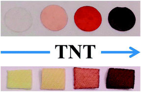
Among the various explosives, TNT (2,4,6-trinitrotoluene) belongs to the nitroaromatic explosive family and is one of the most widely used explosives in civil and military applications as well as by terrorists worldwide. As a chemical, TNT can easily enter groundwater supplies and is consider toxic at concentrations above 2 ppb, presenting harmful effects to all biota.1 For humans, it is a liver toxin that can be absorbed by the gastrointestinal track or even by the skin.2
Accordingly, the detection and quantification of TNT is of the outmost importance and can be performed using a broad set of analytical techniques, such as high performance liquid chromatography (HPLC) and high-resolution gas chromatography (HRGC) paired with different detectors, including mass spectrometry (MS), electrochemical detection (ED), electron capture detectors (ECD), and ultraviolet detectors (UV).1 However, the use of recognition processes in supramolecular chemistry and novel selective and sensitive optical probes have arisen as promising techniques for the detection of explosives by non-specialised personnel in an in situ, rapid and inexpensive manner, both for vapour and in-solution detection; the sensitivity is usually achieved using a quenching strategy based on the use of conjugated polymers or supported solids.3–14
In a recent paper, we demonstrated an insanely simple, novel, and straightforward methodology to prepare handleable solid materials for the naked eye detection of TNT in water environments.15 Using a commercial monomer containing a tertiary amine, 2-(dimethylamino)ethyl methacrylate (AEMA), we prepared the most inexpensive sensory film that changed its colour upon dipping in aqueous media containing a low concentration of TNT. The sensing mechanisms corresponded to the formation, under mild conditions, of colour complexes of Lewis bases, such as the mentioned amine-containing monomer, with electron-deficient aromatic rings, such as TNT, known since the first decade of the former century as Meisenheimer complexes.16–20
The tuning of the amine motif chemical characteristics of sensory monomers permits not only the lowering of the detection limits in aqueous solutions but also the achievement of TNT vapour detection, an especially cumbersome task due to the extremely low vapour pressure of TNT (9.2 ppbv).21 Thus, the use of both commercial and designed sensory monomers with primary and secondary amines, more active towards the formation of Meisenheimer complexes than tertiary amines, would most likely allow for the two-fold detection and quantification of explosives, i.e., as vapours and in solution. From an applied viewpoint, the former is related to homeland security and humanitarian efforts, and the latter is related to forensic and criminal investigations of washed scrap-metals and residues essential in terrorist-strike investigations and environmental control and remediation.
Thus, we have prepared two film-shaped solid sensory membranes: the first one using the commercial monomer 4-{N-(2-(methylamino)-ethyl)aminomethyl}styrene (di-AMS) with two secondary amine groups and the second one using the monomer with 4-(aminomethyl)styrene (AMS), which have a primary amine that was designed and synthesised by us. The objective was to achieve handleable and tractable materials with the highest sensitivity in the colourimetric detection of TNT. Thus, the materials were cut into manageable sensory discs, and the visual detection of TNT was achieved in situ based on clearly visible colour changes of these discs; its quantification was performed using the UV/Vis technique. Moreover, the solid sensory materials also allowed for the direct quantification of the TNT concentration within minutes by processing a digital image taken of these discs using a conventional camera or smartphone using the colour definition, i.e., the RGB parameters, thus avoiding the use of time-consuming techniques operated by skilled and specialised personnel.1,22–24 Furthermore, cotton fibres were coated with the sensory polymers to render wearable TNT sensory intelligent fabrics.
Using this philosophy, numerous chemosensors have been previously described in the literature, most of which are water insoluble organic molecules that work only in organic media. Thus, our objective was to select a well-known chromogenic organic chemosensor, slightly modify its structure by introducing a polymerisable chemical group, and copolymerise it to yield amphiphilic materials that have sufficient hydrophilicity and lipophilicity to be swelled both in water and in organic solvents to allow the target species dissolved in these media to access, by diffusion, the sensory motifs all along the material. In addition, good lipophilicity usually permits the absorption of gases and vapours, thus permitting their detection.
![]() | ||
Scheme 1 Constitution of the sensory film-shaped dense membranes MAMS and Mdi-AMS prepared upon the bulk radical copolymerisation of 2HEA and of the sensory monomers AMS or di-AMS, using ethylene glycol dimethacrylate (EGDMMA) as the cross-linking agent. The structures of the reference membrane (MRef), without sensory moieties and of the membrane with tertiary amines as sensory motifs (MAEMA) are also shown.15 The image of the membranes over a notebook demonstrates the aspect and transparency of the materials. |
The constitution of all of the membranes, as derived by FTIR spectra, corresponds to the proposed structure (νOH, νNH2, or νNH = 3030–3700 cm−1, broad signal; νCO ∼ 1722 cm−1), and its spectral pattern resembles the structure of the well-known poly(2-hydroyethyl methacrylate).25 The spectra of both MAMS and Mdi-AMS show the characteristic bending of the primary and secondary amine groups as two or one medium to weak intensity bands in the range of 700–850 cm−1 and weak bands in the region of 1590–1650 cm−1 (Fig. S3, ESI†).
Thermal resistance is a key parameter of materials for final applications and was evaluated following the weight loss that occurred during the heating of the material at a constant rate (thermogravimetric analysis, TGA). The decomposition temperatures that resulted in a 5% loss under a nitrogen atmosphere (T5) were approximately 180 °C, indicating that the materials have a reasonably good thermal stability (Table 1). However, the higher bond energy of the C–O compared with the C–N linkage can be clearly observed in the diminishment of the T5 at approximately 140 °C of MAMS and Mdi-AMS compared with MRef. This negative result is counteracted by the relevant increase of the char yield at 800 °C of MAMS and Mdi-AMS compared with MRef due to the partial aromatic structure of the former. The TGA curves are presented in Fig. S4, ESI.†
Membrane | T5 (°C) | T10 (°C) | TONSET (°C) | Char yield (%) | SSP (%) | |
---|---|---|---|---|---|---|
Water | Acetone | |||||
MRef | 315 | 349 | 286 | 2 | 56 | 40 |
Mdi-AMS | 171 | 191 | 142 | 14 | 40 | 68 |
MAMS | 180 | 209 | 165 | 14 | 43 | 53 |
The amphiphilic character of the materials was related to the water- and acetone-swelling percentage (SSP); that is, the amphiphilic character was related to the weight percentage of solvent uptake by the films upon soaking until equilibrium in pure solvent at 20 °C. This character is key because the membranes must be hydrophilic materials to regain enough water in aqueous media to allow for the chemicals dissolved in water to enter the membrane by diffusion to reach the sensory motifs and to yield the macroscopic signal, indicating the presence of the target molecules. In addition, the lipophilicity of the sensory materials must be sufficient to be solvent-swelled with the same purpose. In our previous experiments, we observed that a SSP between 40% and 100% is optimal for both the rapid diffusion of chemicals into the membrane and for maintaining the tractability, in terms of mechanical properties, of the solvent-swelled materials. All of the sensory membranes meet this criterion, as demonstrated in Table 1. The data clearly indicate a decrement of the hydrophilicity of Mdi-AMS and MAMS with a parallel increase of the lipophilicity compared with the reference membrane MRef, with this fact being attributed to the hydrophobic nature of the aromatic ring of the former membranes compared with the latter.
In our work, the ability of the monomers AMS and di-AMS to sense TNT in solution was tested before the sensory material preparation. Thus, solutions of the monomers in acetone–water (80:
20, v/v) immediately turned reddish after adding TNT, as observed in Fig. 1. This aqueous system was selected because of two important application fields related to the use of acetone, with water always present, i.e., the detection of TNT in soils for remediation purposes and for the detection of the explosive used by washing the scrap with acetone after a terrorist attack scenario. The measuring media is important because the water affects the Meisenheimer complex formation by altering the kinetics of these reactions.27,28
The interaction of the monomers AMS and di-AMS with TNT was confirmed by 1H NMR. Thus, the addition of TNT to a CDCl3 solution of the monomers caused the low field shift of the primary and secondary amine protons, demonstrating the participation of these groups in the Meisenheimer complexes (Fig. S5 and S6, ESI†). This fact is consistent with the 1H NMR interaction study of TNT with the monomer AEMA with a sensory tertiary amine motif in its structure.15
The coloured Meisenheimer complexes formed upon interaction in aqueous solution of sensory monomers and TNT in solution permitted the titration of the explosive by UV/Vis, as shown in Fig. 2 and S7, ESI.† The limit of detection (LOD) and limit of quantification (LOQ)29 of the titration systems were 1 × 10⁻⁶ and 3 × 10⁻⁶ M, respectively, for the monomers AMS and di-AMS. Notably, these limits were significantly higher for the monomer with a tertiary amine as the sensory motif, AEMA (Table 2), demonstrating the performance increase of the new monomers as sensory probes, as expected.
Monomer | LOD/LOQ × 106 M | Membrane | LOD/LOQ × 105 M | Coated fibre | LOD/LOQ × 104 M |
---|---|---|---|---|---|
AEMA | 19/59 | MAEMA | 14/43 | FAEMA | 11/33 |
di-AMS | 1/3 | Mdi-AMS | 8/24 | Fdi-AMS | 5/16 |
AMS | 1/3 | MAMS | 9/26 | FAMS | 4/11 |
The response time of the materials was evaluated by UV/Vis spectroscopy. Sensory discs of Mdi-AMS and MAMS were immersed in a quartz cuvette containing acetone–water (80:
20, v/v), and the UV/Vis spectra were recorded. Then, TNT was added to the cuvette from a concentrated stock acetone–water (80
:
20, v/v) solution, and UV/Vis spectra were recorded as a function of time (Fig. S8, ESI†). While the absorption maxima due to colour development were noted immediately after the addition of TNT, in our opinion, an better measuring time using the UV/Vis technique would be 20 min because the absorbance maxima is approximately 1. However, for visual and photographic analysis, a dipping time of 1 hour was considered because the colours were more intense and higher sensitivity was envisaged, and longer times were not deemed useful due to the instability of the Meisenheimer complexes under ambient conditions and the decomposition of TNT in solution.
The visual sensing performance of the sensory membranes involved analysing immersed discs of Mdi-AMS and MAMS in solutions of various concentrations of TNT for 1 hour, rendering discs of different colour intensities, as depicted in Fig. 3. A visible colour change was observed at a TNT concentration of 1 × 10⁻⁴ M. After the colour change, digital images of the discs were taken, and the colour definition of each disc was related to the TNT concentration to build the titration curve. As the colour is defined in the RGB system by the three variables red (R), green (G) and blue (B), ranging from 0 to 255, the information regarding these variables were joined in a single result (principal component 1, CP1) using principal components analysis (PCA), accounting for >92% of the information on the three RGB parameters. Using a straightforward mathematical workup, this strategy permits the use of nearly all of the colour information to prepare simple [TNT] vs. CP1 titration curves (the principal component parameters and RGB data are listed in Tables S2–S9, ESI†).
The sensitivity was within the millimolar and micromolar range, improving the performance of the previously described sensory membrane MAEMA (Table 2). Regarding the selectivity, neither the monomers in solution nor the sensory membranes changed their colour with other nitro-aromatics besides TNT and especially remained silent with the related NT and DNT, as visually observed in Fig. 1.
Within this context, intelligent fabrics capable of detecting TNT represent an attractive class of substrates for fabricating wearable chemical polymeric sensors.33 Accordingly, we describe herein the coating of cotton fibres of white fabric pieces cut from a lab coat as smart textiles for the colourimetric sensing of TNT. Squares of 1 × 1 cm of the lab coat were coated with sensory polymers prepared from sensing co-monomers AMS and di-AMS, fabrics FAMS and Fdi-AMS, respectively, as well as 2HEA, EGDMMA and a radical photoinitiator. Thus, following the same behaviour as the sensory membrane discs described in the previous section, the smart textiles turned reddish upon interaction with TNT in solution, as observed in Fig. 4. Noticeably, the detection time was very short, and the colour change was complete in 5 min, which was significantly less time than that required for the sensory membranes, most likely due to the rapid access of the TNT to the sensory motifs of the thin coating compared with the time needed to diffuse inside the solvent-swelled dense membrane.
Following the same procedure used for the sensory membranes, digital images of the sensory fabric dipped in acetone containing various concentrations of TNT permitted the construction of titration curves (Fig. 4) with LOD and LOQ within the micromolar range (Table 2), and this LOD was also clearly visually observed.
Thus, a slight colour change is observed at room temperature for the dense membrane Mdi-AMS and MAMS upon sealing cut sensory discs in small vials containing small quantities of TNT. Noticeably, this behaviour was not observed for the materials with tertiary amines as sensory groups, MAEMA.
Based on this observation, the sensory performance of sensory discs cut from Mdi-AMS and MAMS was analysed. To increase the colourimetric response and to expedite the experimental workup, the experiments were performed at 60 °C, where the vapour pressure of TNT is two orders of magnitude higher than at 25 °C, i.e., 829 ppbv according to the equation logP (ppbv) = (−5481/T) + 19.37, where T is the temperature in K.34 The colour development was similar to that observed in the detection of TNT in solution and was clearly observed by the naked eye and followed by UV/Vis spectroscopy, as depicted in Fig. 5 and in Fig. S9, ESI.† As expected, no change was observed for the reference material MRef.
The vapour sensing phenomena occur due to a diffusion/interaction mechanism, i.e., the vapour enters the membrane by diffusion, and in parallel, the interaction to render the Meisenheimer complexes takes place. The behaviour of the system in terms of diffusion clearly resembles that of gas transport through membranes, where the steady state is characterised by a straight line in pressure vs. time curves and is achieved after a time lag (ϕ) calculated at the intersection of the straight line fit corresponding to the straight line region with the abscissa.35,36 Thus, the flux of TNT molecules inside the membrane keeps the concentration of TNT in the centre of the membrane near zero because the molecules react with amine groups to yield the coloured complexes. The TNT gradient across the membrane is maintained until all of the amine groups interact with TNT, achieving a steady state at a certain time, and this state is maintained at least throughout the experiment, i.e., 11 h. The time lag is similar for both membranes (Fig. 5) and can be related to the diffusion coefficient (D) for Fickian diffusion, with both the solubility coefficient and D independent of concentration, by D = l2/6ϕ, where l is the thickness of the membranes in gas transport experiments, where the pressure on one side of the membrane is negligible. In our systems, as the membrane is in a TNT vapour atmosphere, the concentration gradient is from both sides of the membrane to the centre, where the zero concentration of unreacted TNT could be considered, and l would be half of the experimental membrane thickness.35 Thus, ϕ is 1.75 and 2.01 h for Mdi-AMS and MAMS, respectively, and D is 8.3 × 10⁻¹⁰ and 8.0 × 10⁻¹⁰ cm2 s−1. The higher slope of the relative absorbance increase vs. time, visually observed by a deeper colour development, for Mdi-AMS compared with MAMS may arise from the fact that each di-AMS monomer has two amine groups, while AMS only has one, and the real sensory motif density arising from the same molar feed ratio in each membrane is twice as large in Mdi-AMS compared with MAMS. In addition, the lone pair of the secondary amine is more external than that in the primary amine, giving the former a higher nucleophilic character in absence of the hydrogen bonding contribution of a highly solvating solvent as water.
The need to raise the temperature of the experiments to increase the TNT vapour concentration is a clear disadvantage for any practical application, where the explosive must be detected at ambient temperature. However, by working with membranes and smart fabrics that can transport gases by applying a pressure gradient, it is envisaged that a system could be designed to force rt air by pressure or vacuum on one side of the materials to concentrate the explosive vapours inside the membrane or coating, where the molecules would rapidly undergo the Meisenheimer complex formation.
![]() | ||
Fig. 6 Visual detection of TNT vapours with coated fibres. Conditions: approximately 0.4 × 0.8 cm of each fabric was suspended in a sealed vial with 50 mg of TNT at 60 °C for 11 h. |
Footnote |
† Electronic supplementary information (ESI) available: Monomer synthesis and characterisation, sensory membrane preparation and characterisation, fibre coating procedure, measuring conditions, TNT titration data, principal component analysis data. See DOI: 10.1039/c4ra02716g |
This journal is © The Royal Society of Chemistry 2014 |