Non-covalent modification of thrombolytic agent nattokinase: simultaneous improvement of fibrinolysis activity and enzymatic stability†
Chen Chena,
Haogang Duanab,
Chunmei Gaoa,
Mingzhu Liu*a,
Xin'an Wub,
Yuhui Weib,
Xinyu Zhang*c and
Zhen Liud
aState Key Laboratory of Applied Organic Chemistry and Department of Chemistry, Lanzhou University, Lanzhou 730000, People's Republic of China. E-mail: mzliu@lzu.edu.cn; Fax: +86 931 8912582; Tel: +86 931 8912387
bThe First Hospital, Lanzhou University, Lanzhou 730000, People's Republic of China
cDepartment of Polymer and Fiber Engineering, Auburn University, Auburn, AL 36849, USA. E-mail: xzz0004@auburn.edu; Tel: +1-334-844-5439
dThe Department of Chemical and Biomolecular Engineering, Johns Hopkins University, Baltimore, MD 21218, USA
First published on 10th June 2014
Abstract
Cardiovascular disease (CVD) has become the leading cause of death globally. Nattokinase (NK), as a novel thrombolytic agent, has gained more and more attention. However, NK is susceptible to chemical oxidation, and subsequent inactivation denaturation. Modification of an enzyme to improve its stability usually brings about falling enzymatic activity. In this study, folic acid modified chitosan (CS-FA) was synthesized, and a series of NK/CS-FA complexes with different mass ratios were prepared. In vitro thrombolysis experiments indicated that NK/CS-FA showed obvious advantages in its fibrinolysis activity and stability. With the NK![[thin space (1/6-em)]](https://www.rsc.org/images/entities/char_2009.gif) :CS-FA mass ratio of 100:1 (molar ratio of NK:chitosan pyranose ring = 3:5), NK/CS-FA 100:1 exhibited the best fibrinolysis activity and the thrombi dissolved completely within 17 hours at a constant rate, while the un-modified NK dissolved only 35 wt% of the thrombus in the same duration. NK/CS-FA 100:1 retained more than 53% enzymatic activity at 80 °C or pH 2, as a comparison, only 17% enzymatic activity was left for un-modified NK under the same conditions.
:CS-FA mass ratio of 100:1 (molar ratio of NK:chitosan pyranose ring = 3:5), NK/CS-FA 100:1 exhibited the best fibrinolysis activity and the thrombi dissolved completely within 17 hours at a constant rate, while the un-modified NK dissolved only 35 wt% of the thrombus in the same duration. NK/CS-FA 100:1 retained more than 53% enzymatic activity at 80 °C or pH 2, as a comparison, only 17% enzymatic activity was left for un-modified NK under the same conditions.
1 Introduction
Cardiovascular disease (CVD) has become the number one killer in the world according to the report of the World Health Organization.1 By 2015, almost 20 million people will die from CVDs, mainly from heart disease and stroke.2 Thrombosis is one of the most serious CVDs, and intravascular thrombus, a blood clot in a blood vessel, is one of the main causes for various thrombotic events. It is well-known that in the atherosclerotic coronary artery occlusive thrombus causes a variety of heart diseases such as heart attack or stroke.3 A major component of blood clots is fibrin which is formed from fibrinogen during the proteolysis by thrombin; meanwhile, fibrin clots can be hydrolyzed by plasmin to avoid thrombosis in blood vessels. In a normal situation, these reactions are kept at a balance, however, once the balanced situation is triggered by some disorders, the clots cannot be hydrolyzed and thrombosis occurs.4 Thrombo-embolism is a frequent and often lethal complication of medical diseases and surgical procedure, and thrombolytic therapy has been extensively investigated as a means of medical treatment of this condition. According to their action mechanism, thrombolytic agents are classified as two types: one is plasminogen activator, such as tissue type plasminogen activator (t-PA), urokinase (UK) and streptokinase (SK), the other is the plasmin-like protein, e.g. lumbrokinase, which can directly degrade the fibrin of thrombus, thereby dissolving the thrombus. Although having been widely used, typical thrombolytic agents t-PA, UK or SK, are known to have several challenges such as side effects, short half-life after intravenous administration and high cost in the intestinal tract. For example, SK, one of the first clinically used intravenous thrombolytic agents, is a relatively inexpensive and effective thrombolytic agent, however, its clinical use is limited due to its short in vivo half-life, immunogenicity, and high incidence of systemic bleeding, which undermines its therapeutic efficacy.5 Over the past decade, a number of studies have concentrated on improving the efficacy, potency, ease of administration, and duration of action of thrombolytic agents.6,7Since the incidence of CVDs in Asia is relatively low, traditional foods from Asia have been the subject of much attention. For more than 1000 years, people throughout Asia have been consuming soybeans in a variety of traditional soy food products. NK, a potent fibrinolytic enzyme, was primarily isolated from a typical and popular soybean food in Japan “natto” by Sumi et al.8 NK was of particular interest as soon as was discovered due to their effective biological thrombolysis of fibrin and thrombus in blood vessels,9 which is approximately four-times stronger fibrinolytic activity than plasmin. Nattokinase is less sensitive to the cleavage of fibrinogen, but is more sensitive to the cleavage of cross-linked fibrin compared to plasmin.5 Based on its food origin and relatively strong fibrinolytic activity, NK has advantages over other commercially used medicine in preventative and prolonged effects. The catalytic center of nattokinase, with 275 amino acids, contains three conserved residues, Asp-32, His-64, and Ser-221, while the molecular mass and isoelectric point were 27.7 kDa and 8.6, and there is no disulfide bonds in its secondary structure.10 Oral administration of NK can enhance fibrinolysis in dogs with experimentally induced thrombosis.11 These observations, together with the fact that it can be absorbed across the intestinal tract after oral administration and subsequentially induce fibrinolysis, make NK a potential thrombolytic agent for the treatment of cardiovascular disease.6,12 Dietary supplementation with NK-related foods have been considered to be safe and healthy for circulation system in human body.5 Accordingly, NK is currently used as a nutrient supplement to improve the blood circulation in humans.6 However, NK is sensitive to variations in temperature and pH, and its activity decreases in stomach due to the acidic condition.13 Moreover, although soy foods have been consumed for more than 1000 years, they have not been introduced into Western diet until the past two decades. Obviously, acceptance of the soy foods will take some time. Dietary supplementation is more effective on intimal thickening of arteries after vessel endothelial denudation, while for acute thrombus formation, timely therapy is necessary.
Development of new or modified thrombolytic agents with higher clot specificity, longer circulation time, and lower antigenicity remains still a challenge. NK was immobilized onto magnetic Fe3O4 nanoparticles in order to target the drug-bearing magnetic particles to the specific part of the body. However, the thrombolytic activity of NK decreased after immobilization (48–91% activity retained).4 The encapsulation improved the temperature and pH stability of NK over those free forms, but the enzymatic activity decreased significantly after encapsulation. Another effective method to obtain high-performance NK is gene mutations, as reported by Weng et al. Two mutant (T220S and M222A) were obtained after site-directed mutagenesis, the oxidative stability of NK was substantially increased while the relative activity decreased (70% and 53% retained, respectively) compared with the wild-type.14 In general, simultaneous enhancement of both the enzymatic activity and stability is critical to improve the therapeutic efficacy of NK. Timely therapy is crucial for achieving adequate reperfusion to salvage the affected organs. As mentioned above, modification of enzyme with synthetic or natural polymers, covalent attachment or bounded in liposome, enzymatic stability was increased to varying degrees, but the enzymatic activity obviously decreased and the curative effect dropped as a result. Folic acid (FA) have been widely used as an targeting molecule in anticarcinogen drug delivery systems, we accidently observed that complexion of NK with FA modified chitosan (CS-FA) may increase its enzymatic stability and enzymatic activity simultaneously. In this study, NK with folic acid modified chitosan (NK/CS-FA) conjugates with varying NK:CS-FA mass ratios were prepared. In vitro enzymatic activity, stability and thrombolysis were tested and compared against native NK.
2 Experimental part
2.1 Materials
Chitosan (CS, deacetylation degree 88.0%) was obtained from Sinopharm Chemical Reagent Co., Ltd. Nattokinase (NK, 12000 U) was purchased from Shanxi Sciphar Hi-Tech Industry Co., Ltd. Folic acid (FA) was supplied by Tianjin Guangfu fine chemical research institute. N-Hydroxysuccinimide (NHS) was purchased from Shanghai Medpep Co., Ltd. 1-Ethyl-3-(3-dimethyllaminopropyl)-carbodiimide hydrochloride (EDC·HCl) was supplied by Aladdin Chemistry Co., Ltd. Other reagents were of analytical pure grade and used without further purification. Deionized water was used for preparing all the solutions of the experiment.
2.2 Preparation of chitosan oligomers
Linear chitosan oligomers with number-average degrees of polymerization in the range of 20–40 were prepared by nitrous acid depolymerization of CS as described earlier.15 In brief, CS (5.0 g) was dissolved in 130 ml of acidic solution (containing 37 mM HCl and 66 mM CH3COOH). The reaction mixture was degassed by repeated (four) freeze–pump–thaw cycles. After cooling to 4 °C, a freshly prepared solution of NaNO2 (18 mM in 20 ml deionized water) was added, and the reaction was allowed to proceed for 12 h at 4 °C in darkness without stirring. The product was centrifuged (4 min, 12000 rpm) to remove the insoluble fractions before lyophilisation, the dialyzed solution was freeze-dried to get the final product.
2.3 Synthesis of folic acid modified chitosan derivative (CS-FA)
CS-FA was synthesized via the reaction between the amino group of chitosan and the primary carboxyl group of FA in the presence of EDC·HCl and NHS as carboxyl activating agent.16–18 The resultant mixture was dialyzed (Mw 1000 cut off) first against pH 7.4 buffer solution and finally deionized water. The dialyzed solution was freeze-dried to get the final product. The degree of substitution (DS, %) was defined as the average number of FA molecules bound per glucose unit.2.4 In vitro thrombolysis
In vitro thrombolysis characterization was performed using the method reported elsewhere with partial modifications.19 In brief, fresh chicken whole blood was obtained without any medication. Chicken whole blood was collected in a LDPE hose (Φ 5 mm) and allowed to form a stable thrombus at room temperature for 6 h and then stored at 4 °C. After a certain time interval, the hose was cut into about 6 cm pieces and set upright to let the thrombus out without disturbing. The thrombus was weighed (W0) and immersed into solutions which were prepared by mixing different amounts of NK solution (0.5 mg ml−1 in saline), CS-FA solution (8.3 μg ml−1 in saline) and saline together. The exact amount in the solutions were shown in Table 1.| Sample name | NK (mg) | CS (μg) | CS-FA (μg) | FA (μg) | H2O (ml) |
|---|---|---|---|---|---|
| a Prepared through a mass ratio NK:CS-FA of 100:0.33 in 100 ml deionized water.b FA in this sample is equal to that of sample 100:1. |
|||||
| Control 1 | 0 | 0 | 0 | 0 | 100 |
| NK | 25 | 0 | 0 | 0 | 100 |
| NK/CS-FA 00:0.33a |
25 | 0 | 83 | 0 | 100 |
| NK/CS-FA 100:0.67 |
25 | 0 | 166 | 0 | 100 |
| NK/CS-FA 100:1 |
25 | 0 | 249 | 0 | 100 |
| NK/CS-FA 100:1.33 |
25 | 0 | 332 | 0 | 100 |
| NK/CS-FA 100:1.67 |
25 | 0 | 415 | 0 | 100 |
| NK/FAb | 25 | 0 | 0 | 72 | 100 |
| NK/CS | 0 | 75 | 0 | 0 | 100 |
| Control 2 | 0 | 0 | 415 | 0 | 100 |
At set time interval, the clot was weighed (Wt) to observe the difference in weight after clot disruption. The extent of thrombolysis was measured by the decrease of the wet weight of thrombi at set time intervals. The thrombi weight (%) was calculated by eqn (1)
 | (1) |
2.5 Nattokinase activity
Nattokinase activity was measured according to a fibrin degradation assay developed by Japan Bio Science Laboratory Co., Ltd. (JBSL) using previous method.5,20 In brief, 1.3 ml Tris–HCl (50 mM, pH 7.5) and 0.4 ml of 1.4% (w/v) fibrinogen solution were mixed in vials and kept in water bath (37 °C) for 5 min. Then 0.1 ml thrombin (20 U ml−1) was added and kept in water bath (37 °C) for 10 min. To this thrombus, 0.l ml of enzyme was added. After incubation (37 °C, 20 min), 2 ml of 0.2 M tri-chloroacetic acid (TCA) was added. Vials were kept for 20 min and centrifuged at 3000 rpm for 5 min. One unit enzymatic activity is defined as the amount of enzyme required to produce an increase in absorbance equal to 1.0 in 60 min at 275 nm. Enzymatic activity of NK or NK/CS-FA 100:1 at different temperature/pH was collected after the samples were kept at the set conditions for 40 min. The pH was adjusted by sodium hydroxide (NaOH) or hydrochloric acid (HCl).
2.6 Cytotoxicity test
Human Hepatic Sinusoidal Endothelial Cells (HHSEC) were grown in Dulbecco's modified Eagle's medium (DMEM) supplemented with 10% fetal bovine serum at 37 °C, at 5% CO2 and 95% relative humidity. Cells were seeded into a 96-well TCPS plate at a density of 4 × 10⁴ cells per well. After 24 h, the culture medium was replaced with CS-FA, NK, NK/CS-FA 100:1 solutions with different concentrations (μg ml−1). After incubation for 24 or 48 h, 20 μl 3-(4,5-dimethylthiazol-2-yl)-2,5-diphenyltetrazolium bromide (MTT; 5 mg ml−1 in PBS) was added. After an incubation time of 4 h, the medium was removed and the cell culture plate was washed with PBS before the addition of 200 μl dimethyl sulfoxide (DMSO). And then the value of optical density was measured at 570 nm to calculate the relative growth rate.
2.7 In vivo animal study and histology
All animal experiments were performed under a protocol approved by Gansu laboratory animal committee. Male rats were anesthetized with chloral hydrate (5 ml kg−1) and tied on an operating table during the period of experimentation. The carotid artery was isolated carefully. The thrombosis was induced by topically applying a piece of filter paper, soaked in FeCl3 (20% w/v) to the exposed rat abdominal aorta as described previously.21 A small piece of tinfoil was kept below the artery prior to the topical application of FeCl3 so that it should not come in contact with the adjacent tissue. The filter paper was applied and the animal was monitored for 20 min. After thrombi formed, 0.5 ml saline, 0.5 ml NK or NK/CS-FA 100:1 was injected through caudal vein. Animals were executed after 3 hours, about 1 cm of the experimental vessel was cut off and immersed in formalin. To examine the thrombolysis efficacy of the thrombolytic agents, we examined histological slices of the experiment sites. Samples were dehydrated through a series of graded alcohols, embedded in paraffin and sectioned at 5 μm thickness and stained with hematoxylin and eosin (H&E).
The UV-Vis absorbance spectra were recorded at 298 K in the range of 200–800 nm using a Perkin-Elmer Lambda 35 UV-Vis spectrophotometer.
3 Results and discussion
Chitosan (CS), which has a repeated structure unit of β-(1-4)-2-amino-deoxy-β-D-glucose, is a fully or partial N-deacetylated derivative of chitin, the second-most abundant natural resource next to cellulose.22 Due to its biocompatibility, biodegradability, low immunogenicity and economical advantages, CS has been reported to be a promising polymer not only in the chemical field but also in biomedical and industrial areas.23,24 It has shown potential for use as scaffolds in tissue-engineered medical products, as an encapsulating matrix for immobilization of living cells and for drug delivery. However, extensive applications of native chitosan are limited by its insolubility in water, and various techniques have been used to enhance the solubility of chitosan.25 In this study, chitosan is depolymerized by nitrous acid to improve its solubility. Water soluble chitosan reacted with FA through its amino groups and the primary carboxyl group of FA in the presence of EDC·HCl and NHS and the chemical structure of CS-FA is shown in Fig. S1.†FTIR spectra, UV-Vis absorption and XRD pattern were utilized to characterize the synthesized CS-FA. The FTIR spectra of CS, CS-FA and FA are shown in Fig. 1A. In the FTIR spectrum of CS, the strong broad absorption at 3443 cm−1 is corresponding to the N–H stretching vibration of –NH2 group and O–H stretching vibration of –OH group in chitosan, and the C–O stretching peak of the pyranose ring is at 1029–1071 cm−1.26 The carbonyl absorption band C![[double bond, length as m-dash]](https://www.rsc.org/images/entities/char_e001.gif) O, is known as the amide I band, and N–H is termed the amide II band, their position depends on the degree of hydrogen bonding. Absorption of amide I is typically around 1720–1740 cm−1 and shifts to lower frequencies by hydrogen bonding. Absorption of amide II is typically around 1500–1550 cm−1 and shifts higher upon hydrogen-bond formation. There are two absorption peaks for the CO group, the peak at 1635 cm−1 represented the bonded CO stretching and the peak at about 1716 cm−1 represented the free CO stretching. The amide II band is observed at 1558 cm−1 due to the hydrogen-bond formation. Peaks of FA at 1453, 1518, and 1607 cm−1 are also observed in the spectrum of CS-FA. The absorption bands at 1453 and 1607 cm−1 are assigned to the stretching vibrations of CC in the backbone of the aromatic ring of FA.27 Their appearance in the FTIR spectra of CS-FA, confirms the grafting. Successful synthesis of CS-FA conjugates was also confirmed by 1H NMR spectroscopy (Fig. S2†).
O, is known as the amide I band, and N–H is termed the amide II band, their position depends on the degree of hydrogen bonding. Absorption of amide I is typically around 1720–1740 cm−1 and shifts to lower frequencies by hydrogen bonding. Absorption of amide II is typically around 1500–1550 cm−1 and shifts higher upon hydrogen-bond formation. There are two absorption peaks for the CO group, the peak at 1635 cm−1 represented the bonded CO stretching and the peak at about 1716 cm−1 represented the free CO stretching. The amide II band is observed at 1558 cm−1 due to the hydrogen-bond formation. Peaks of FA at 1453, 1518, and 1607 cm−1 are also observed in the spectrum of CS-FA. The absorption bands at 1453 and 1607 cm−1 are assigned to the stretching vibrations of CC in the backbone of the aromatic ring of FA.27 Their appearance in the FTIR spectra of CS-FA, confirms the grafting. Successful synthesis of CS-FA conjugates was also confirmed by 1H NMR spectroscopy (Fig. S2†).
 | ||
| Fig. 1 (A) FTIR spectra, (B) UV-Vis absorption spectra and (C) XRD pattern of native CS, the synthesized CS-FA, CS and FA; (D) possible structure drawing of the “transition zone”. | ||
Fig. 1B shows the UV-Vis absorption spectra of FA and CS-FA. As we all know, natural polysaccharide has no obvious absorption from 200 to 500 nm, whereas FA has two distinct absorption peaks at 290 and 350 nm, respectively. The absorption peak at 290 and 350 nm in the UV absorption spectra of CS-FA confirmed the successful conjugation of FA with CS.28 Moreover, according to the calibration curve of FA, DS (degree of substitution) of CS-FA is calculated to be 0.135.
The XRD pattern of CS, CS-FA and FA are shown in Fig. 1C. The diffraction pattern exhibited two broad diffraction peaks with different intensities at 2θ = 20° and a relatively weak reflection centering at 2θ = 10° associated with the most ordered region involving the acetamide group and its intensity reflects the hydrated crystal content of native CS and the low molecular weight CS which was degraded by nitrous acid in this work.29 Chitosan has some ordered regions inside its structure and the crystallinity decreases as its molecular weight decrease,30 the reason is that, mobility of CS chains were enhanced as the molecular weight decreased, ordered regions were destroyed and showed lower diffraction peak intensities. CS-FA, prepared through the formation of amide bond between CS and FA, showed certain crystallinity as shown in Fig. 1C with a weak reflection at 2θ = 6° and 2θ = 10°, which are associated with the crystallinity of the grafted FA. After grafted onto CS, the ordered molecular structure of FA was destroyed and its crystallinity reduced with only two strong reflection peaks can be distinguished (2θ = 6° and 10°). This also indicated that, FA has been grafted onto chitosan successfully.
Fibrin is a primary protein component of blood clots, which is formed from fibrinogen by thrombin. The insoluble fibrin fiber is hydrolyzed into fibrin degradation products by thrombolytic agents and the thrombus dissolved. In vitro thrombolysis experiment was carried out to examine the thrombolytic efficiency of NK/CS-FA complexes with the aged thrombi (72 h). As shown in Fig. 2 A, NK/CS-FA 100:1 shows the best thrombolytic efficiency. We can see from the curve that, the weight loss kinetics of thrombi in NK and various NK/CS-FA complexes met with the zero-order equation, which means that the thrombi weight decreased in a constant rate. The thrombolytic efficiency of Control 1 and Control 2 were also depicted in Fig. 2A, and there is no significant weight loss of Control 1 and Control 2 in the experiment, which indicated that CS-FA improved the thrombolytic efficiency of NK apparently. One point to be mentioned is that, the same phenomenon was observed for all the samples except for control 1 and NK, that the thrombi weight increased and reached the maximum after 2 h, from then on, the thrombi weight began to decrease and equaled with W0 after another 2 h, and the thrombi dissolve kinetics were met with the zero-order equation.
 | ||
| Fig. 2 (A) In vitro thrombolysis experiment with various NK:CS-FA mass ratios; (B) photographs of thrombi which have been dissolved for 10 and 15 h, respectively (the size of the grid is 2 mm × 2 mm). | ||
Nattokinase is water soluble, while thrombus is in-soluble, so that this hydrolysis reaction is a solid liquid two-phase reaction. FA is a small molecule which contains hydrophilic –COOH and hydrophobic aromatic ring, it can improve the interfacial compatibility, thus the fibrinolysis activity was improved. We can see from Fig. 2A (data named “NK/FA”) that, FA can improve the fibrinolysis activity. In comparison with FA, CS-FA has a larger hydrophilic group and is more stable in aqueous solutions, so that CS-FA behaves better than FA.
As shown in Fig. 2A, very small amount of CS-FA can hardly form a transition zone between the two phases, while excess CS-FA may block the contacts of enzyme and the substrate, so that the mass ratio of NK:CS-FA = 100:1 (NK/CS-FA 100:1) has the best fibrinolysis activity. The possible structure drawing of the “transition zone” was shown in Fig. 1D.
It has been widely accepted that the age of thrombi adversely affects the outcome of thrombolytic treatment.31 “Fresh” thrombi have the highest proportion of platelets, whereas the proportion of fibrin fibers increases over time, as the level of thrombin increases, leading to “older” fibrin-rich thrombi.32 Someone suggested that thrombolytic therapy should be used only in recent venous thrombosis and when treatment is carried out in the first 2 h after symptom onset, the so-called “golden hour,” survival rate dramatically increases.33 The poor solubility of older arterial and venous thrombi has been attributed to the organization of thrombi during which substitution of fibrin with fibrous tissue takes place. It is generally believed that the dissolubility of thrombi substantially decreases over a period of a few days.
The age of thrombi was defined as the time interval from collected to dissolve. The thrombi were distributed into two groups: fresh (24 h) and aged (72 h). Fresh arterial and venous thrombi dissolved somewhat better than the aged ones with all activators, and the difference is obvious. The in vitro thrombolysis characterization of fresh and aged thrombi for 12 h is shown in Fig. S3.† Six fresh and aged thrombi were subjected to dissolve in a series of milieus with different NK:CS-FA mass ratios, where fresh thrombi dissolved much faster than aged ones. Although not dissolved so well as fresh thrombi, the aged thrombi also showed significant weight loss within 12 h, which indicated that, un-modified NK and CS-FA modified NK all have excellent thrombus dissolubility on both the two thrombi. Another point to be mentioned is that, NK/CS-FA 100:1 showed the best thrombolytic efficiency for both fresh and aged thrombi. Thrombolysis experiment with newly formed thrombus was also carried out and the data were shown in Fig. S4.†
In Fig. 2B, the thrombi, which have been dissolved in enzyme milieus for 10 and 15 h, respectively, are shown on a glass platform. After immersed in the CS-FA/NK complex solutions for 10 h, a significant decrease in its size can be obtained from the photograph, and this phenomenon became obvious after 15 h. In the picture, we can see that the thrombi have no obvious loss in its length during the thrombolysis experiment, but its diameter had a noticeable decrease over time.
As shown in the digital photograph of the thrombolytic milieus (Fig. 3A), we can see the milieus turned to be blood colour after 2 h in the thrombolysis experiment. The reason is that thrombolytic agents hydrolyzed the fibrin, and blood cells were released from the fibrin network to present the blood colour. As demonstrated in Fig. 2A, the thrombus weight increased continuously during the first 2 h, and the milieus turned into light blood colour (Fig. 3A). This could be due to minor release of the blood cells. To explain this phenomenon, a “fishing net modal” was proposed as shown in Fig. 3B on the basis of a previous work.31 As we all know, fibrin was formed and strengthens the platelet aggregation in the early stage, the resulting red thrombus is comprised of erythrocytes and inflammatory cells entrapped by the fibrin network. The thrombolysis experiment can be divided into two stages: Stage I (0–2 h), a small portion of fibrin network was cut off by hydrolysis, blood cells were released and water molecules crowded into the empty space appeared after the release of blood cells, resulting in thrombi weight increased to about 104–114%, that means more water gained than blood cells lost. Stage II (2 h after), with the action of thrombolytic agent, more fibrin network were decomposed, more holes in fibrin net work were formed, blood cells release became the predominant issue, and the weight decreased continuously. The initial weight gain (Stage I) could also be due to binding of NK/CS-FA to thrombi, this weight gain phenomenon does not happen in NK sample because of the absence of CS-FA.
 | ||
| Fig. 3 (A) Photographs of thrombolytic milieus at 2 and 6 h; (B) schematic representation of a “fishing net modal”. | ||
The main objective of the present work was to increase the fibrinolysis activity of NK. Short in vivo half-life leading to higher doses and continuous infusion of NK is often required for clinical treatment; this in turn increases the risk of bleeding and immune reactions. Over the past decade, a number of studies have concentrated on improving the stability of NK, to reduce the dosing frequency. In this work, NK kept a constant thrombolysis rate as CS-FA was added. Our long-term goal is to utilize this CS-FA modified NK to achieve effective thrombolysis. It is possible that any nonspecific attachment of polymer to NK may occupy or shield the active sites of NK, and prevent NK from inactivating.
Fig. 4A shows the fluorescence spectra of NK and NK/CS-FA 100:1 at room temperature and 80 °C, respectively. The fluorescence spectrum of tryptophan residue in protein would change as its conformation changed. Small molecules which interacted non-covalently with proteins via hydrophobic or electrostatic interactions may lead to the change of fluorescence spectrum. The optimum temperature of NK is 40 °C, and relatively stable below 50 °C, however, remarkable loss activity will be observed at 60 °C.11 In Fig. 4A, we can see that intensity and maximum emission wavelength of NK at room temperature are approximately the same whether mixed with CS-FA or not. While at 80 °C, the fluorescence intensity decreased and the emission peak shifted to higher wavelength, which indicated that, the environment for the amino-acid residues of NK has changed. The secondary and tertiary structures of NK at various conditions were investigated by CD spectra (Fig. 4B and D). As compared to the un-modified NK, the peak at 215 nm, which attributed to β-sheet, has disappeared in the CD spectrum of NK/CS-FA 100:1. And the near-UV CD spectra (250–320 nm) detects no obvious change in tertiary structure,34 although these peaks and the shoulder are less pronounced than those observed in the native NK. Thus, the specific tertiary packing structures of NK was retained in NK/CS-FA 100:1.35 It can be inferred that, the interaction between CS-FA and NK molecules resulted in the β-sheet decomposed without thrombolytic efficiency lost. After kept at 80 °C for 40 min, CD spectra of NK/CS-FA 100:1 complex showed no obvious change, but the natural NK turned out to be out-of-order and the secondary and tertiary structures were completely changed.
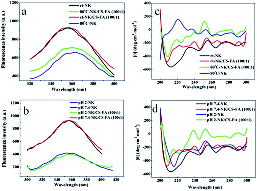 | ||
| Fig. 4 Effect of (A) temperatures and (C) pH on the fluorescence spectra of NK and NK/CS-FA 100:1; corresponding CD spectra of the samples in (A) and (C) are illustrated in (B) and (D) respectively. | ||
To investigate the influence of the pH values on the NK systems, fluorescence and CD spectra of NK and NK/CS-FA 100:1 were obtained under different pH values (Fig. 4C and D). The fluorescence intensity of NK/CS-FA 100:1 decreased sharply in acidic environments (Fig. 4C), while in CD spectrum (Fig. 4D), there is no obvious secondary structure change at different pH values. However, un-modified NK made a more obvious transition in secondary structure to β-sheet at pH 2. The result is consistent with the lowest in vitro enzymatic activity (103 U) of NK at pH 2 as shown in Table 2. The above experiments indicated that, after mixed with CS-FA, the secondary structure of NK was changed, but the fibrinolysis activity was enhanced. Moreover, the enzyme stability was improved after CS-FA was added. Synchronous fluorescence spectroscopy was measured to evaluate the effect of CS-FA on NK systems (Fig. 5). Apparently, the addition of CS-FA results in fluorescence quenching of NK and the maximum emission wavelength has slight red shift, which indicates that the polarity around tryptophan residues has changed. This is in agreement with the CD spectra of NK/CS-FA 100:1 in Fig. 4B and D, and the corresponding enzymatic activity showed that, NK/CS-FA 100:1 has higher fibrinolysis activity (Table 2), so it is natural to guess that change of polarity around tryptophan residues accelerated the fibrinolysis, some specific problems are going to be solved.
| Room temperature | Temperature | pH | |||||
|---|---|---|---|---|---|---|---|
| 37 °C | 80 °C | 2 | 4 | 8 | 10 | ||
| NK(U g−1) | 596 | 518 | 195 | 103 | 493 | 330 | 333 |
| NK/CS-FA 100:1 (U g−1) |
672 | 598 | 339 | 315 | 444 | 475 | 459 |
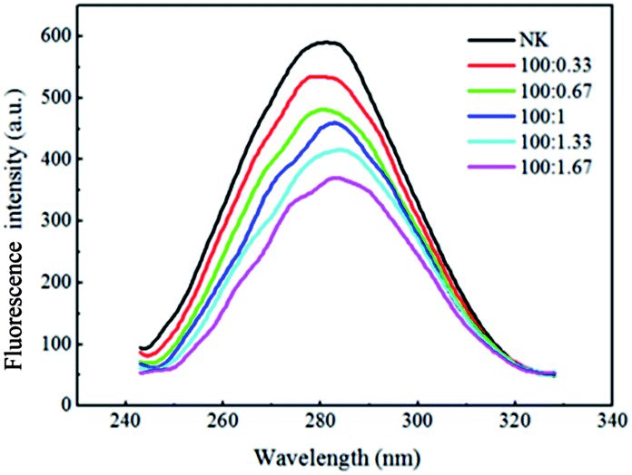 | ||
| Fig. 5 Fluorescence quenching spectra of NK/CS-FA with different mass ratios. | ||
To evaluate the cytotoxicity of CS-FA, NK and NK/CS-FA 100:1, in vitro cytotoxicity tests of these three samples against Human Hepatic Sinusoidal Endothelial Cells (HHSEC) were conducted as shown in Fig. 6, which illustrates the viability of HHSEC cells that have been treated by NK, CS-FA and NK/CS-FA 100:1 for 24 and 48 h, respectively. The cytotoxicity of all these three samples was both time and concentration-dependent.
 | ||
| Fig. 6 MTT assays of HHSEC cells treated with CS-FA, NK and NK/CS-FA 100:1 at various concentrations for 24 or 48 h. | ||
In vitro thrombolysis experiments showed that, NK/CS-FA 100:1 behaves best in thrombolysis test, so that NK/CS-FA 100:1 with different concentrations were also tested and illustrated in Fig. 6. The results indicated that, NK/CS-FA has no inhibition upon HHSEC cells during 24 h and low concentration of NK/CS-FA has promotion on cellular growth (cell viability above 100%). Although long-incubation with NK/CS-FA showed slightly cell viability decrease, the HHSEC cell viability is remain above 80%, which proved that NK/CS-FA has no obvious cell cytotoxicity and can be used as biomedical materials. In this work, concentration of NK/CS-FA 100:1 is 250 μg ml−1 in the in vivo/vitro experiment, cells which were incubated with NK/CS-FA 100:1 at 250 μg ml−1 is 98% and 82% for 24 and 48 h, respectively. The cytotoxicity test showed that, NK/CS-FA 100:1 (250 μg ml−1) has no obvious cytotoxicity.
Histological slices were prepared from the veins of the experimental animals and the results were showed in Fig. 7. Both control, NK and NK/CS-FA 100:1 treated vessels revealed signs of mild edema and inflammation.36,37 The magnitude of vessel injury was the same in the NK or NK/CS-FA 100:1 treated vessels as in the control one. Vessels treated with saline (control) turned out to be density homogeneous thrombi (Fig. 7A), whereas in NK treated vessels, the thrombi is uneven and light-colored (Fig. 7B), in NK/CS-FA 100:1 treated vessels, large sized thrombi become porous due to the thrombolysis (Fig. 7C). During the 3 h treatment, NK/CS-FA 100:1 dissolved much more thrombi than NK did, indicating NK/CS-FA 100:1 has great advantage over NK in the animal experiment.
 | ||
| Fig. 7 Histological pictures of thrombi in abdominal aortas (A: saline injected; B: NK injected; C: NK/CS-FA 100:1 injected; stars: density homogeneous thrombi, arrows: partly dissolved thrombi.). | ||
4 Conclusions
Demonstrated for the first time, CS-FA modified NK was used for thrombolytic treatment, with enhanced stability and fibrinolytic activity, which reveals great potential opportunities for treating/preventing cardiovascular diseases. Both in vivo and in vitro experiment results indicated that, the fibrinolytic activity and the stability of NK were strengthened by CS-FA. NK/CS-FA complex with the mass ratio of NK:CS-FA 100:1 showed the best thrombolytic efficiency, which is about 2 times of pure NK. Fluorescence and CD spectra showed that, the secondary structure of NK has changed in NK/CS-FA, but the fibrinolysis activity and stability was strengthened. NK/CS-FA has great potential for applications in the treatment of acute thrombotic patients.
Acknowledgements
The authors gratefully acknowledge the financial support of the National Natural Science Foundation of China (grant no. 51273086) and Special Doctorial Program Fund from the Ministry of Education of China (grant no. 20130211110017).Notes and references
- T. Tillin, A. D. Hughes, J. Mayet, P. Whincup, N. Sattar, N. G. Forouhi, P. M. McKeigue and N. Chaturvedi, J. Am. Coll. Cardiol., 2013, 61, 1777 CrossRef PubMed.
- Z. Deng, S. Wang, Q. Li, X. Ji, L. Zhang and M. Hong, Bioresour. Technol., 2010, 101, 1954 CrossRef CAS PubMed.
- X. Ren, G. Cui, M. Zhao, C. Wang and S. Peng, J. Phys. Chem. B, 2008, 112, 8174 CrossRef CAS PubMed.
- L. Ren, X. Wang, H. Wu, B. Shang and J. Wang, J. Mol. Catal. B: Enzym., 2010, 62, 190 CrossRef CAS PubMed.
- V. Deepak, K. Kalishwaralal, S. Ramkumarpandian, S. B. Venkatesh, S. R. Senthilkumar and G. Sangiliyandi, Bioresour. Technol., 2008, 99, 8170 CrossRef CAS PubMed.
- L. Yin, H. Lin and S. Jiang, J. Agric. Food Chem., 2010, 58, 5737 CrossRef CAS PubMed.
- X. Wei, M. Luo, L. Xu, Y. Zhang, X. Lin, P. Kong and H. Liu, J. Agric. Food Chem., 2011, 59, 3957 CrossRef CAS PubMed.
- H. Sumi, H. Hamada, H. Tsushima, H. Mihara and H. Muraki, Experientia, 1987, 43, 1110 CrossRef CAS.
- J. Liu, J. Xing, T. Chang, Z. Ma and H. Liu, Process Biochem., 2005, 40, 2757 CrossRef CAS PubMed.
- J. Liu, J. Xing, R. Shen, C. Yang and H. Liu, Biochem. Eng. J., 2004, 21, 273 CrossRef CAS PubMed.
- C. Wang, M. Du, D. Zheng, F. Kong, G. Zu and Y. Feng, J. Agric. Food Chem., 2009, 57, 9722 CrossRef CAS PubMed.
- C. Chiang, H. Chen, Y. Chao and J. T. C. Tzen, J. Agric. Food Chem., 2005, 53, 4799 CrossRef CAS PubMed.
- T. Ku, R. Tsai and T. Pan, J. Agric. Food Chem., 2009, 57, 292 CrossRef CAS PubMed.
- M. Weng, Z. Zheng, W. Bao, Y. Cai, Y. Yin and G. Zou, Biochim. Biophys. Acta, Proteins Proteomics, 2009, 1794, 1566 CrossRef CAS PubMed.
- S. Strand, M. Issa, B. Christensen, K. Vårum and P. Artursson, Biomacromolecules, 2008, 9, 3268 CrossRef CAS PubMed.
- C. H. Kuo, Y. C. Liu, C. M. J. Chang, J. H. Chen, C. Chang and C. J. Shieh, Carbohydr. Polym., 2012, 87, 2538 CrossRef CAS PubMed.
- T. Takei, H. Nakahara, H. Ijima and K. Kawakami, Acta Biomater., 2012, 8, 686 CrossRef CAS PubMed.
- G. Tronci, H. Ajiro, S. J. Russell, D. J. Wood and M. Akashi, Acta Biomater., 2014, 10, 821 CrossRef CAS PubMed.
- B. Vaidya, M. Nayak, D. Dash, G. P. Agrawal and S. Vyas, Int. J. pharm., 2011, 403, 254 CrossRef CAS PubMed.
- B. Naveena, K. P. Gopinath, P. Sakthiselvan and N. Partha, Bioresour. Technol., 2012, 111, 417 CrossRef CAS PubMed.
- W. R. Surin, P. Prakash, M. K. Barthwal and M. Dikshit, J. Pharmacol. Toxicol., 2010, 61, 287 CrossRef CAS PubMed.
- J. P. Wang, Y. Z. Chen, Y. Wang, S. J. Yuan, G. P. Sheng and H. Q. Yu, RSC Adv., 2012, 2, 494 RSC.
- D. Hua, J. Jiang, L. Kuang, J. Jiang, W. Zheng and H. Liang, Macromolecules, 2011, 44, 1298 CrossRef CAS.
- K. Kurita, H. Ikeda, Y. Yoshida, M. Shimojoh and M. Harata, Biomacromolecules, 2002, 3, 1 CrossRef CAS PubMed.
- W. Wu, J. Shen, P. Banerjee and S. Zhou, Biomaterials, 2010, 31, 8371 CrossRef CAS PubMed.
- C. Li, H. Tian, N. Rong, K. Liu, F. Liu, Y. Zhu, R. Qiao and Y. Jiang, Biomacromolecules, 2011, 12, 298 CrossRef CAS PubMed.
- S. Yang, F. Lin, K. Tsai, M. Wei, H. Tsai, J. Wong and M. Shieh, Bioconjugate Chem., 2010, 21, 679 CrossRef CAS PubMed.
- J. Shi, H. Zhang, L. Wang, L. Li, H. Wang, Z. Wang, Z. Li, C. Chen, L. Hou, C. Zhang and Z. Zhang, Biomaterials, 2013, 34, 251 CrossRef CAS PubMed.
- V. K. B. Acharya, M. Varadaraj and R. Tharanathan, Biomacromolecules, 2007, 8, 566 CrossRef PubMed.
- A. R. Fajardo, L. C. Lopes, A. F. Rubira and E. C. Muniz, Chem. Eng. J., 2012, 183, 253 CrossRef CAS PubMed.
- E. Boersma, Eur. Heart J., 2006, 27, 779 CrossRef PubMed.
- J. Silvain, J. Collet, C. Nagaswami, F. Beygui, K. E. Edmondson, A. A. Bellemain, G. Cayla, A. Pena, D. Brugier, O. Barthelemy, G. Montalescot and J. W. Weisel, J. Am. Coll. Cardiol., 2011, 57, 1359 CrossRef PubMed.
- P. G. Steg, E. Bonnefoy, S. Chabaud, F. Lapostolle, P. Y. Dubien, P. Cristofini, A. Leizorovicz and P. Touboul, Circulation, 2003, 108, 2851 CrossRef PubMed.
- C. D. A. Chalton and J. H. Lakey, Anal. Chem., 2010, 82, 3073 CrossRef PubMed.
- M. Nakao, K. Maki, M. Arai, T. Koshiba, K. Nitta and K. Kuwajima, Biochemistry, 2005, 44, 6685 CrossRef CAS PubMed.
- J. S. Michael, F. Victor, D. Sergio, T. Peter, P. L. Ryan, C. P. L. King, H. McDonald and J. W. Bradford, Thromb. Res., 2007, 121, 193 CrossRef PubMed.
- L. Huai, B. Yochai, C. F. Michael, M. P. Thomas, N. Tomoo, N. Toshihiko and J. S. Robert, Thromb. Res., 1998, 89, 171 CrossRef.
Footnote |
| † Electronic supplementary information (ESI) available. See DOI: 10.1039/c4ra02626h |
| This journal is © The Royal Society of Chemistry 2014 |