DOI:
10.1039/C4RA02175D
(Paper)
RSC Adv., 2014,
4, 19613-19621
Luminous block copolymer–quantum dots hybrids formed by cooperative assembly in a selective solvent†
Received
12th March 2014
, Accepted 7th April 2014
First published on 8th April 2014
Abstract
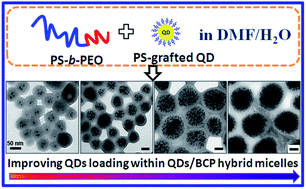
The formation of well-defined polymer/inorganic nanoparticle (NP) hybrid micelles is of considerable interest to the development of nanomaterials with desired optical, electric, magnetic, and mechanical properties. Herein, we introduce the efficient encapsulation of monodisperse polystyrene (PS)-grafted semiconductor quantum dots (QDs) into the PS cores of PS-b-PEO micelles through a “solution phase self-assembly” approach. We demonstrated that the size and the QD loading number of the coassemblies can be thermodynamically or kinetically tuned by varying the concentration of QDs and/or block copolymer (BCP) in the initial solution. Moreover, diverse morphologies of QD/BCP hybrid assemblies can be obtained by tuning the experimental parameters, such as the size of QDs, stirring time and many others. Interestingly, a novel Janus complex comprising a vesicular part without QDs and a spherical part with lots of QDs was firstly reported in our work. The Janus complexes were unstable and tended to transform their morphologies to the predominantly spherical QD-loaded micelles. The QD/BCP hybrid assemblies can present good water solubility, stability, lower toxicity and high luminous performance, indicating their potential applications in the biological field, especially for use as fluorescence probes and labels.
1. Introduction
Block copolymers (BCPs), consisting of two or more chemically different polymer segments or blocks connected by covalent linkages, have been widely recognized as particularly important materials because they can self-assemble into a wide range of periodically nanoscopic morphologies (e.g., spheres, cylinders, double gyroids, and lamellae).1 Interestingly, among various types of block copolymers, when placed in selective solvents, amphiphilic diblock copolymers composed of a hydrophobic block and a hydrophilic block have the propensity to form a large number of aggregate structures, such as spheres, cylinders, vesicles, among others.2–6 Novel functions of these aggregates can be achieved by decoration of functional nanoparticles (NPs). The incorporation of NPs in polymer matrices offers a powerful and adaptable tool for the fabrication of composite materials with desired optical, electrical, magnetic, and mechanical properties through the choice of incorporated NPs and their dispersion in polymer matrices.7–13 In solution, NPs/BCP hybrid micelles have been prepared by incorporating one or multiple hydrophobic NPs into the hydrophobic domains of amphiphilic BCP micelles.12–36 The morphology of the hybrids and the location of NPs are quite important for realizing the full potential applications of NPs/polymer hybrid materials. Hitherto, main focus has been on the coassembly of amphiphilic BCPs with gold NPs, including spherical particles,12–20 rods,21–25 wires,26,27 etc. and magnetic NPs.28–31 The coassembly behaviors of semiconductor NPs and amphiphilic BCPs were seldom reported.32–36 There are still many scientific problems that need to be solved, including the QDs loading and location control, the ergodicity of the hybrid micelle morphologies and the mechanisms for the morphological transition. Moreover, the properties and applications of semiconductor NPs/BCP hybrid assemblies are urgent to be investigated.
Cadmium sulphide (CdS) and cadmium selenide (CdSe) NPs are the most common types of semiconductor nanomaterials.37 They are recognized as materials perfectly suited to replace organic dyes currently used in experiments involving fluorescent labeling. In addition to their better photostability, semiconductor nanocrystals are superior to organic dyes in other aspects, such as broader excitation and narrower emission spectra, which can be easily tuned.38–40 Cooperative self-assembly of amphiphilic BCPs and semiconductor NPs offers a powerful route to fabricate functional hybrid materials. Semiconductor NPs can impart unique optical properties to the hybrid nanomaterials, meanwhile the flexible polymers provide morphological controllability and processability.32–34,41 Taton and co-workers reported the encapsulation of semiconductor quantum dots (QDs) within PS-b-PAA micelles of which the shells were crosslinked by a diamine.34 Their QDs/BCP hybrid micelles were stable to heat and pH and behaved good biocompatibility. Park et al.36,42,43 obtained unique cavity-like structures in core–shell type micelles through the cooperative self-assembly of alkyl-terminated QDs and PS-b-PAA. They demonstrated that QDs could play an active role in the self-assembly process rather than being passively incorporated as a solute. Their results indicated that both the enthalpic interaction and the polymer stretching energy were important factors in the formation of coassemblies. Winnik et al.33,44 used the structures of the polystyrene-b-poly(4-vinylpyridine) (PS-b-P4VP) micelles as motifs for organizing the location of QDs. The QDs/BCP hybrid micelle with CdSe QDs bound to the corona adjacent to the core was extremely sensitive to solvent composition and was affected by the stirring rate. It's well known that the interaction between NPs and BCP plays an important role for the coassembly behavior.45 Despite the considerable progress in the assembly of semiconductor QDs with block copolymer micelles, previous work mainly focused on the assembly of QDs (e.g. TOPO-capped QDs) with BCPs by virtue of unfavorable interaction, the cooperative assembly behaviors of QDs by using enthalpically favorable interaction with BCP have seldom reported.46–48 In our previous work, high luminescent PS-grafted QDs were encapsulated within the central PS cores of block copolymer cylindrical and toroidal micelles by “solution phase self-assembly” approach.46 Moffitt et al. prepared the PS-brushes coated CdS QDs through the H2S treatment of PS-b-poly(cadmium acrylate) (PACd) reverse micelles in an organic solvent, followed by their assembly with PS-b-PAA or PS-b-PEO to form QDs-containing micelles.47–50
In this paper, we are interested in the micellization of polystyrene-block-poly(ethylene oxide) (PS-b-PEO) block copolymers which are so sensitive to the water content that can self-assemble into different morphological structures, such as spheres, rods and vesicles.5 Herein, in order to improve the enthalpic attraction between the semiconductor QDs and the PS block of BCP, we modified the pre-synthesized QDs with PS-SH (thiol terminated polystyrene) through the “ligand exchange” procedure. Incorporation of PS-grafted CdS or CdSe/CdS core–shell QDs within PS-b-PEO micelles was achieved using the “solution phase self-assembly” approach. Thermodynamic and kinetic effects, including QDs weight fraction and BCP initial concentration in cosolvent, played important roles on the morphology, the size and the QDs loading of hybrid assemblies. A first example of the formation of novel Janus complex micelles comprising of vesicular parts without QDs and spherical parts containing lots of QDs was illustrated. The Janus complexes were unstable and tended to transform their morphologies to the predominant spherical QDs-loaded micelles. The hybrid assemblies can present good water solubility, stability, less toxicity and high luminous performance, showing their potential applications in fluorescence labeling, probing, tracing and so on.
2. Experimental section
2.1. Synthesis of PS-grafted CdS and CdSe/CdS QDs
Details for the preparation of TOPO-coated CdS or CdSe/CdS core–shell QDs can be seen in the ESI.† PS-grafted CdS QDs were synthesized through a phase transfer ligand-exchange approach.46,51 Typically, PS-SH (10 mg, Mn = 2000, PDI = 1.15) dissolved in 1 mL of DMF under magnetic stirring for overnight. TOPO-coated CdS QDs in chloroform (2.8 mg mL−1, 1 mL) was dried by N2 bubbling. Then, the QDs redispersed in hexane and PS-SH dissolved in DMF were mixed together to give a two-phase system. After ultrasonication for 2 hours, the QDs were transferred to DMF phase by displacing TOPO with PS-SH. After keeping stationary at room temperature for one day, the PS-grafted CdS QDs were precipitated in methanol three times to remove displaced TOPO and were washed with hexane to eliminate unmodified QDs (capped with TOPO) which might be accidentally transferred to the DMF phase during the ultrasonication process. The PS-grafted CdSe/CdS QDs were obtained by the similar procedure as described above and were also reported in our previous work.46
2.2. Preparation of QDs/PS-b-PEO hybrid micelles
In typical experiments, 0.2 mL of PS-grafted CdS QDs in CHCl3 (2.5 mg mL−1) were firstly dried by N2 bubbling. Then, 0.02 mL of PS-b-PEO in DMF (50 mg mL−1, Mn = 37
000 for the PS block and Mn = 6500 for the PEO block, PDI = 1.06) and additional 0.48 mL of DMF were added in the vial. The mixture, with the BCP initial concentration (C0) of 0.2 wt% and QDs weight fraction (f) of 0.33, was stirred at room temperature for overnight to completely dissolve the BCP. To induce the assembly of the block copolymer and QDs, water (30 μL, 6 wt%) was slowly added to the mixture at the rate of 10 μL per 30 s. The aliquots of the solution (0.01 mL) were taken out and then quenched by a lot of water (2 mL) to completely frozen the morphological structures. Finally, the solution was dialyzed against water for two days to remove the DMF. In all experiments, the initial concentration of PS-b-PEO (C0) and the water content were both the mass concentration relative to DMF. The initial weight fraction of PS-grafted QDs (f) is defined here by the weight of QDs over the combined weight of QDs and block copolymer. For the hybrid micelles consisting of PS-grafted CdSe/CdS QDs and BCP, the preparation method was made similarly.
For some QDs/BCP hybrid micelles, we made the purification by several centrifugations to improve the QDs loading and minimize the size distribution of the coassemblies. The colloidal aqueous solution of QDs/BCP micelles was firstly centrifugated at 10
000 rpm for 10 min. The supernatant was discarded and the sediment was dissolved in equal amount of water. Then, the centrifugation was resumed at 8000 rpm for 10 min. The same procedure was done as described above. Finally, the sediment was redispersed in water and the solution was centrifugated at 6000 rpm for 15 min to completely remove the micelles with no or few QDs encapsulated.
3. Results and discussion
3.1. The PS-grafted QDs and their properties
The enthalpic attraction of QDs and PS block of the copolymer can be improved by modification of the pre-synthesized QDs (e.g. TOPO-coated CdS QDs) with PS-SH. In this study, TOPO-coated CdS QDs with the average diameter of 7.1 nm (Fig. 1a and b) were synthesized through a “two-phase” approach.39 The TOPO-coated QDs are soluble in chloroform (CHCl3), tetrahydrofurane (THF) and n-hexane but are insoluble in DMF and water. For the synthesis of PS-grafted QDs, the ligand exchange in a two phase system of n-hexane (a good solvent for TOPO-coated QDs) and DMF (a good solvent for PS-SH) was used. The modification of QD surfaces can be evidenced by the phase transfer of QDs from n-hexane to DMF. Moreover, the PS-grafted QDs had ever been characterized by FTIR and 1H NMR measurements for the further proof of ligand exchange.46 Here, the PS-grafted CdS QDs with the average diameter of 7.6 nm (Fig. 1c and d and S1, ESI†) can be easily dispersed in the good solvents of PS, like CHCl3, DMF, THF, 1,4-dioxane, etc. The absorption bands of TOPO-coated and PS-grafted CdS QDs were both lay at 507 nm (Fig. S2†), indicating almost no size change of the QDs before and after surface modification.
 |
| Fig. 1 (a and b) TEM images and size distribution of TOPO-coated CdS QDs with the average diameter of 7.1 nm; (c and d) TEM images and size distribution of PS-grafted CdS QDs with the average diameter of 7.6 nm. | |
The emission properties and fluorescence efficiency of QDs can be changed over a wide range of the spectrum by varying of the size and the composition therein.38,52 With adding CdS layers, the TOPO-coated CdSe/CdS QDs presented brighter emission color and higher fluorescent efficiency than single CdSe or CdS QDs. After modification, the PS-grafted CdSe/CdS QDs can still possess the high fluorescent properties as compared to the original TOPO-capped QDs (Fig. S3†).46 For the convenience to figure out the QDs structural parameters, the PS-grafted CdS QDs (7.6 nm) were mainly used in the following section, while the PS-graftd CdSe/CdS QDs were preferred in terms of the properties of QDs/BCP hybrid micelles.
3.2. The effects of QDs weight fraction
In the following sections, the PS-grafted CdS QDs with 7.6 nm are all simplified as QDs for convenience unless specially noted. The cooperative assembly of QDs and PS-b-PEO can be readily achieved given the favorably enthalpic interaction of QDs and the PS block of block copolymers. Briefly, PS-b-PEO and QDs were firstly dispersed in a good solvent of DMF, then water was added to simultaneously desolvate the QDs and PS block of the copolymer, consequently, confining the QDs within the micelles. In this section, we firstly investigated the effects of QDs weight fraction on the size, QDs loading and morphologies of QDs/BCP hybrid micelles.
The PS-b-PEO block copolymer with initial concentration of 0.2 wt% self-assembled into predominant vesicles and lamellas at the water content of 6 wt% (Fig. S4a†). Most of the vesicles contain one or two cavities. When small amount of the QDs (f = 0.06) were added in the system, besides lots of the small spherical micelles and vesicles all without QDs encapsulated within them, some extraordinarily huge complexes with QDs mainly dispersing in the spherical parts and the other parts being giant vesicles were observed (Fig. S4b and c†). With the addition of more QDs (f = 0.20), besides many QDs/BCP hybrid micelles, we found lots of bare micelles which had no QDs encapsulation within them in Fig. 2a. We found that the monodisperse micelles with QDs uniformly incorporated in the cores can only be obtained when the QDs weight fraction was in the range of 0.25 to 0.70 (Fig. 2b–e). The QDs-loaded micelles are thermodynamic stable structures with no morphological transitions along the time course of stirring. When the QDs weight fraction (f = 0.75) was larger than the threshold value, BCPs and QDs were macroscopically phase-segregated and precipitated out of solution upon water addition (Fig. 2f).43
 |
| Fig. 2 (a–f) Bright-field TEM images of hybrid assemblies formed from PS-b-PEO and PS-grafted CdS QDs (7.6 nm) with the same experimental conditions, including BCP initial concentration of 0.2 wt%, DMF volume of 0.5 mL, water content of 6 wt%, and stirring time of 1 day, but with different QDs weight fraction: (a) f = 0.20; (b) f = 0.27; (c) f = 0.33; (d) f = 0.43; (e) f = 0.67; (f) f = 0.75. The insert in (f) is the photograph of the corresponding sample dispersed in water, as can be seen the precipitate. All the scale bars are 100 nm. | |
It has been demonstrated that localizing polymer-coated NPs near the center of the compatible polymer domain sacrifices the translational entropy of the particles but avoids an even larger chain stretching penalty incurred by distributing particles throughout the domain.14,19 Several groups have demonstrated that NPs can play active roles in the assembly process rather than being passively incorporated in the polymer matrix.14,36,43 It should be noted that, due to the solvent evaporation and hydrogen bond of PEO blocks, the micelles on the TEM grid appear close to each other and in some cases the micelles are connected to each other (Fig. 2a–f). This is a common phenomenon in the self-assembly of amphiphilic BCPs.47 Actually, the micelles in the solution are all well-dispersed, which can be characterized by the dynamic light scattering (DLS) test.41
Additionally, with increasing the QDs weight fraction from 0.27 to 0.67, we further did statistics on the micelles diameter and PS shell thickness by measuring at least 200 spherical QDs/BCP micelles from 3–5 TEM images. Also, the QDs number and QDs volume fraction per micelle were calculated (Fig. 3 and Table S1†). Both the micelle diameter and the PS shell thickness measured by TEM exclude the outmost thin PEO layer because the PEO layer is not visible by TEM. The results presented in Fig. 3a showed that the micelles diameter and PS shell thickness increased with the QDs weight fraction (f) when f < 0.43. Both of them reached a maximum when f = 0.43, and they decreased with a further increase of the f until to 0.67. The increases of the micelle diameter and PS shell thickness agreed with the typical trend observed for homogeneous incorporation of NPs,19,28 as well as the decreases of both the parameters were also illustrated by Park and coworkers.43 For a fixed polymer concentration, a higher number of QDs can be accommodated by increasing either the micelle diameter or the number of coassemblies.43 For homogeneous incorporation of the QDs when f < 0.43, the sizes of the block copolymer micelles became larger with the addition of QDs because of the volume taken up by the QDs. Correspondingly, the PS shell thickness increased with the f because the volume variation of QDs domains in hybrid micelles lagged far behind that of the micelles diameter. Above the QDs weight fraction of 0.43, we found that the penalty from PS chains stretching in the block copolymers prevented further increase in the micelle diameter, and the addition of QDs must instead be accommodated by an increase in the number of coassemblies.43 Consequently, we can also readily understand the variation of the QDs number per micelle with the f as depicted by the black curve in Fig. 3b. The QDs number per micelle increased to the maximum when f = 0.43 then decreased and finally kept constant with increasing the f. However, the QDs volume fraction per micelle (φQDs) increased linearly with the QDs weight fraction (Fig. 3b, the blue curve), which attributed to either the increment of the QDs number per micelle or the size decrescence of total micelles (i.e. the escalation of the number of total micelles). The rationale can also be given based on the equation of φQDs19 (see the ESI†). Actually, the φQDs is more critical than the QDs weight fraction to affect the structural parameters of the hybrid assemblies. Numerous early reports have shown that the volume fraction of NPs is an important parameter that considerably affects the location of NPs and the morphologies of NPs/BCP hybrid micelles.8,19,43 Generally, NPs can disperse well in the BCP micelles at low NPs volume fraction. Otherwise, the high NPs volume fraction triggered the swelling of the host domains, the order–order morphological transitions of BCP, or even macrophase separation.14,16,19 Herein, owing to the simplicity and feasibility, the QDs weight fraction (f) was used in our experiments for convenience. The size and QDs loading within QDs/BCP hybrid micelles can be readily tuned by varying the f.
 |
| Fig. 3 (a) A plot of statistic overall diameters (■) and PS shell thicknesses (●) of QDs/BCP hybrid micelles versus QDs weight fraction (f), showing that both the parameters increase first then decrease with increasing the QDs weight fraction; (b) a plot of calculated QDs number (□) and QDs volume fraction (○) per micelle versus QDs weight fraction (f). The statistical values were obtained by measuring at least 200 spherical QDs/BCP micelles. The lines were added to guide the eyes. | |
3.3. Improving the QDs loading in hybrid micelles
The formation of well-defined QDs/BCP hybrid micelles with high QDs loading is crucial to the development of functional nanomaterials.19,53 However, in most previous reports, the NPs density in polymer hybrid micelles was usually low. The reason is that the incorporation of NPs with high loading in host polymer domain leads to high chain conformation entropic penalty and drastically affects the self-assembly structure by changing the relative volume ratio between the two different blocks.19 In our work, besides the procedures described in Section 3.2 with increasing the QDs weight fraction while keeping BCP initial concentration constant, the loading density of QDs can also be improved to a large extent by changing the BCP initial concentration (Fig. 4). Fig. 4a depicted the well dispersed QDs/BCP hybrid micelles with small amount of QDs that were formed at the BCP initial concentration (C0) of 2.0 wt%, QDs weight fraction (f) of 0.10, water content of 6 wt% and the stirring time of one day. Besides the predominant spherical hybrid micelles, there were few hybrid vesicles as indicated by arrows in Fig. 4a. The average diameter of the coassemblies in Fig. 4a was 63.4 ± 6.5 nm with one to ten QDs per micelle. The f can be minimized to 0.10 for the efficient incorporation of QDs within the QDs/BCP hybrid micelles when C0 = 2.0 wt%, while it must be above 0.25 to achieve the resultful encapsulation of QDs at C0 = 0.2 wt% (Section 3.2). Keeping the QDs weight fraction (f = 0.30) constant, the loading number of QDs was greatly improved while maintaining the spherical micellar morphology by increasing the BCP initial concentration from C0 = 0.2 wt% (Fig. 4b) to C0 = 0.5 wt% (Fig. 4c) and finally to C0 = 1.0 wt% (Fig. 4d). Correspondingly, the sizes of the QDs/BCP hybrid micelles increased from 79.0 ± 8.3 nm (Fig. 4b) to 131.8 ± 10.2 nm (Fig. 4c) and to 157.9 ± 17.2 nm (Fig. 4d). Given the PS chain end-to-end distance in the unperturbed state and fully stretching length in PS356-b-PEO148 (the subscripts indicate the number of repeat units) to be 5.2 nm and 89.0 nm respectively,5 the aggregates in Fig. 4c and d are large compound micelles generally with large polydispersities,2,47,48 rather than the regular spherical micelles. It is well known that the aggregation number (Nagg) of polymer assemblies increases with the BCP concentration, which can cause the observed change in the assembly size.3,43 Increasing the initial BCP concentration led to an earlier onset of QDs/BCP hybrid micelles formation as water is added at a given rate and provided an effective route to kinetically control the aggregates sizes.48 We obtained the nearly monodisperse QDs/BCP hybrid micelles that can be well dispersed in water. However, the hybrids with high loading of QDs and low size distribution at higher BCP initial concentration of 0.5 wt% (Fig. 4c) and 1.0 wt% (Fig. 4d) were produced by purification through several centrifugations. Before purification, the corresponded samples directly used for TEM tests were polydisperse, i.e. with broad size distributions (Fig. S5†). This was in accordance with the general polydispersities of large compound micelles. Some small bare micelles without QDs even appeared at C0 = 1.0 wt% (Fig. S5b†). The broader size distribution of hybrid micelles at higher BCP concentration was ascribed to the following reasons: (i) the incorporation of high-content QDs disrupts the order of BCP structures due to the loss of the polymer conformational entropy; (ii) the formation of larger QDs domains causes further excessive stretching of polymer chains.19 (iii) As Nagg increases further, the polymer stretching energy becomes too high. This can cause the inclusion of QDs and small micelles in the polymer aggregates, which leads to the polydispersity of the assemblies.43
 |
| Fig. 4 Bright-field TEM images of QDs/BCP hybrid assemblies with improved QDs loading formed at the same water content of 6 wt% and stirring time of one day but different QDs weight fraction (f) and BCP initial concentration (C0): (a) f = 0.10, C0 = 2.0 wt%; (b) f = 0.30, C0 = 0.2 wt%; (c) f = 0.30, C0 = 0.5 wt%; (d) f = 0.30, C0 = 1.0 wt%. TEM images of (c and d) are the corresponding QDs/BCP hybrid assemblies after purification by several centrifugations. All the scale bars in TEM images are 100 nm. | |
3.4. Diverse morphologies and morphological transition
The cooperative assembly of QDs and PS-b-PEO was extremely sensitive to the effects of QDs size, stirring time and rate, among others. Diverse morphologies of QDs/BCP hybrid assemblies were generalized by changing the QDs size or prolonging the stirring time. We used the PS-grafted CdSe QDs with average diameter of 4.3 nm to achieve their assembly within PS-b-PEO aggregates at the BCP initial concentration of 0.2 wt%, QDs weight fraction of 0.30, water content of 6 wt% and prolonging the stirring time for 1 day to 10 days. To our interests, an intriguing and novel structure of QDs/BCP hybrid coassembly with elongated shape was obtained at t = 1 day (Fig. 5a). The aggregate, termed the Janus complex, mainly consist of two parts of which one side is the vesicle without QDs and the other side is the incomplete spherical micelle containing lots of QDs. The Janus complexes are anisotropic and unstable given the thermodynamic aspects. The unequal stretching degree between the PS chains coated on QDs and the PS chains covalently bonded with PEO chains in block copolymer resulted in non-uniform density within the core of spherical side, which caused an inhomogeneous distribution of free energy in the Janus complex. In order to form a thermodynamic equilibrium state, the adjustment of PS chains conformation took place, thus a morphological transition can easily occur. With prolonging the stirring time to 5 days, the complexes can still exist with slight shape variation, as well as more separate QDs-loaded spherical micelles appeared (Fig. 5b). This indicated the morphological transition from the predominant Janus complexes to the spherical micelles, which can be further evidenced by continuing the stirring time until to 10 days (Fig. 5c). Indeed, the separate spherical micelles with lots of QDs encapsulated within them (Fig. 5c, indicated by the blue arrows) and rarely the separate vesicles with no or few QDs incorporated were observed after 10 days. The number of the spherical micelles was evidently lager than that of vesicles. Also, the intermediates with spherical morphology (Fig. 5c, indicated by the black arrows), which are different from the elongated Janus complexes, integrated by small faction of cavities and large PS domains with QDs encapsulated. The vesicular parts of the Janus complexes were squeezed from the bodies and the vesicles walls were used to average the QDs loading in the spherical parts (hints as indicated by the red dash circles in Fig. 5c), which both led to the effects that the cavities of intermediates became smaller or even broke away from the intermediates as well as the morphology of the intermediates became spherical to reduce their free energies. This elucidation was also supported by comparing the statistical values of the equivalent diameter and area of QDs-loaded domains before and after the morphological transition. The equivalent diameter of QDs-loaded domains increased from 118 ± 20 nm in the Janus complexes in Fig. 5a to 123 ± 22 nm in the QDs-loaded spherical intermediates (indicated by the black arrows) and micelles (indicated by the blue arrows) in Fig. 5c. Moreover, the statistical area increased from 11
271 ± 3812 nm2 to 12
249 ± 4493 nm2, correspondingly. The size of NPs plays an important role to the morphologies of hybrid assemblies,14,28 the locations of NPs within aggregates,16 and many others.35,43 Xu and coworkers16 demonstrated that the location of Au NPs in vesicle wall or in spherical micelle is an entropy-driven process and is heavily size dependent. Whether the Au NPs enter the vesicle wall or not is determined by a ratio of the diameter of Au NPs (D0) to the thickness of the vesicle wall (dw0). For the PS-grafted Au NPs, they can be located in the vesicle wall when D0/dw0 < 0.5, otherwise, they prefer to locate in spherical micelles. However, the PS-grafted QDs in our work seem preferentially to disperse in the spherical micelles despite of their sizes (the QDs with diameter from 3.8 nm in Fig. S6a† to 7.6 nm in Fig. 2 were used). To the best of our knowledge, this is the first example of the formation of Janus complex micelles comprising of vesicles and spherical micelles at two separate sides.
 |
| Fig. 5 Bright-field TEM images of hybrid QDs/BCP assemblies formed from PS-grafted CdSe QDs (4.3 nm, f = 0.30) and PS-b-PEO (C0 = 0.2 wt%) at the water content of 6 wt% with prolonging the stirring time from (a) t = 1 day to (b) t = 5 days to (c) t = 10 days, indicating the morphological transition from elongated Janus complexes to the QDs-loaded spherical micelles. All the scale bars in TEM images are 100 nm. | |
Additionally, using the PS-grafted CdS QDs (7.6 nm, f = 0.33) at the BCP initial concentration of 0.2 wt% and water content of 6 wt%, an uncommon morphological transition of QDs/BCP hybrid micelles occurred with solely prolonging the stirring time. The monodisperse spherical micelles with QDs uniformly encapsulated within them were formed at the stirring time of one day (Fig. 6a). The PS-grafted QDs were selectively incorporated into PS central domains of the micelles due to the entropic contributions and the preferentially enthalpic interaction between the PS brushes of the QDs and the PS block of the copolymer.46,54,55 Interestingly, the quasi vesicles with cavities in the centers of the micelles became predominant at the stirring time of 5 days (Fig. 6b). The QDs in the vesicles inclined to aggregate at the inner sides of the walls. We speculate the quasi vesicles are not stable in virtue of the high interface energies that are always proportional to the interfacial areas.13 Indeed, with continuous stirring until to 10 days, the cavities in the vesicles disappeared and the QDs aggregated and fused into a condensed core to eventually form the stable core–shell structures (Fig. 6c). Despite of the interesting phenomena, we are not certain for the reason of morphological transformation. Maybe, the thermodynamic and kinetic aspects both played important roles in the process. The equilibrium size and morphology of the resulting hybrid micelles will be determined by a free energy minimum, attributed to a balance between the interfacial tension between the core and the solvent and the entropic penalty arising from the stretching of PS chains.3 It's well known that stirring time is a vital parameter that considerably affects the morphologies of BCP micelles. Winnik et al.44,56 have ever reported the morphological transition of QDs/BCP hybrid micelles containing PS cores and QDs bound to the P4VP coronas from spheres to wormlike networks and finally to clusters of oval-shaped vesicles by only prolonging the stirring time. They indicated the morphological transition was extremely sensitive to solvent composition and was also affected by the stirring rate.
 |
| Fig. 6 Bright-field TEM images of hybrid QDs/BCP assemblies formed from PS-grafted CdS QDs (7.6 nm, f = 0.33) and PS-b-PEO (C0 = 0.2 wt%) with prolonging the stirring time, indicating the morphological transformation from simple spherical micelles with QDs uniformly dispersed within the central cores at t = 1 day (a), to quasi vesicles with QDs aggregated at the inside walls at t = 5 days (b), to ultimately core–shell structures with QDs aggregated as the cores at t = 10 days (c). Inserts are the schematic illustrations of the corresponding morphological structure of QDs/BCP hybrids. | |
3.5. The properties of QDs/BCP hybrid micelles
Considering the properties of QDs/BCP hybrid micelles, the PS-grafted CdSe/CdS QDs are used here because such core–shell nanocrystals typically show bright photoluminescence (PL) and are stable against photooxidation.52 In this section, the PS-grafted CdSe/CdS QDs with average diameter of 3.8 nm (Sample I) and 4.9 nm (Sample II) were used to assemble with the PS-b-PEO, as well as the corresponding aqueous solution of hybrid QDs/BCP micelles were labeled as Sample III and Sample IV, respectively. The black dotted lines in Fig. 7a and b are the UV-vis absorption spectra of the Sample I and Sample II well dispersed in CHCl3. It can be seen that the absorption peaks are located at 515 nm and 565 nm for the PS-grafted CdSe/CdS QDs with 3.8 nm and 4.9 nm, respectively. After the cooperative assembly of PS-grafted CdSe/CdS QDs and the PS-b-PEO for one day, the absorption peaks of the QDs/BCP hybrid micelles are so indistinguishable that we can't clearly identify whether the shifts of the peaks occurred or not (Fig. 7a and b, the red dotted lines). Absorption spectra after encapsulation show that the polymer shell contributed some scattering to the overall extinction of the suspension.34 However, the PL spectra peaks of the QDs/BCP hybrid micelles (the red solid lines) show distinct red shifts34 compared to that of the PS-grafted CdSe/CdS QDs (the black solid lines) in Fig. 7a and b. The red shifts of the PL spectra peaks from 538 nm (Sample I) to 543 nm (Sample III) and 579 nm (Sample II) to 584 nm (Sample IV) were clearly observed. This was ascribed to the confinement and assembly of the QDs within the BCP micelles. The PL intensities of the PS-grafted CdSe/CdS QDs changed a little after being encapsulated within the PS-b-PEO micelles. It is difficult to measure the actual PL quantum yield of the QDs inside the hybrid micelles because of the turbidity of the aqueous sample solution.57 The TEM images of Sample III and Sample IV are given in the Fig. S6.† Both the QDs/BCP hybrid micelles are spherical with QDs uniformly incorporated. Fig. 7c shows the photographs of the corresponding Sample I to Sample IV under visible light and 365 nm UV light irradiation, respectively. The PS-grafted CdSe/CdS QDs with 3.8 nm (Sample I) and 4.9 nm (Sample II) are soluble in the organic solvents, like CHCl3 (Fig. 7c, A1–A2 and B1–B2), THF, DMF and 1,4-dioxide but are insoluble in water.46 When they are encapsulated into the PS-b-PEO micelles, the QDs/BCP hybrid micelles can be well dispersed in water (Fig. 7c, A3–A4 and B3–B4). The luminescence of the Sample I to the Sample IV under 365 nm UV light irradiation (Fig. 7c, B1–B4) presented the purely bright colors, namely, green (B1), yellow (B2), green (B3) and orange (B4), respectively, which were all consistent with the peak locations of the PL spectra of the corresponding samples. Furthermore, although with some slight decreases of the luminescence brightness, the QDs/BCP hybrid micelles well dispersed in water can still exhibit highly emissive properties and good stability. Besides the good biocompatibility of the PEO block, the QDs/BCP hybrid micelles which segregate the QDs from the exterior environment can reduce the toxicity of the QDs,48 as well as they can present good water solubility, stability and high luminous performance, all the advantages leading to their potential applications in fluorescence labeling,11,34,41 chemical sensing,58 light-emitting devices51 and many others.8
 |
| Fig. 7 (a) UV-vis absorption spectra (dotted lines) and PL spectra (solid lines) of the Sample I (black) and the Sample III (red); (b) UV-vis absorption spectra (dotted lines) and PL spectra (solid lines) of the Sample II (black) and Sample IV (red); (c) A1–A4 and B1–B4 are the photographs of the corresponding Sample I to Sample IV under visible light and 365 nm UV light irradiation, respectively. Sample I and Sample II are the PS-grafted CdSe/CdS QDs with average diameter of 3.8 nm and 4.9 nm well dispersed in CHCl3, respectively; Sample III and Sample IV are the aqueous QDs/BCP hybrid micelles formed from the PS-b-PEO and PS-grafted CdSe/CdS QDs with average diameter of 3.8 nm and 4.9 nm, respectively. | |
4. Conclusion
We demonstrate a facile and effective approach to prepare QDs/BCP hybrid micelles by the cooperative assembly of PS-grafted QDs and PS-b-PEO in DMF–H2O. The size and the QDs loading of QDs/BCP hybrid micelles can be tuned by changing the QDs weight fraction thermodynamically or the initial concentration of PS-b-PEO in DMF kinetically. We found that the monodisperse spherical micelles with QDs uniformly and effectively incorporated in the cores can only be obtained when the QDs weight fraction was in the range of 0.25 to 0.70 at the BCP initial concentration of 0.2 wt%. However, the QDs weight fraction can be minimized to be 0.10 for their efficient incorporation in BCP micelles at the BCP initial concentration of 2.0 wt%. Furthermore, using PS-grafted QDs with 4.3 nm, the first example of the formation of novel Janus complex micelles comprising of vesicular parts without QDs and spherical parts with lots of QDs was illustrated. The Janus complexes were unstable and tended to transform their morphologies to the predominant spherical QDs-loaded micelles. An additional uncommon morphological transition of QDs/BCP hybrids was demonstrated: from the spherical micelles with QDs uniformly encapsulated in the cores to the quasi vesicles with QDs aggregated at the inner side walls to the eventually stable core–shell micelles with QDs condensed as intact cores. The PS-grafted QDs in our work seem preferentially to disperse in the spherical micelles despite of their sizes. The hybrid assemblies can express good water solubility, stability, less toxicity and high luminous performance, all the advantages laying the foundation for their potential applications in the biological field, especially being used as fluorescence probes and labels.
Acknowledgements
This work was financially supported by the National Natural Science Foundation of China for General Program (21374118).
Notes and references
- C. J. Hawker and T. P. Russell, MRS Bull., 2005, 30, 952 CrossRef CAS.
- L. F. Zhang and A. Eisenberg, Science, 1995, 268, 1728 CAS.
- L. F. Zhang and A. Eisenberg, J. Am. Chem. Soc., 1996, 118, 3168 CrossRef CAS.
- Y. S. Yu, L. F. Zhang and A. Eisenberg, Macromolecules, 1998, 31, 1144 CrossRef CAS.
- P. Bhargava, J. X. Zheng, P. Li, R. P. Quirk, F. W. Harris and S. Z. D. Cheng, Macromolecules, 2006, 39, 4880 CrossRef CAS.
- Y. Mai and A. Eisenberg, Chem. Soc. Rev., 2012, 41, 5969 RSC.
- H. Sun, J. He, S. Xing, L. Zhu, Y. J. Wong, Y. Wang, H. Zhai and H. Chen, Chem. Sci., 2011, 2, 2109 RSC.
- H. Zhang, Y. Liu, D. Yao and B. Yang, Chem. Soc. Rev., 2012, 41, 6066 RSC.
- T. N. Ribeiro, T. J. V. Prazeres, M. Moffitt and J. P. S. Farinha, J. Phys. Chem. C, 2013, 117, 3122 CAS.
- W. Dong, Y. Li, D. Niu, Z. Ma, J. Gu, Y. Chen, W. Zhao, X. Liu, C. Liu and J. Shi, Adv. Mater., 2011, 23, 5392 CrossRef CAS PubMed.
- J. H. Park, G. V. Maltzahn, E. Ruoslahti, S. N. Bhatia and M. J. Sailor, Angew. Chem., Int. Ed., 2008, 47, 7284 CrossRef CAS PubMed.
- Y. Mai and A. Eisenberg, Acc. Chem. Res., 2011, 45, 1657 CrossRef PubMed.
- H. Wang, L. Chen, Y. Feng and H. Chen, Acc. Chem. Res., 2013, 46, 1636 CrossRef CAS PubMed.
- W. K. Li, S. Q. Liu, R. H. Deng and J. T. Zhu, Angew. Chem., Int. Ed., 2011, 50, 5865 CrossRef CAS PubMed.
- Y. Mai and A. Eisenberg, J. Am. Chem. Soc., 2010, 132, 10078 CrossRef CAS PubMed.
- J. Xu, Y. Han, J. Cui and W. Jiang, Langmuir, 2013, 29, 10383 CrossRef CAS PubMed.
- Y. J. Kang and T. A. Taton, Angew. Chem., Int. Ed., 2005, 44, 409 CrossRef CAS PubMed.
- H. Y. Chen, S. Abraham, J. Mendenhall, S. C. Delamarre, K. Smith, I. Kim and C. A. Batt, ChemPhysChem, 2008, 9, 388 CrossRef CAS PubMed.
- W. K. Li, S. Q. Liu, R. H. Deng, J. Y. Wang, Z. H. Nie and J. T. Zhu, Macromolecules, 2013, 46, 2282 CrossRef CAS.
- Y. Y. Mai and A. Eisenberg, Macromolecules, 2011, 44, 3179 CrossRef CAS.
- W. K. Li, P. Zhang, M. Dai, J. He, T. Babu, Y. L. Xu, R. H. Deng, R. J. Liang, M. H. Lu, Z. H. Nie and J. T. Zhu, Macromolecules, 2013, 46, 2241 CrossRef CAS.
- K. Liu, Z. Nie, N. Zhao, W. Li, M. Rubinstein and E. Kumacheva, Science, 2010, 329, 197 CrossRef CAS PubMed.
- C. C. Liu, G. Chen, H. Sun, J. Xu, Y. H. Feng, Z. Zhang, T. Wu and H. Chen, Small, 2011, 7, 2721 CrossRef CAS.
- Y. Mai, L. Xiao and A. Eisenberg, Macromolecules, 2013, 46, 3183 CrossRef CAS.
- D. Fava, Z. Nie, M. A. Winnik and E. Kumacheva, Adv. Mater., 2008, 20, 4318 CrossRef CAS.
- J. Xu, Y. Zhu, J. Zhu and W. Jiang, Nanoscale, 2013, 5, 6344 RSC.
- J. Xu, H. Wang, C. C. Liu, Y. M. Yang, T. Chen, Y. W. Wang, F. Wang, X. G. Liu, B. G. Xing and H. Y. Chen, J. Am. Chem. Soc., 2010, 132, 11920 CrossRef CAS PubMed.
- B. S. Kim, J. M. Qiu, J. P. Wang and T. A. Taton, Nano Lett., 2005, 5, 1987 CrossRef CAS PubMed.
- A. Pavía-Sanders, S. Zhang, J. A. Flores, J. E. Sanders, J. E. Raymond and K. L. Wooley, ACS Nano, 2013, 7, 7552 CrossRef PubMed.
- R. J. Hickey, A. S. Haynes, J. M. Kikkawa and S. J. Park, J. Am. Chem. Soc., 2011, 133, 1517 CrossRef CAS PubMed.
- R. J. Hickey, X. Meng, P. J. Zhang and S. J. Park, ACS Nano, 2013, 7, 5824 CrossRef CAS PubMed.
- J. Bae, J. Lawrence, C. Miesch, A. Ribbe, W. Li, T. Emrick, J. Zhu and R. C. Hayward, Adv. Mater., 2012, 24, 2735 CrossRef CAS PubMed.
- M. F. Wang, S. Kumar, A. Lee, N. Felorzabihi, L. Shen, F. Zhao, P. Froimowicz, G. D. Scholes and M. A. Winnik, J. Am. Chem. Soc., 2008, 130, 9481 CrossRef CAS PubMed.
- Y. Shibasaki, B. S. Kim, A. J. Young, A. L. McLoon, S. C. Ekker and T. A. Taton, J. Mater. Chem., 2009, 19, 6324 RSC.
- K. H. Ku, M. P. Kim, K. Paek, J. M. Shin, S. Chung, S. G. Jang, W. S. Chae, G. R. Yi and B. J. Kim, Small, 2013, 9, 2667 CrossRef CAS PubMed.
- B. L. Sanchez-Gaytan, W. H. Cui, Y. J. Kim, M. A. Mendez-Polanco, T. V. Duncan, M. Fryd, B. B. Wayland and S. J. Park, Angew. Chem., Int. Ed., 2007, 46, 9235 CrossRef CAS PubMed.
- Y. M. Chen and X. B. Nie, Chin. J. Polym. Sci., 2013, 31, 1284 CrossRef CAS PubMed.
- S. Flamee, M. Cirillo, S. Abe, K. De Nolf, R. Gomes, T. Aubert and Z. Hens, Chem. Mater., 2013, 25, 2476 CrossRef CAS.
- D. C. Pan, Q. Wang, S. C. Jiang, X. L. Ji and L. J. An, J. Phys. Chem. C, 2007, 111, 5661 CAS.
- T. Pazhanivel, V. P. Devarajan, G. Bharathi, K. Senthil, V. Ganapathy, K. Yong and D. Nataraj, RSC Adv., 2013, 3, 26116 RSC.
- R. D. Corato, N. C. Bigall, A. Ragusa, D. Dorfs, A. Genovese, R. Marotta, L. Manna and T. Pellegrino, ACS Nano, 2011, 5, 1109 CrossRef PubMed.
- A. C. Kamps, B. L. Sanchez-Gaytan, R. J. Hickey, N. Clarke, M. Fryd and S. J. Park, Langmuir, 2010, 26, 14345 CrossRef CAS PubMed.
- B. L. Sanchez-Gaytan, S. Li, A. C. Kamps, R. J. Hickey, N. Clarke, M. Fryd, B. B. Wayland and S. J. Park, J. Phys. Chem. C, 2011, 115, 7836 CAS.
- M. Zhang, M. F. Wang, S. He, J. S. Qian, A. Saffari, A. Lee, S. Kumar, Y. Hassan, A. Guenther, G. Scholes and M. A. Winnik, Macromolecules, 2010, 43, 5066 CrossRef CAS.
- J. Cui, W. Li and W. Jiang, Chin. J. Polym. Sci., 2013, 31, 1225 CrossRef CAS PubMed.
- X. Nie, J. Xu, J. Cui, B. Yang and W. Jiang, RSC Adv., 2013, 3, 24625 RSC.
- M. Moffitt, H. Vali and A. Eisenberg, Chem. Mater., 1998, 10, 1021 CrossRef CAS.
- H. Yusuf, W. G. Kim, D. H. Lee, Y. Guo and M. G. Moffitt, Langmuir, 2007, 23, 868 CrossRef CAS PubMed.
- R. B. Cheyne and M. G. Moffitt, Macromolecules, 2007, 40, 2046 CrossRef CAS.
- G. Schabas, H. Yusuf, M. G. Moffitt and D. Sinton, Langmuir, 2008, 24, 637 CrossRef CAS PubMed.
- M. Zorn, W. K. Bae, J. Kwak, H. Lee, C. Lee, R. Zentel and K. Char, ACS Nano, 2009, 3, 1063 CrossRef CAS PubMed.
- J. J. Li, Y. A. Wang, W. Z. Guo, J. C. Keay, T. D. Mishima, M. B. Johnson and X. G. Peng, J. Am. Chem. Soc., 2003, 125, 12567 CrossRef CAS PubMed.
- Z. Liu, T. Chang, H. Huang and T. He, RSC Adv., 2013, 3, 20464 RSC.
- R. B. Thompson, V. V. Ginzburg, M. W. Matsen and A. C. Balazs, Science, 2001, 292, 2469 CrossRef CAS PubMed.
- M. R. Bockstaller, Y. Lapetnikov, S. Margel and E. L. Thomas, J. Am. Chem. Soc., 2003, 125, 5276 CrossRef CAS PubMed.
- M. Zhang, Y. Hu, Y. Hassan, H. Zhou, K. Moozeh, G. D. Scholes and M. A. Winnik, Soft Matter, 2013, 9, 8887 RSC.
- M. F. Wang, M. Zhang, C. Siegers, G. D. Scholes and M. A. Winnik, Langmuir, 2009, 25, 13703 CrossRef CAS PubMed.
- Y. Ning, H. Zhang, J. Han, C. Yang, Y. Liu, D. Zhou and B. Yang, J. Mater. Chem., 2011, 21, 6837 RSC.
Footnote |
† Electronic supplementary information (ESI) available: Experimental materials, the preparation of TOPO-coated CdS and CdSe/CdS core–shell quantum dots, additional TEM images, photographs, UV-vis absorption spectra, as well as calculations on the characteristics of QDs/PS-b-PEO hybrid micelles, including the micelles diameter, the PS shell thickness, the number of QDs and QDs volume fraction per micelle. See DOI: 10.1039/c4ra02175d |
|
This journal is © The Royal Society of Chemistry 2014 |
Click here to see how this site uses Cookies. View our privacy policy here.