DOI: 
10.1039/C4RA02117G
(Paper)
RSC Adv., 2014, 
4, 21891-21898
Sensitive sandwich electrochemical immunosensor for human chorionic gonadotropin using nanoporous Pd as a label
Received 
18th March 2014
, Accepted 2nd May 2014
First published on 7th May 2014
Abstract
A novel sandwich-type electrochemical immunosensor for sensitive detection of human chorionic gonadotropin (hCG) was designed. The nanoporous Pd (NP-Pd) was prepared by a dealloying method and used as an electrochemical label due to its wonderful conductivity, good biocompatibility and strong electrocatalytic activity toward antigen–antibody reaction. Results proved that the immunosensor fabricated using the label based on NP-Pd loaded with horseradish peroxidase (HRP) and secondary anti-hCG antibody (Ab2) (NP-Pd–HRP–Ab2) had high sensitivity, and the sensitivity of the label NP-Pd–HRP–Ab2 was much higher than that of HRP–Ab2. Gold nanoparticles (GNPs), Prussian blue (PB) and GNPs as immobilization matrix were not only used to immobilize anti-hCG (Ab1) but also took part in the signal amplification. Under optimized conditions, the amperometric signal increased linearly with hCG concentration in the range of 0.5 ng mL−1 to 200 ng mL−1 (γ = 0.9986) with a low detection limit (3σ) of 9.2 pg mL−1 (0.093 mIU mL−1). The immunosensor displayed good sensitivity and selectivity. In addition, the immunosensor was successfully used for the determination of hCG in human serum.
Introduction
Human chorionic gonadotropin (hCG), a highly glycosylated protein (37.5 kDa), is produced in pregnancy.1 During pregnancy, the variation of hCG in the urine or serum of humans can cause some diseases, such as ovarian cancer, chorioepithelioma and male sterility. Therefore, there is a pressing need to develop a device that can rapidly measure the hCG concentration.
Currently, many analytical techniques were described in the literature, such as radioimmunoassay,2–4 luminescence immunoassay,5–7 fluoroimmunoassay8–12 spectrometry,13 resonance scattering spectral assay14 and electrochemical immunosensors.15–17 Among these techniques, electrochemical immunosensors18–21 have been proved to be ideal methods due to their inherent advantages, such as the high sensitivity, low cost, easy of miniaturization, simple pretreatment procedures, fast analytical time, precise measurement, and small-sized instruments. Moreover, sandwich-type electrochemical immunosensors22–24 have gained much attention because of their high specificity and sensitivity. In the process of the design and fabrication of highly sensitive electrochemical immunosensors, signal amplification, antibody (Ab)–antigen (Ag) immobilization and noise reduction are the crucial steps. In general, noise can be reduced by reducing the background current of the as-prepared immunosensor. Signal amplification and Ag immobilization can be improved by using nanoparticles.
Nowadays, particular attention has been paid to nanoparticles25 with the rapid development of nanotechnology. Nanoparticles are used as labels or to modify electrode to amplify signal. Various nanomaterials including gold nanoparticles (GNPs),26–28 graphite,29 carbon nanotube30–34 have been reported in immunosensors. Meanwhile, it has been demonstrated that nanoporous metals with three-dimensional pore-ligament structure obtained by simple dealloying have special physicochemical properties35,36 and Pt (Pd)-based catalysts are very important in green-energy technologies and industrial catalysis. The catalytic activity of Pd nanomaterials has been extensively investigated.37–39 In addition, Pd is cheaper compared with other materials, such as Au and Pt. Nanoporous Pd (NP-Pd), prepared using dealloying method, not only provides the higher surface area for the conjugation of antibody but also facilitates the electron transfer. The surface of NP-Pd prepared in alkaline solution (without any surfactants) is extremely clean, and NP-Pd can be easily employed. Therefore, NP-Pd is believed to offer unprecedented benefits in catalysis design.
In this study, a novel sandwich-type immunosensor was successfully prepared to detect hCG. Firstly, GNPs/Prussian blue (PB)/GNPs nanocomposites were modified onto the electrode surface in order to immobilize hCG Ab1 then capture Ag, based on which the amount of the immobilized Ab1 was enlarged and the current response of the electrode enhanced. Secondly, the synthesized NP-Pd was employed as the carrier for HRP–Ab2. HRP–Ab2–NP-Pd bioconjugates were used as a second signal amplification strategy. The aim of this work is to employ advanced nanoporous materials (NP-Pd) as a carrier to fabricate a highly sensitive, selective, and simple sandwich-type immunosensor for detecting hCG. The as-prepared immunosensor may provide potential applications for the ultrasensitive detection of different biomolecule.
Reagent and materials
Materials and methods
Reagents. Ag, hCG antibody (Ab1, HRP–Ab2), carcinoembryonic antigen (CEA) and alfa fetoprotein (AFP) were purchased from Shanghai Lingcao Biotechnology Co., Ltd (China). Gold chloride (HAuCl4·4H2O), bovine serum albumin (BSA), folic acid, ascorbic acid (Vc) and L-glutamic acid were purchased from Sinopharm Chemical Reagent Co., Ltd (China). Other chemicals and solvents were of guaranteed analytical grade. Ultrapure water was used throughout this study.
Apparatus. All electrochemical measurements were performed on Zennium electrochemical workstation (Zahner, Germany). The micro-structures of NP-Pd were characterized on a QUANTA PEG 250 scanning electron microscope (SEM) and a JEM-2100 high resolution transmission electron microscopy (HRTEM). The Brunauer–Emmett–Teller (BET) analyses were using nitrogen adsorption instrument (America, Quantachrome). Grain size distribution of NP-Pd were tested by using laser particle size analyzer (Britain, Malvern).Electrochemical measurements were performed using a three-electrode system composed of a modified glassy carbon electrode (GCE) as working electrode, a platinum wire as counter electrode, and Ag/AgCl as reference electrode. All electrochemical measurements were done in an unstirred electrochemical cell at room temperature.
 
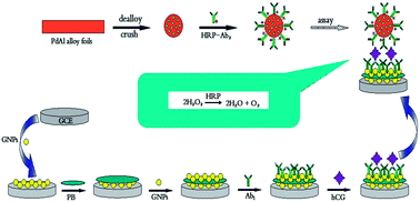
Synthesis of NP-Pd. The NP-Pd was prepared according to literature.40 PdAl alloy foils were made by refining pure (>99.9%) Pd and Al under the protection of argon atmosphere in a furnace, followed by melt-spinning. Then the NP-Pd was gained by dipping PdAl alloy foils in NaOH solutions at room temperature for 24 h. During the dealloying process, Pd atoms left behind would self-organize into an interconnected network of pores and ligaments. After dealloying, the foils were crushed to uniformed grain. 
Bioconjugation of nanoporous Pd with HRP–Ab2. 5 mg NP-Pd was initially added into 1 mL PBS (0.1 M, pH 7.2) and sonicated 5 min to obtain a homogeneous dispersion. 100 μL HRP–Ab2 (1 mg mL−1) was injected into the mixture and incubated for 12 h at 4 °C. The residual antibody was removed by centrifugation. Then the precipitate was washed with 0.1 M PBS for three times and immersed in 1% BSA solution for 1 h at 4 °C to block the remaining active groups and eliminate nonspecific binding effect. The resulting mixture was centrifuged and washed with PBS for several times. The prepared HRP–Ab2–NP-Pd was then stored in PBS (0.1 M, pH 7.2) at 4 °C until use.
Preparation of the modified electrode. The GCE was polished to a mirror finish with 0.3 and 0.05 μm alumina slurry, and then thoroughly washed ultrasonically in ethanol and ultrapure water in turn. The cleaned electrode was soaked in HAuCl4 aqueous solution (1%, w/w) and electrodeposited at −0.2 V for 300 s to get a GNPs layer. The preparation of PB/GNPs/GCE was accomplished in aqueous solution containing 2.5 mM FeCl3, 2.5 mM K3[Fe(CN)6], 0.1 M HCl and 0.1 M KCl by applying a constant potential of 0.4 V for 400 s. Following that, electrodeposition was performed in HAuCl4 solution (1%, w/w) at potential of −0.2 V for 5 min again. After that, the GNPs/PB/GNPs/GCE was obtained.
Preparation of electrochemical immunosensor. The modified working electrode was incubated with Ab1 for 12 h, followed by washing with PBS to remove nonspecific physically adsorption. Then the modified electrode was immersed in 1% BSA solution for 1 h at 4 °C to block nonspecific binding sites between the Ab1 and the electrode surface. Subsequently, the electrode was incubated in a varying concentration of hCG solution for 2 h. Finally, 10 μL of the prepared HRP–Ab2–NP-Pd solution was dropped onto the electrode surface and incubated for an additional 1 h. Each step was washed by PBS thoroughly for several times. The schematic diagram of the stepwise procedure of the immunosensor was shown in Scheme 1.
|  | 
|  | Scheme 1  Illustration of the stepwise immunosensor fabrication process. |  | 
 
Results and discussion
Characterization of NP-Pd
NP-Pd sample was tablet for SEM (Fig. 1(A)). It is clear that the dealloyed sample consists of a number of ultrafine nanoporous. Meanwhile, NP-Pd sample was dispersed in solvent, and then it was further confirmed by HRTEM. Fig. 1(B) showed HRTEM image of NP-Pd sample. The clear contrast between the bright regions and the dark skeletons further confirms the formation of a three-dimensional (3D) interconnected network structure on the nanoscale, which is beneficial for the mass and electron transport during electrochemical sensing. It is consistent with the SEM observation. The energy dispersive X-ray spectrum (EDS) (Fig. 1(C)) proved that the nanoporous material is composed of Pd with a small amount of residual Al. Pore size distributions were determined by BET analyses for NP-Pd. The pore diameter of NP-Pd was 5.1 nm as shown in Fig. 1(D). Meanwhile, the grain size distribution were tested (Fig. 1(E)). The average size of grain was 88.6 nm. These results indicate that NP-Pd was fabricated by means of a simple dealloying procedure. This method is in sharp contrast to the traditional approach, and this method can achieve a nearly 100% yield.
|  | 
|  | Fig. 1  (A) SEM and (B) HRTEM images showing the microstructure of NP-Pd; (C) EDX spectrum of NP-Pd; (D) pore size distribution of NP-Pd; (E) grain size distribution of NP-Pd. |  | 
Characterization of immunosensor
The characteristics of the differently modified electrodes were measured by CVs after each assembly step. In Fig. 2, after coated with the mixture of GNPs and PB, the current response of the electrode in PBS containing K3[Fe(CN)6] was apparently larger than that of the bare electrode. The higher current response might be attributed to dual-amplification effects of the GNPs and PB. GNPs with good conductivity increased the effective surface area of the electrode. PB, which has the formula Fe3+[Fe2+(CN)6]3, is a prototype of metal hexacyanoferrates and could be used as the electron transfer mediator due to electrochemical activity, fine redox reversibility and stability. Moreover, the addition of PB could prevent the leakage of GNPs. On the contrary, when Ab1 was immobilized on the GNPs/PB/GNPs modified electrode surface, the peak currents decreased. Subsequently, the peak currents decreased after the Ab1/GNPs/PB/GNPs/GCE reacted with hCG. When Ag/Ab1/GNPs/PB/GNPs/GCE reacted with HRP–Ab2–NP-Pd, the peak current further decreased, which demonstrated that hCG and antibody were successfully immobilized on the electrode surface. The reason might be the fact that the fact that hCG antigen and anti-hCG, as biomacromolecules, have weak conductivity, so as to hinder the diffusion of the redox marker toward the electrode surface, hinder electron transfer and insulate the electrode. It could be deduced that the proposed sandwich-type immunosensor was successfully fabricated from the above results.
|  | 
|  | Fig. 2  (A) CVs of (a) bare GCE; (b) GNPs/GCE; (c) PB/GNPs/GCE; (d) GNPs/PB/GNPs/GCE; (B) CVs of (c) Ab1/GNPs/PB/GNPs/GCE; (b) Ag/Ab1/GNPs/PB/GNPs/GCE; (a) NP-Pd–HRP–Ab2/Ag/Ab1/GNPs/PB/GNPs/GCE. CV conditions: pH 7.2 PBS solution containing 0.2 M KCl and 5 mM K3[Fe(CN)6]; scan rate: 50 mV s−1. |  | 
Signal amplification via catalytic reduction of H2O2 by NP-Pd
To further investigate whether the as-prepared NP-Pd could improve the analytical property of the electrochemical immunoassay, 100 ng mL−1 and 150 ng mL−1 of hCG antigen were analyzed using the GNPs/PB/GNPs modified electrodes by immunoreacting with either HRP–Ab2 or HRP–Ab2–NP-Pd. Fig. 3 displayed the obvious redox peaks of the modified electrodes in PBS (pH 7.2) containing 0.8 mM H2O2. The redox peaks might be attributed to the labeled HRP toward the reduction of H2O2 and catalytic activity of NP-Pd because of the non-electrochemical activity of the supporting electrode (PBS). The peak current of HRP–Ab2–NP-Pd/Ag/Ab1/GNPs/PB/GNPs modified electrode was increased compared with that of HRP–Ab2/Ag/Ab1/GNPs/PB/GNPs modified electrode. The changes of current were 21.67 and 27.3 μA at 100 ng mL−1 and 150 ng mL−1 hCG. The increase was ascribed to the synergistic action between HRP–Ab2 and NP-Pd. Firstly, NP-Pd with lots of porous could display a high surface-to-volume ratio, which could enhance the immobilized quantity of HRP–Ab2. Then it provided more chance of the Ag–Ab interaction in turn. Meanwhile, NP-Pd has higher capability of electron transfer. When numerous HRP–Ab2 were loaded with NP-Pd, NP-Pd could effectively transfer electrons from the base electrode surface to the redox center of HRP. Secondly, the immunosensor exhibited higher catalytic efficiency to the H2O2 due to large amounts of HRP. In addition, NP-Pd could catalyze the reduction of H2O2, which further enhanced the current response. The results indicated that NP-Pd nanostructures could improve the sensitivity of the electrochemical immunosensors and could be used as an efficient label for ultra-sensitive electrochemical immunoassay. In addition, Fig. 3 displayed the catalytic current increased with the increase of hCG concentration. The results provided a convenience for detecting hCG.
|  | 
|  | Fig. 3  The CV response after the sandwich format immunoreaction of the proposed immunosensor in the presence of (A) 100 ng mL−1 hCG (B) 150 ng mL−1 hCG by using various labels: (a) HRP–Ab2–NP-Pd, (b) HRP–Ab2 in pH 7.2 PBS containing 0.8 mM H2O2. |  | 
HRP–Ab2 adsorbed on NP-Pd was quantified by plotting absorbance (A) against the concentration of HRP–Ab2 by ultraviolet spectrophotometry. The linear regression equation: A = 0.02585 + 0.1617c (mg L−1)−1, r = 0.9995. The concentration of HRP–Ab2 in the supernatant (∼1 mL) was 10.31 mg L−1. The results also demonstrated that HRP–Ab2 was successfully loaded with NP-Pd.
Optimisation of the immunoassay conditions
The goal of this study was to obtain the excellent performance of the proposed immunosensor for hCG detection under optimized condition. The current response of the as-prepared immunosensor was directly affected by the quantity and thickness of GNPs and PB. The quantity and thickness of GNPs and PB were decided by the time of electrochemical reaction. The films could be thick, and then hinder the electronic transmission, when the time of electrochemical reaction exceeded a certain value. The CV measurements were carried out in pH 7.2 PBS solution containing 0.2 M KCl and 5 mM K3[Fe(CN)6] at a scan rate of 50 mV s−1. As shown in Fig. 4(A), after achieving the maximum current at 300 s, the current response decreased, indicating that the optimal reduction time of HAuCl4 was 300 s. The time of forming PB film was also studied and the results were shown in Fig. 4(B). The current response reached a maximum at 400 s. Therefore, 400 s was selected as an optimal time.
|  | 
|  | Fig. 4  Effect of (A) electrochemical reduction time of HAuCl4; (B) the time of forming PB film; (C) pH of PBS; (D) incubation time between Ab and Ag; (E) the substrate (H2O2) concentration on the electrochemical response. |  | 
In the sandwich-type immunoassays, the pH of the detection solution, the incubation time for the antigen–antibody interaction and the concentration of H2O2 also influenced the sensitivity of the as-synthesized immunosensor. The influence of pH was investigated and the results were shown in Fig. 4(C). The current response decreased at strong acidic and alkaline solution. The optimal response was obtained at pH 7.2. The reason is that the pH of the detection solution might influence not only the catalytic potential of activity of HRP and the bioactivity of immobilized immunoproteins, but also the electrochemical performance of GCE. Therefore, a pH 7.2 of the working buffer was applied for further experiments.
The incubation time between Ab and Ag was an important parameter for capturing hCG antigen and NP-Pd–HRP–Ab2 and the results were shown in Fig. 4(D). The current response increased with the increasing of the incubation time and then leveled off slowly after 60 min, showing an equilibration state. This may be because the interaction between Ab and Ag in the binding sites had reached saturated. Then further increasing incubation time will not affect the change of the current signal. Thus, 60 min was selected as the incubation time for the determination of hCG antigen in this study.
The concentration of H2O2 was another important parameter for the immunoassays. As shown in Fig. 4(E), when the H2O2 concentration was less than 0.8 mM, the reduction peak currents increased with increasing H2O2 concentration, and then started to level off. The reason is that all the enzyme molecules become saturated with H2O2, so the increasing of H2O2 concentration does not make much difference. Therefore, 0.8 mM was chosen for the optimal concentration of H2O2.
Performance of the immunosensor
A sandwich-type assay format was applied for the detection of hCG by using the synthesized NP-Pd as molecular tags under optimal condition. After hCG molecules were immobilized, they specifically bound to Ab1, followed by reaction with HRP–Ab2. In the presence of H2O2, HRP (Fe2+) is efficiently converted to its oxidized form, HRP (Fe3+). HRP (Fe3+) is then reduced at the electrode surface. Under a certain condition of H2O2 concentration, the reduction current is proportional to the concentration of HRP, thereby indirectly proportional to the concentration of hCG. CV measurements were recorded in PBS containing 0.8 mM H2O2 at a scan rate of 50 mV s−1. The immunosensor was incubated in hCG solution of different concentration. As shown in Fig. 5, the linear relationship between the peak current and the hCG concentration was obtained from 0.5 ng mL−1 to 200 ng mL−1 with a correlation coefficient of 0.9986. The limit of detection was 9.3 pg mL−1. Therefore, the use of HRP–Ab2–NP-Pd as detection antibody could significantly amplify the signal through the enzymatic signal amplification of high-content HRP and the presence of NP-Pd on immunosensor surface.
|  | 
|  | Fig. 5  (A) The CVs of the immunosensor at different concentration of hCG; (B) calibration plots for hCG solution. |  | 
Selectivity, reproducibility and stability of the immunosensor
To evaluate the selectivity of the immunosensor, the effect of interferents was studied on the current responses of the immunosensor in the presence of 20.0 ng mL−1 hCG. The interference effects of AFP, CEA, and Vc, etc, were investigated by adding them into the incubation solution. The 20.0 ng mL−1 hCG solution containing 50.0 ng mL−1 interfering substances was measured by the immunosensor. As shown in Fig. 6, when hCG coexisted with the interferents, no apparent signal change took place in comparison with that of only hCG, which indicated that the system exhibited sufficiently selectivity for the determination of hCG.
|  | 
|  | Fig. 6  CVs peak currents of the immunosensor for the detection of (a) hCG, (b) hCG + glucose, (c) hCG + BSA, (d) hCG + L-Cys, (e) hCG + L-glutamic acid, (f) hCG + folic acid, (g) hCG + Vc, (h) hCG + CEA, (i) hCG + AFP. |  | 
Reproducibility of the immunosensor is a key factor for developing a practical immunosensor. The inter-assay precision was estimated by determining the same hCG level with five immunosensors made using the same conditions independently. The relative standard deviations (RSDs) were 4.9%, 4.1%, and 4.6% for 5.0, 20.0 and 50.0 ng mL−1 hCG. Similarly, the intra-assay precision was estimated by testing three hCG levels for five replicate measurements. The RSDs were 5.1%, 4.3% and 3.9% for 5.0, 20.0 and 50.0 ng mL−1 hCG. The results obtained were satisfactory.
The stability, another important parameter of immunosensor, was examined by the property of HRP–Ab2–NP-Pd bioconjugates. When the bioconjugates were not in use, they were stored in PBS (pH 7.2) at 4 °C. After 3 weeks, the current response of the immunosensor was retained 86% of the initial response. The slow decrease in response might be on account of the gradual deactivation of the immobilized antibody incorporated in the composite. It conformed that the developed immunosensor had excellent stability.
Real sample analysis
To evaluate the applicability of the proposed method, it was used for hCG assay by standard addition methods in human serum. The analytical results were shown in Table 1. The recovery efficiency was in the range of 95.2%–103.45%. Therefore, the proposed immunosensor could be considered as an optional method for detection of hCG in real sample.
Table 1 Determination results of real samples (n = 9)
		
| Amount added (ng mL−1) | This method (ng mL−1 ± RSD%) | Recovery (%) | 
| 2.0 | 1.92 ± 4.87 | 96 | 
| 5.0 | 4.76 ± 3.38 | 95.2 | 
| 10.0 | 10.49 ± 3.25 | 104.9 | 
| 50.0 | 48.76 ± 4.18 | 97.52 | 
| 100.0 | 103.45 ± 2.18 | 103.45 | 
Comparison of methods
The proposed method for the determination of hCG was compared with the previously reported methods in Table 2. Compared with previous methods, the proposed method shows relatively higher sensitivity and lower detection limit. The low detection limit might be ascribed to two possible explanations: (i) as discussed earlier, NP-Pd with high surface-to-volume ratio and the high-content HRP exhibited high catalytic efficiency toward the reduction of H2O2; (ii) a relatively large amount of HRP–Ab2 had been conjugated to NP-Pd label. Usually, when the hCG concentration was low, the amount of hCG captured by Ab1 immobilized onto the electrode surface was also low. However, the large amount of HRP–Ab2 immobilized onto the label increases the probability of interactions between HRP–Ab2 and antigen.
Table 2 Comparison of analytical parameters for the determination of hCG
		
| Methods | Reagents or condition | Linear range | Detection limit | Ref. | 
| Sandwich electrochemical immunosensor | GNPs, PB, NP-Pd | 0.5–200 ng mL−1 (5–2000 mIU mL−1) | 9.3 pg mL−1 (0.093 mIU mL−1) | This paper | 
| Photoluminescence | Hierarchical ZnO arrays | 2–160 ng mL−1 | — | 7 | 
| Fluoroimmunoassay | Polymer nanoparticles | 1.25–300 mIU mL−1 | 0.3 mIU mL−1 | 11 | 
| Fluoroimmunoassay | Magnetic particles | 0.16–450 ng mL−1 | 0.08 ng mL−1 | 12 | 
| Spectrometry | An inductively coupled plasma mass spectrometry | 5.0–170 μg L−1 | 1.7 μg L−1 | 13 | 
| Resonance scattering spectral assay | Gold nanoparticles | 2.5–208.3 mIU mL−1 | 0.83 mIU mL−1 | 14 | 
| Immunosensor | Nanoporous gold/graphene sheets | 0.5–40.00 ng mL−1 | 0.034 ng mL−1 | 15 | 
| Immunosensor | Gold nanoparticles/poly-(2,6-pyridinediamine)/multiwall carbon nanotubes | 1–160.0 mIU mL−1 | 0.3 mIU mL−1 | 16 | 
| Immunosensors | Gold nanoparticles/methylene blue/thiourea | 1.0–100.0 mIU mL−1 | 0.3 mIU mL−1 | 17 | 
Conclusions
In this study, a new sandwich-type immunoassay protocol for sensitive electrochemical determination of hCG was devised. The NP-Pd with catalytic activity was used for labeling HRP–Ab2, which could load numerous HRP–Ab2 to enhance the sensitivity of the immunosensor. The proposed immunosensor showed a wide linear range and satisfactory selectivity, reproducibility and stability. In addition, the proposed strategy provides a biocompatible immobilization and sensitized recognition platform for analytes as small antigens and possesses promising potential in clinical diagnosis.
Acknowledgements
This project was financially supported by Shandong Provincial Natural Science Foundation, China (Grant no. ZR2012BL11), Shandong Provincial Science and Technology Development Plan Project, China (Grant no. 2013GGX10705) and the National Natural Science Foundation of China (Grant no. 51003040 and 51102114).
References
- J. F. Wang, R. Yuan, Y. Q. Chai, S. R. Cao, S. Guan, P. Fu and L. G. Min, Biochem. Eng. J., 2010, 51, 95–101 CrossRef CAS PubMed.
- W. Schmidt, K. Klinga, K. Neudeck, B. Runnebaum and F. Kubli, Geburtshilfe Frauenheilkd., 1983, 43, 664–669 CrossRef CAS PubMed.
- A. Banerjee, N. S. Srilatha and G. S. Murthy, Biochim. Biophys. Acta, 2002, 1569, 21–30 CrossRef CAS.
- S. Madersbacher and P. Berger, Antibodies and immunoassays, methods,  2000, vol. 21, pp. 41–50 Search PubMed.
- H. Sasamoto, M. Maeda, A. Tsuji and H. Manita, Anal. Chim. Acta, 1995, 309, 221–225 CrossRef CAS.
- L. X. Zhao, J. M. Lin and F. Qu, Acta Chim. Sin., 2004, 62, 71–77 CAS.
- X. Yan, Z. B. Huang, M. He, X. M. Liao, C. Q. Zhang, G. F. Yin and J. W. Gu, Colloids Surf., B, 2012, 89, 86–92 CrossRef CAS PubMed.
- N. Nakamura, T. K. Lim, J. M. Jeong and T. Matsunaga, Anal. Chim. Acta, 2001, 439, 125–130 CrossRef CAS.
- Q. P. Qin, M. Christiansen, T. Lovgren, B. Norgaard-Pedersen and K. Pettersson, J. Immunol. Methods, 1997, 205, 169–175 CrossRef CAS.
- S. Scsuková, M. Jezová, J. Vranová, M. Tatara and J. Kolena, Gen. Physiol. Biophys., 1996, 15, 451–462 Search PubMed.
- Y. H. He, Y. P. Li and X. Hun, Microchim. Acta, 2010, 171, 393–395 CrossRef CAS.
- J. Y. Hou, T. C. Liu, Z. Q. Ren, M. J. Chen, G. F. Lin and Y. S. Wu, Analyst, 2013, 138, 3697–3704 RSC.
- S. C. Zhang, C. Z. Zhang, Z. Xing and X. R. Zhang, Clin. Chem., 2004, 50, 1214–1221 CAS.
- A. H. Liang, M. J. Zou and Z. L. Jiang, Talanta, 2008, 75, 1214–1220 CrossRef CAS PubMed.
- R. Li, D. Wu, H. Li, C. X. Xu, H. Wang, Y. F. Zhao, Y. Y. Cai, Q. Wei and B. Du, Anal. Biochem., 2011, 414, 196–201 CrossRef CAS PubMed.
- J. F. Wang, R. Yuan, Y. Q. Chai, S. R. Cao, S. Guan, P. Fu and L. G. Min, Biochem. Eng. J., 2010, 51, 95–101 CrossRef CAS PubMed.
- R. Chai, R. Yuan, Y. Q. Chai, C. F. Ou, S. R. Cao and X. L. Li, Talanta, 2008, 74, 1330–1336 CrossRef CAS PubMed.
- K. Kerman, N. Nagatani, M. Chikae, T. Yuhi, Y. Takamura and E. Tamiya, Anal. Chem., 2006, 78, 5612–5616 CrossRef CAS PubMed.
- J. Chen, J. H. Tang, F. Yan and H. X. Ju, Biomaterials, 2006, 27, 2313–2321 CrossRef CAS PubMed.
- J. Chen, F. Yan, Z. Dai and H. X. Ju, Biosens. Bioelectron., 2005, 21, 330–336 CrossRef CAS PubMed.
- J. M. Liu, X. M. Huang, M. L. Cui, L. P. Lin, L. H. Zhang and Z. Y. Zheng, Anal. Biochem., 2012, 431, 19–29 CrossRef CAS PubMed.
- Y. L. Cui, H. F. Chen, L. Hou, B. Zhang, B. Q. Liu and G. N. Chen, Anal. Chim. Acta, 2012, 738, 76–84 CrossRef CAS PubMed.
- C. L. Hong, R. Yuan, Y. Q. Chai, Y. Zhuo and X. Yang, J. Electroanal. Chem., 2012, 664, 20–25 CrossRef CAS PubMed.
- B. Zhang, D. P. Tang, B. Q. Liu, Y. L. Cui, H. F. Chen and G. N. Chen, Anal. Chim. Acta, 2012, 711, 17–23 CrossRef CAS PubMed.
- J. P. Lei and H. X. Ju, Chem. Soc. Rev., 2011, 41, 2122–2134 RSC.
- Z. H. Wen, S. Q. Ci and J. H. Li, J. Phys. Chem. C, 2009, 113, 13482–13487 CAS.
- G. Pampalakis and S. O. Kelley, Analyst, 2009, 134, 447–449 RSC.
- D. M. Kim, M. A. Rahman, M. H. Do, C. G. Ban and Y. B. Shim, Biosens. Bioelectron., 2010, 25, 1781–1788 CrossRef CAS PubMed.
- C. S. Shan, H. F. Yang, J. F. Song, D. X. Han, A. Ivaska and L. Niu, Anal. Chem., 2009, 81, 2378–2382 CrossRef CAS PubMed.
- T. W. Tsai, G. Heckert, L. F. Neves, Y. Q. Tan, D. Y. Kao, R. G. Harrison, D. E. Resasco and D. W. Schmidtke, Anal. Chem., 2009, 81, 7917–7925 CrossRef CAS PubMed.
- J. F. Wang, R. Yuan, Y. Q. Chai, S. R. Cao, S. Guan, P. Fu and L. G. Min, Biochem. Eng. J., 2010, 51, 95–101 CrossRef CAS PubMed.
- W. Sun, Y. Y. Zhang, X. M. Ju, G. G. Li, H. W. Gao and Z. F. Sun, Anal. Chim. Acta, 2012, 752, 39–44 CrossRef CAS PubMed.
- S. Chawla, R. Rawal, S. Sharma and C. Shekhar Pundir, Biochem. Eng. J., 2012, 68, 76–84 CrossRef CAS PubMed.
- S. Chawla, R. Rawal and C. S. Pundir, J. Biotechnol., 2011, 156, 39–45 CrossRef CAS PubMed.
- L. F. Liu, O. Albrecht and U. Gosele, Electrochem. Commun., 2010, 12, 835–838 CrossRef CAS PubMed.
- J. S. Yu, Y. Ding and M. W. Chen, Chem. Mater., 2008, 20, 4548–4550 CrossRef CAS.
- X. G. Wang and Z. H. Zhang, Electrochem. Commun., 2009, 11, 1896–1899 CrossRef CAS PubMed.
- A. H. Liu, H. R. Geng, C. X. Xu and H. J. Qiu, Anal. Chim. Acta, 2011, 703, 172–178 CrossRef CAS PubMed.
- C. X. Xu, Y. Q. Liu, J. P. Wang, H. R. Geng and H. J. Qiu, J. Power Sources, 2012, 199, 124–131 CrossRef CAS PubMed.
- C. X. Xu, A. H. Liu, H. J. Qiu and Y. Q. Liu, Electrochem. Commun., 2011, 13, 766–769 CrossRef CAS PubMed.
| 
 | 
| This journal is © The Royal Society of Chemistry 2014 | 
Click here to see how this site uses Cookies. View our privacy policy here.