IM-17: a new zeolitic material, synthesis and structure elucidation from electron diffraction ADT data and Rietveld analysis

Yannick Lorgouillouxab, Mathias Dodina, Enrico Mugnaiolic, Claire Marichala, Philippe Caulleta, Nicolas Batsd, Ute Kolbc and Jean-Louis Paillaud*a
aEquipe Matériaux à Porosité Contrôlée (MPC), Institut de Science des Matériaux de Mulhouse (IS2M), UMR CNRS 7361 – UHA, ENSCMu, 3b rue Alfred Werner, 68093 Mulhouse Cedex, France. E-mail: jean-louis.paillaud@uha.fr
bLMCPA, EA 2443, UVHC, Pôle Universitaire de Maubeuge, Boulevard Charles de Gaulle, 59600 Maubeuge, France
cJohannes Gutenberg-Universität Mainz, Institut für Physikalische Chemie, Jakob-Welder-Weg 11, 55128 Mainz, Germany
dIFP Energies nouvelles, Direction Catalyse et Séparation, Rond-point de l'échangeur de Solaize – BP 3, 69360 Solaize, France

Received 17th February 2014 , Accepted 2nd April 2014

First published on 8th April 2014


Abstract

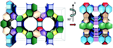
The synthesis and the structure of IM-17, a new germanosilicate with a novel zeolitic topology, prepared hydrothermally with decamethonium as the organic structure directing agent, are reported. The structure of calcined and partially rehydrated IM-17 of chemical formula per unit cell |(H2O)14.4|[Si136.50Ge39.50O352] was solved ab initio using electron diffraction ADT data in the acentric Amm2 (setting Cm2m) space group and refined by the Rietveld method. This new zeolite framework type contains a 3D pore system made of intersecting 12, 10 and 8-ring channels.


Introduction

Among the strategies adopted to synthesize new zeolite structures, besides the use of organic structure directing agents (OSDAs) and the replacement of the hydroxide anion by the fluoride anion as a mineralizing agent, the partial or complete substitution of the traditional aluminium and silicon framework atoms by other elements such as phosphorus,1 titanium,2 zinc,3 gallium,4 boron,5 or germanium6 has proven very efficient. In particular germanium gave very interesting results. On one hand, the fact that germanium can adopt various coordinations is indeed a property that allows the formation of many new frameworks. Microporous materials with germanium not only in tetrahedral coordination, but also inside square pyramids or trigonal bipyramids (5-coordination) and octahedra (6-coordination) were thus obtained. This is the case of the majority of the ICMM-n family materials (e.g. ICMM-77) and of many solids of the SU-n (e.g. SU-M8 and SU-749), ASU-n (e.g. ASU-1210), UCSB-n (e.g. UCSB-4011) and IM-n (e.g. IM-1412) families. On the other hand, even in germanates or germanium-containing silicates where germanium is only in tetrahedral coordination (e.g. as in CCUT-9,13 IM-1014 (UOZ), ASU-715 (ASV), FOS-516 (BEC), ITQ-3317 (ITT) or IM-1218 and ITQ-1519 (both of UTL topology)) and that can thus be considered as real zeolitic materials, new structures can be obtained as the T–O–T angles are much smaller (∼130°) than in silicates (∼145°). Because of these smaller T–O–T angles the formation of germanates displaying highly strained 3-membered rings (3MRs) is also possible.17,20 Recently, two zeolitic structures with double 3-membered rings (d3r), ITQ-4021 (IRY) and ITQ-4422 (IRR), were even synthesized. These new germanosilicates, like a lot of other germanium-containing silicates, hold double 4-membered ring (d4r) units. It is well-known that the d4r unit can be stabilized not only by the presence of fluoride anions that locate inside this unit, but also by the substitution of silicon by germanium.23–25 Whereas there are very few examples of silicate or aluminosilicate zeolites containing d4rs that can be synthesized without fluoride anions, many Ge-containing silicates are characterized by the presence of d4rs that do not need to be stabilized by fluoride anions.17–19,26–28

In our search for new large pore zeolites in the (Si,Ge) system, we decided to engage decamethonium (Fig. 1) as OSDA by varying several synthesis parameters. This diquaternary ammonium cation has a quite long chain and is known to lead to the aluminosilicates Nu-8729 (NES) and ZSM-1230 (MTW), which are 10 and 12MR zeolites, respectively. Note that the magnesium-containing aluminophosphate DAF-1 (DFO), a 12, 10 and 8MR molecular sieve is also hydrothermally synthesised with the same OSDA.31 Our investigations led to the discovery of a new germanosilicate zeolite framework containing d4r units, IM-17,32 that was obtained among known zeolites. Here we present the synthesis, characterization and structure of this new molecular sieve. The structure was determined by a combination of automated electron diffraction tomography (ADT) and Rietveld refinement on powder X-ray diffraction data (PXRD). Electrons have a strong interaction with matter and can be easily focused in nanoprobes. These characteristics allow to perform single-crystal diffraction on single nanometric particles. Since the late 1980s, electron diffraction has been used together with other techniques for structure determination of porous materials. In particular, electron diffraction assisted PXRD for space group determination and peak deconvolution.33–37 Early encouraging examples also demonstrated that it is in principle possible to perform ab initio structure solution based on electron diffraction data alone.38,39 Nevertheless, due to dynamical scattering and experimental difficulties in data acquisition, only a partial structure can be normally achieved using conventional in-zone electron diffraction data, and there is a major concern in recognizing the correct phase scheme among the several proposals automatically determined by direct methods.40 In the last years, ADT method has allowed to collect more complete and off-zone (less dynamical) electron diffraction data and to reduce the exposure time and the beam damage on the sample. Electron diffraction is therefore becoming a routine method for structure determination of porous materials and diverse nanocrystalline phases that have been solved and refined with a combination of ADT and PXRD methods.41–44


image file: c4ra01383b-f1.tif
Fig. 1 Diquaternary ammonium cation decamethonium.

Experimental details

Synthesis

Hydrothermal syntheses were performed at 170 °C for 14 days unless clearly stated otherwise, in a homemade multi-autoclave containing sixteen 2 mL Teflon-lined reactors. Gels were prepared both in OH and F media by mixing Aerosil 200 (>98%, Degussa) or TEOS (>98%, Fluka), amorphous germanium oxide GeO2 (>99.99%, Aldrich), HF acid (40%, Carlo Erba), deionized water, and decamethonium dihydroxide obtained from the bromide form (>98%, Fluka) by ion exchange in water from anion resin (Dowex SBR LC NG, OH Form (Supelco)) as OSDA. The OSDA solutions were concentrated by lyophilization when necessary. The chemical composition of a typical starting gel leading to the new IM-17 zeolite was: 0.6SiO2: 0.4GeO2: 0.25R(OH)2: 10H2O, where R represents the decamethonium cation. The initial and final pH values of the mixtures were about 14 and 12, respectively. The final products were recovered by filtration, washed several times with deionized water and then dried at 70 °C for 24 h.

Scanning electron microscopy

The morphologies and sizes of the crystals were determined by scanning electron microscopy (SEM) using a Philips XL30 FEG SEM microscope.

Chemical analysis

The AAS bulk chemical analysis (Si,Ge) of three IM-17 samples after dissolution in HF were performed on a Varian AFS240 apparatus. The exact amount of occluded OSDA molecules was determined from quantitative 1H liquid nuclear magnetic resonance (NMR) spectroscopy. For that, a known amount of the as-synthesised IM-17 (∼50 mg) was dissolved into 2 mL of HF (40% in water). Thereafter, 300 μL of a 0.282 M 1,4-dioxane–D2O solution were added as an internal standard to the dissolved material. After homogenization, 0.5 mL of the liquid was transferred with an equivalent volume of pure D2O in a Teflon® tube for the NMR analysis. The spectra were recorded on a Bruker AC 400 spectrometer. The recording conditions were: frequency = 400.17 MHz; recycle time = 1 s; pulse width = 2.1 μs; pulse angle = π/6, 1H chemical shifts being referenced to TMS.

Crystallographic study

The powder X-ray diffraction (PXRD) data of calcined IM-17 samples were collected on a PANalytical MPD X'Pert Pro diffractometer in a Debye–Scherrer geometry equipped with a capillary sample holder, a hybrid mirror monochromator (λ = 1.5406 Å) which gives the monochromatic parallel beam geometry and a X'Celerator Real-Time Multiple Strip detector (active length = 2.122°(2θ)). For the structure elucidation, sample no. 14 was used. After calcination (550 °C under air), the ground powder of IM-17 was introduced in a Mark-tube made of special glass (no. 14, outside diameter 0.3 mm, Hilgenberg Gmbh). Then the capillary tube was sealed and mounted on a precise goniometric head, which is screwed on a rotary sample stage, the spinning rate of which was 1 rotation per second. The powder pattern was collected at 295 K in the range 4° < (2θ) < 90°, step = 0.017°(2θ), time/step = 3970 s, the total collecting time being about 61 h.

The PXRD pattern of calcined IM-17 was indexed in a C-centred orthorhombic unit cell with the parameters a = 39.109(8) Å, b = 22.242(5) Å, c = 12.680(4) Å and V = 11029.6(45) Å3 by the Louër's DICVOL9145 indexing routine of the STOE WinXPow program package.46 The observed systematic extinctions suggested C222 (#21), Cmm2 (#35), C2mm (Amm2 (#38)) and Cmmm (#65) as possible space groups. For the structure determination the extracted intensities of the reflections up to 60°(2θ) were taken. From this set of data, the structure of calcined IM-17 was mainly solved by direct methods with the highest symmetry, i.e. in space group Cmmm using the EXPO2009 software.47 Some oxygen and two missing disordered framework silicon T sites were deduced from model building using Cerius2 environment together with difference Fourier maps during the Rietveld refinement.48 The Rietveld49 refinement was performed using the GSAS package50 on the complete pattern via the interface EXPGUI.51 The atomic coordinates of the whole framework atoms (i.e. T and O atoms with T = Si or Ge) were used as the starting model. All the atoms were refined isotropically. Soft restraints were placed on the bond lengths and angles of the framework (T–O = 1.68(4) or 1.62(4) Å and O–T–O = 109.5(30)°). Initially, for the Rietveld refinement, on each T atom site, a silicon atom and a germanium atom were both placed at the same position with the sum of their occupancy factors constrained to be 1. Then, as the refining progressed, some T sites were set as purely siliceous. After refinement of the framework, successive calculated Fourier difference maps revealed a scattering density inside the void volume attributed to the presence of adsorbed water molecules. Then, one oxygen atom has been placed on these positions and refined (position and occupancy factor). Further details on crystallographic and Rietveld refinement data are given in ESI, Table S1 and Fig. S1. The final atomic parameters, selected bond distance and bond angles are listed in ESI, Tables S2 and S3.

In order to verify if the space group Cmmm was the true one, a study by ADT (Automated electron Diffraction Tomography) has been undertaken. Thus, calcined sample no. 14 (Table 1) was dispersed in ethanol, sonified and sprayed on a carbon-coated cupper grid using a modified UIS250v Hielscher sonifier.52 ADT acquisition was performed with a FEI TECNAI F30 S-TWIN transmission electron microscope equipped with field emission gun and working at 300 kV, using the semi-automatic module described by Kolb et al.53 Crystal position was tracked in microprobe STEM mode and nano electron diffraction (NED) patterns were acquired sequentially in steps of 1°. STEM images were collected by a FISHIONE high angular annular dark field (HAADF) detector and acquired by Emispec ESVision software. NED patterns were taken with a CCD camera (16-bit GATAN ULTRASCAN4000, 4096 × 4096 pixels) and acquired by the Gatan Digital Micrograph software. A condenser aperture of 10 μm and mild illumination setting were used in order to produce a semi-parallel beam of 70 nm in diameter on the sample. ADT was coupled with electron beam precession in order to improve reflection intensity integration.52 Precession was performed with a NANOMEGAS DigitStar device keeping the precession angle at 1.2°.

Table 1 Selection of the most representative syntheses in the (Si,Ge) system with the decamethonium cation as OSDAa
Samples Molar synthesis mixture compositions Materials
SiO2 GeO2 OSDA HF H2O
a All syntheses were performed at 170 °C for 14 days.b Silica source is TEOS.c Silica source is Aerosil 200.d No solid synthesized.e Very small amount of MFI-type zeolite synthesized.f GeO2 dense phase argutite.g Pure phase but lower yield.
1b 0.6 0.4 0.25 0.5 8 BEC
2b 0.6 0.4 0.5 0.5 8 AST
3c 0 1 0.25 0 20 d
4c 0.2 0.8 0.25 0 20 IM-17 + MFI + 122B
5c 0.4 0.6 0.25 0 20 IM-17 + MFI + 122B
6c 0.5 0.5 0.25 0 20 IM-17 + 122B + εMFIe
7c 0.6 0.4 0.25 0 20 IM-17 + 122B + εMFIe
8c 0.75 0.25 0.25 0 20 122B
9c 0.9 0.1 0.25 0 20 122B
10c 1 0 0.25 0 20 122B
11c 0.6 0.4 0.5 0 20 IM-17 + MFI
12c 0.6 0.4 0.5 0 40 IM-17 + 122B
13c 0.6 0.4 0.125 0 10 MFI + argf + IM-17
14c 0.6 0.4 0.25 0 10 IM-17
15c 0.6 0.4 0.25 0 5 IM-17
16c 0.6 0.4 0.125 0 5 IM-17
17c 0.8 0.2 0.25 0 3 IM-17
14bisb 0.6 0.4 0.25 0 10 IM-17g


ADT data elaboration, including three-dimensional diffraction volume reconstruction and visualization, cell parameter determination and reflection intensity extraction, was performed with ADT3D software, according with the algorithms developed by Kolb et al.52–55 Ab initio structure solution was performed by direct methods implemented in the program SIR2011,56 assuming the kinematic approximation IFhkl2. The structure model obtained from ADT, (see IM-17 structure paragraph of the results and discussion part below), was refined by Rietveld refinement using GSAS as described above.

Tiling

The tiling data were calculated with TOPOS 4.0.57 The embedding of the tiling visualization was performed by 3dt58 a programme of the GAVROG project.

Thermal analyses

Thermogravimetric (TGA) and differential thermal (DTA) analyses were performed under air on a Setaram Labsys thermoanalyser with a heating rate of 5 °C min−1 up to 1000 °C.

29Si solid state MAS NMR spectroscopy

29Si (I = 1/2) Magic Angle Spinning (MAS) NMR spectra were recorded on a Bruker Avance II 300WB spectrometer (B0 = 9.4 T) operating at a Larmor frequency of 59.6 MHz. A 4 mm double-channel Bruker probe was used and the sample spun at a spinning frequency of 8 kHz. 29Si single-pulse MAS NMR experiments were performed with a π/6 pulse duration of 2.3 μs and a 80 s recycling delay. These recording conditions ensure the quantitative determination of the different Si species.59 1H–29Si CPMAS NMR experiments were acquired with a contact time of 4 ms and a recycle delay of 4 s. The radiofrequency field strength used for 1H decoupling was set to 50 kHz. 29Si chemical shifts are reported relative to tetramethylsilane (TMS).

N2 adsorption isotherm measurement

After calcination (550 °C under air) the sample was placed in a glass measurement cell, and was then degassed at 350 °C under vacuum prior to the measurement. The microporous volume of IM-17 was determined from nitrogen adsorption isotherms obtained at 77 K on a Micromeritics ASAP 2010 porosimeter using the t-plot method.60

Results and discussion

Synthesis parametric study

As shown in the first rows of Table 1, use of the fluoride medium led to the synthesis of a silicogermanate corresponding to the polymorph C of beta zeolite (BEC) when decamethonium was used as OSDA (sample 1). When the amount of OSDA was doubled, a zeolite of the AST framework type was obtained (sample 2). Considering the size of this OSDA, the real template for the AST framework must have been a degradation product of the decamethonium cation. It should be noted that these two frameworks contain d4r units, which once again shows that these units are stabilized not only by germanium but also by fluoride anions. Although the synthesis of these two materials with the decamethonium cation was previously unseen, similar silicogermanates having been extensively studied in the past,61,62 they were not characterized in detail in the present work and no further syntheses in the fluoride medium were conducted. Most of the samples that were synthesized in hydroxide medium (Tables 1 and 2) contained three different materials. One of them is a germanosilicate zeolite of the well-known MFI-type. The two other solids are new and seem to be related. One is well crystallized and was named IM-17, whereas the other one, that was called 122B, shows a XRD pattern with very wide peaks (Fig. 2), which is the sign of a poorly crystalline phase and/or the consequence of a strain broadening. The crystals of the 122B material are of nanometric sizes and can not be distinguished from an amorphous phase by SEM (not shown). On the contrary, as shown on the SEM picture in Fig. 3, the IM-17 crystals are well-defined rhombus shaped plated-like.
Table 2 Influence of the synthesis conditions (time and stirring) on the crystallization of IM-17 and 122Ba
Samples SiO2 GeO2 Synthesis time (day) Stirringb Materials
a All samples synthesized at 170 °C, with OSDA/(Si + Ge) = 0.25, H2O/(Si + Ge) = 20 and Aerosil 200 as the silica source.b Autoclave stirred at 15 rpm during thermal treatment.c Very small amount of MFI-type zeolite synthesized.
7 0.6 0.4 14 No IM-17 + 122B + εMFIc
18 0.6 0.4 3 No 122B
19 0 1 7 No 122B + IM-17
20 0.2 0.8 14 Yes 122B + IM-17
8 0.75 0.25 14 No 122B
21 0.75 0.25 30 No 122B
22 0.75 0.25 14 Yes 122B


image file: c4ra01383b-f2.tif
Fig. 2 XRD patterns of samples 8 and 14 showing the very broad peaks of the 122B material and the sharp peaks corresponding to pure IM-17, respectively.

image file: c4ra01383b-f3.tif
Fig. 3 SEM pictures of sample 14 showing the rhombus shaped plate-like crystals of IM-17.

As shown in Table 1, the three obtained phases were present in sample 7, the first sample that was synthesized in hydroxide medium. A parametric synthesis study was thus conducted in order to obtain a pure IM-17 sample. A selection of the most representative results is given in Table 1. Modification of the Si/Ge ratio had a great effect on the synthesized materials (samples 3 to 10). When the synthesis mixture was Si rich, i.e. when Si/Ge ≥ 3 and even in purely siliceous system (samples 8, 9 and 10), samples only contained the poorly-crystalline 122B material. IM-17 was the main product for intermediate Si/Ge ratios: 1 ≤ Si/Ge ≤ 1.5 (samples 6 and 7). When the GeO2 content was further increased, an MFI-type zeolite crystallized in a quite large amount along with IM-17 and small quantities of 122B.

For the syntheses leading to samples 11 to 16, the water and decamethonium amounts were varied. The results of these syntheses showed that IM-17 could be obtained as a pure material only when the water amount was decreased (samples 14, 15 and 16). However, the H2O/OSDA ratio also seems very important, as a concentrated synthesis mixture with a low amount of decamethonium (H2O/OSDA = 80, sample 13) did not lead to pure IM-17, but to a mixture containing also MFI-type zeolite and the GeO2 dense phase argutite. To synthesize pure IM-17, the reaction mixture thus needs water and OSDA contents such that H2O/(Si + Ge) ≤ 10 and H2O/OSDA ≤ 40. Sample 17 showed that with highly concentrated medium, the Si/Ge ratio in the mixture could be increased up to 4 and still lead to the pure IM-17 material, which shows that these media very poor in water are favourable for the synthesis of IM-17.

A few syntheses not described in detail here were conducted with TEOS as the silica source and/or with seeds, but they did not allow getting better results. Indeed, as reported in Table 1, sample 14bis prepared with TEOS instead of Aerosil 200 as silica source used for sample 14 leads also to IM-17 but with a yield three times lower as it was already reported for IM-16 (UOS).63

The influence of the synthesis conditions was also studied (Table 2). Samples 18, 19 and 7 showed that for the same synthesis mixture composition, the 122B material was obtained first (3 days). After longer time (7 days), IM-17 was also visible on the XRD pattern of the product, and this material became predominant after 14 days. It is thus possible that the 122B material transformed into IM-17 when the synthesis time increased, and 122B might be an early-stage disordered IM-17 analogue. Presence of a large amount of 122B solid in sample 20 may have been due to a much higher nucleation rate of 122B under stirring. As sample 8 contained only 122B, samples 21 and 22 were prepared to see if the same mixture composition could lead to IM-17 under other synthesis conditions, but no improvement was observed.

Bulk chemical analysis of sample 14, 15 and 14bis gave a mean Si/Ge molar ratio of 3.5, 2.8 and 5.3, respectively. These results prove also the influence of the water content and the chemical source of silica on the chemical composition of IM-17. According to the quantitative 1H liquid NMR analysis performed on sample 14, the OSDA is intact inside the structure of IM-17 (in ESI, Fig. S2). A total weight loss of about 11.9% is observed on the TGA curve (not shown) of as-made sample 14. Accordingly, from quantitative 1H liquid NMR, TGA and chemical analyses and the present structural study on the calcined same sample, the chemical formula per unit cell of as-made IM-17 (sample 14) is close to |(C16H38N2)5.5 (OH)11 (H2O)2|[Si136.50Ge39.50O352].

Solid state NMR

The preliminary PXRD study of calcined IM-17 led to a proposed structure belonging to Cmmm space group. The asymmetric unit contains 14 crystallographically non equivalent T-sites from which 2 are disordered (Si13 and Si14, ESI, Table S2, Fig. S3). From this disorder, three models may be generated. Two ordered structures in sub-space groups Pmma and Amm2 and one zeolitic structure with an interrupted framework in the same space group Cmmm (see explanation in ESI, Fig. S4–S6). The interrupted framework should lead to a large number of silanol groups for IM-17. Solid state NMR was performed to evidence these silanols. 29Si MAS NMR spectrum of calcined IM-17 (sample no. 14) is given on Fig. 4a. At least two resonances are detected between −100 and −122 ppm that could correspond to Si(OSi)4, Si(OSi)3OH, Si(OSi)3O or Si(OSi)(4−n)Gen species.6
image file: c4ra01383b-f4.tif
Fig. 4 1H decoupled 29Si MAS (a) and 1H–29Si CPMAS (b) NMR spectra of calcined IM-17 (sample 14).

According to the preliminary structural study, 14 inequivalent crystallographic sites are present but are not resolved on the spectrum due to a distribution of environments. In order to enhance the signal corresponding to silanols, a 1H–29Si CPMAS experiment was performed. This experiment based on magnetization transfer from 1H to 29Si thanks to dipolar interaction is shown on Fig. 4b. A resonance is observed at −101.5 ppm corresponding to silanol species. Surprisingly, despite a very long acquisition time (34[thin space (1/6-em)]156 scans) a low signal to noise ratio was obtained suggesting that the sample contains only a very small amount of silanol groups. Decreasing the contact time to 1 ms (spectrum not shown) did not improve the signal to noise ratio. Furthermore, this resonance can not be detected on the 29Si MAS NMR spectrum (Fig. 4a) that was recorded in quantitative conditions suggesting that indeed only traces of silanol are present in this sample. As a consequence, the interrupted framework model is not possible.

Structure of IM-17

IM-17 crystallizes in platelet-like crystals with sizes ranging from 200 nm to a few microns (Fig. 3). Reconstructed ADT diffraction volumes showed that each platelet produces a single crystal spotty diffraction. Considering ADT uncertainties, about 2% in cell lengths and 1° in angles, diffraction volumes were consistent with a metrically hexagonal cell with a = 22.6(5) Å and c = 12.4(2) Å. As already observed for several materials analyzed by ADT method,64,65 reflection intensity distribution revealed that the real symmetry correspond to a lower crystal system. Considering a resolution limit of 1.1 Å, Rsym associated with trigonal Laue class −3 was 49.52%, while Rsym associated with orthorhombic Laue class mmm was just 19.99%. On this basis, a C-centered orthorhombic cell with parameters a = 22.5(5) Å, b = 39.2(8) Å and c = 12.4(2) Å was determined (Fig. 5a–c). This cell is in agreement with the model proposed by the preliminary PXRD study.
image file: c4ra01383b-f5.tif
Fig. 5 Three-dimensional ADT reconstructed diffraction volume for IM-17: (a) view along a*, (b) view along b*, (c) view along c*. Space group Cm2m.

No further extinction was detected in the ADT reconstructions, leading to extinction symbol ‘C---’ and related space groups C222, C2mm, Cm2m, Cmm2 and Cmmm. The short direction of growth of the platelets is always associated with the main crystallographic direction c*. As a consequence, this direction stands always vertical and falls in the missing cone of conventional ADT acquisitions.66,67 Because 00l reflections could not be sampled, extinction symbol ‘C--21’ could not be excluded, and therefore space group C2221 was also taken into account for structure solution.

Structure solution of IM-17 was obtained ab initio on the basis of electron diffraction intensities extracted from the best ADT acquisition. Structure refinement converged in acentric space group Cm2m (Amm2) with R = 27.19% (ESI, Table S4), while it did not converge in all other considered space groups. Remarkably, in contrast with the initial PXRD study (in space group Cmmm), ab initio structure solution was not possible assuming the centrosymmetric space group Cmmm. The fact that the solution converged in this space group, whereas it did not converge in all the other considered space groups, suggests that this is the real symmetry of the structure and that IM-17 nano-domains crystallize coherently in Cm2m (Amm2) symmetry.

The potential map of the solution could be interpreted as a framework of 25 silicon and 32 oxygen positions. The 23 strongest peaks in the potential maps corresponded nicely to 12 equivalent silicon or germanium positions of the model refined in Cmmm space group (Si,Ge1-12 in ESI, Table S2). The remaining 2 silicon atoms are associated with significantly weaker potentials, listed as positions Si17 and Si18 in ESI, Table S5. These last two positions correspond to the two disordered silicon atoms in the Cmmm model (Si13 and Si 14 in ESI, Table S2). It is important to note that these two Si atoms associated with disorder in the Cmmm model correspond in fact to weak and smeared peaks in the potential map obtained ab initio.

This is a strong hint that this part of the structure is affected by a certain disorder, probably associated with alternating nano-domains that crystallize with opposite orientation. The ADT acquisition performed with a nano-probe of 70 nm was able to sample a particle that was “mostly” made by a coherent domain (in ESI, Fig. S7) allowing ab initio solution with a single crystal approximation.

The presence of one or more smaller domains differently oriented, whose reflections geometrically coincide with the ones of the main domain, produced an increment of the structural residual R values of the solution (ESI, Table S4) and smeared the peaks associated with the part of the structure that more prominently violate centrosymmetric geometry. This kind of disorder was recognized and described by electron diffraction in the case of several microporous materials.44,68,69

Consequently, the starting model used for the definitive Rietveld refinement of IM-17 was then the complete structure generated in sub-space groups Amm2 after the PXRD study which corresponds to the incomplete ADT model. The final Rietveld refinement is plotted on Fig. 6 and details on crystallographic and Rietveld refinement data are given in Table 3. The atomic parameters are listed in Table S6. In Table S7 are reported selected bond distances and bond angles, respectively. A CIF file is also available in ESI.


image file: c4ra01383b-f6.tif
Fig. 6 Final Rietveld plot of calcined IM-17 (sample 14), experimental (×) and calculated (solid red line) XRD patterns. Vertical ticks are the positions of the theoretical reflections for space group Amm2. The lowest trace is the difference plot.
Table 3 Crystal and Rietveld refinement data of calcined and partially rehydrated IM-17
a After the Rietveld refinement, the total number of extra framework oxygen atoms (Ow1–Ow5) is about 19.2 (see Table S6†) per unit cell i.e. 2.4 per asymmetric unit. However, the refinement did not take into account the scattering power of the hydrogen atoms of each water molecule. Consequently, the true number of water molecules is about 25% lower than the total number of extra framework oxygen atoms refined, i.e. about 1.8 water molecules per asymmetric unit as reported in the chemical formula of the table.b The definition of these residual values are given in the GSAS manual.50
Chemical formula per asymmetric unit |(H2O)1.8|[Ge4.94Si17.06O44]a
Space group Amm2
λ (Å) 1.5406
a (Å) 12.679(2)
b (Å) 22.217(4)
c (Å) 39.058(6)
V3) 11[thin space (1/6-em)]002(5)
Z 8
Temperature (K) 293
Number of data points 5057
Number of observed reflexions 3066
Number of structural parameters 240
Number of profile parameters 12
Number of restraints (bonds, angles) 251 (101, 150)
Number of constraints 133
Rpb 0.0532
wRpb 0.0739
wRexpb 0.0207
RFb 0.0410
RF2b 0.0488
χ2b 9.64
Largest difference peak and hole (ē Å−3) 0.761, −0.786


The asymmetric unit of calcined IM-17 contains 25 crystallographically non equivalent T-sites and 60 bridging oxygen atoms (ESI, Table S6 and Fig. S8). The Rietveld refinement indicates that 14 T-sites are purely siliceous (Si10 and Si13 correspond to sites Si17 and Si18 of the ADT model or Si13 and Si14 of the Cmmm model), one is fully occupied by germanium (Ge24) and 10 are Si, Ge mixed sites. The refined chemical composition of the inorganic framework per unit cell is [Si136.5Ge39.50O352] (Si/Ge ≈ 3.46), which closely matches that obtained by chemical analysis (Si/Ge ≈ 3.5 for sample no. 14).

The refined total amount of adsorbed water molecules converged to about 14.4 molecules per unit cell (see Table 3 and S6). They are distributed on 5 non equivalent crystallographic sites (Table S6). Ow3 and Ow5 interact strongly with the framework (Fig. S9 and Table S7), the shortest Ow–O distances being 2.19(12) and 2.33(12) Å, respectively.

The structure of the new zeolite IM-17 can be described in a manner already used for IM-1663 (UOS) and IM-2070 (UWY) which consists in using convenient tertiary building units which are made of composite building units or CBUs. Thus, as illustrated on Fig. 7a, T49-units containing 49 T atoms (T = Si, Ge) and consisting in 6 fused CBUs (…ats-mor-stf-mel-bre-mel…) sharing 4- and 5-rings with four additional T atoms connected to the mor CBU are stacked along the c-axis to form columns, each T49-unit is rotated by 180° relative to its neighbours according to a twofold-axis parallel to the c-axis (Fig. 7a). After connection via edges in the b,c-plane (Fig. 7b), each column being a mirror image relative to its neighbours, a periodic building unit or PerBU layer is formed (Fig. 7c). This connection mode generates new mel and pairs of fused lau and mtw CBUs. The full structure is obtained by connecting these layer PerBUs through the 4-rings of the lau and mtw CBUs via bridging oxygen atoms in the b,c-plane along the a-axis as illustrated in Fig. 7d. Three types of d4r CBUs are thus created which differ in their Si/Ge molar ratio. It is worthy to note that, as expected, 83.4% of the germanium atoms are concentrated at the vertices of these d4r CBUs. A tile representation of the IM-17 framework is given on Fig. 8.


image file: c4ra01383b-f7.tif
Fig. 7 Stacking modes for IM-17: (a) two T49-units that fuse to form column of CBUs after rotation by 180° around a twofold-axis, (b) connection mode between two columns in the b,c-plane, (c) the resulting PerBU parallel to the b,c-plane showing the generated mel and pairs of fused lau and mtw CBUs, (d) connection between the PerBUs with the d4r CBUs formed, the oxygen atoms have been omitted for clarity.

image file: c4ra01383b-f8.tif
Fig. 8 Tiling of IM-17 along (a) [100] and (b) [001]. Only the eight different CBUs are represented, light blue mtw, light brown ats, blue lau, yellow bre, dark green mel, purple stf, red mor and pink d4r.

The three-dimensional pore system of IM-17 is constituted of large 12MRs and small 8MRs pores along [100] (Fig. 9a) that intersect medium 10MRs pores along [001] (Fig. 9b). Due to the presence of intercalated d4r CBUs, no channel is visible along the b-axis (Fig. 9c). This channel system, for a better visualisation, has been computed with ZEOMICS71 and its representation by spheres and cylinders is reported on Fig. 10.


image file: c4ra01383b-f9.tif
Fig. 9 Projections of the IM-17 structure along (a) [100], (b) [010] and (c) [001]. The oxygen atoms have been omitted for clarity.

image file: c4ra01383b-f10.tif
Fig. 10 The 3D channel system of IM-17 as computed by ZEOMICS.71 The orange cylinders are large pore, the yellow ones are medium pores and the blue spheres are medium cages. The small 8MRs pores have been omitted for clarity.

As illustrated on Fig. 11, two types of large cages connect the large and medium channels. The [4464104122] cage is crossed by the 12MRs channel along [100] and two orthogonal 10MRs channels along [001] and the small 8MRs channel passes through the [44586682104] cage along [100] which is also crossed by the same 10MRs channels. Assuming a van der Waals radius for oxygen of 1.35 Å, the free diameter of the tri-axial ellipsoidal [4464104122] and [42586682104] cages are 5.3 × 9.9 × 15.7 Å and 6.1 × 7.8 × 15.8 Å, respectively. The free diameters of the elliptical 12MRs and 10MRs channels are 6.0 × 7.7 Å and 4.6 × 6.0 Å, respectively.


image file: c4ra01383b-f11.tif
Fig. 11 Details of the net and tiling construction of IM-17 showing principally the large [4464104122] (green) and [44586682104] (dark pink) cages that connect 12MRs and 10MRs channels and 10MRS and 8MRs channels, respectively. In blue are [42102] (t-bal) tiles and in yellow are the d4r CBUs. The other tiles have been omitted for clarity.

Finally, the measured pore volume of calcined IM-17 from N2 adsorption at 77 K (used kinetic diameter for N2 = 3.86 Å) is 0.22 cm3 g−1 (in ESI, Fig. S10). It corresponds to a large-pore zeolite and to the computed accessible volume value of 0.222 cm3 g−1 by ZEOMICS71 from a probe diameter of 4 Å. These micropores volumes are intermediate to those obtained for the germanosilicates IM-2070 (0.16 cm3 g−1) and IM-1218 (0.26 cm3 g−1). The coordination sequences and vertex symbols calculated from TOTOPOL72 for IM-17 are listed in ESI, Table S8.

Conclusions

In summary, we have reported the synthesis and structure determination of IM-17, a new thermally stable zeolitic material prepared with decamethonium as OSDA. The present work shows the role of solid state NMR in probing the structure suggested by PXRD and how ADT and PXRD can be coupled advantageously for structure characterization of nanocrystalline porous materials, even when affected by disorder and pseudosymmetry. ADT can be used for determining the real symmetry and obtain an ab initio starting model useable for the Rietveld refinement. The structure of IM-17 presents a three dimensional pore system with 12-, 10- and 8-ring channels. Eight different CBUs appear in the framework of IM-17 and, according to the Atlas of zeolite framework types,73,74 this is a record. This new material is thermally stable and some investigations are under progress in order to substitute the framework germanium atoms by silicon75 or aluminum76 after degermanation for eventual application in catalysis or adsorption. This new topology has been submitted to the Structure Commission of the International Zeolite Association with the proposed code letters UOV.

Acknowledgements

The authors acknowledge the financial support of the French research institutions: Centre National de la Recherche Scientifique (CNRS) and IFP Energies nouvelles for a Doctoral Grant to Y. L. (Agreement no. 50163400) and for support and the Ministère de la Recherche for a Doctoral Grant to M. D. (Agreement no. 28314).

Notes and references

  1. S. T. Wilson, B. M. Lok, C. A. Messina, T. R. Cannan and E. M. Flanigen, J. Am. Chem. Soc., 1982, 104, 1146 CrossRef CAS.
  2. D. M. Chapman and A. L. Roe, Zeolites, 1990, 10, 730 CrossRef CAS.
  3. M. J. Annen, M. E. Davis, J. B. Higgins and J. L. Schlenker, J. Chem. Soc., Chem. Commun., 1991, 1175 RSC.
  4. G. Giannetto, J. Papa, J. Perez, L. Garcia, R. Monque and Z. Gabelica, Zeolites, 1994, 14, 549 CrossRef CAS.
  5. S. Vortmann, B. Marler, H. Gies and P. Daniels, Microporous Mater., 1995, 4, 111 CrossRef CAS.
  6. T. Blasco, A. Corma, M. J. Díaz-Cabañas, F. Rey, J. A. Vidal-Moya and C. M. Zicovich-Wilson, J. Phys. Chem. B, 2002, 106, 2634 CrossRef CAS.
  7. M. E. Medina, E. Gutierrez-Puebla, M. A. Monge and N. Snejko, Chem. Commun., 2004, 2868 RSC.
  8. X. Zou, T. Conradsson, M. Klingstedt, M. S. Dadachov and M. O'Keeffe, Nature, 2005, 437, 716 CrossRef CAS PubMed.
  9. A. K. Inge, S. Huang, H. Chen, F. Moraga, J. Sun and X. Zou, Cryst. Growth Des., 2012, 12, 4853 CAS.
  10. H. Li, M. Eddaoudi, D. A. Richardson and O. M. Yaghi, J. Am. Chem. Soc., 1998, 120, 8567 CrossRef CAS.
  11. X. Bu, P. Feng and G. D. Stucky, Chem. Mater., 2000, 12, 1505 CrossRef CAS.
  12. Y. Lorgouilloux, J.-L. Paillaud, P. Caullet and N. Bats, Solid State Sci., 2008, 10, 12 CrossRef CAS PubMed.
  13. Y.-f. Li, W.-y. Gao, X.-l. Qin, J.-j. Lu and Y. Liu, Inorg. Chem. Commun., 2014, 40, 15 CrossRef CAS PubMed.
  14. Y. Mathieu, J.-L. Paillaud, P. Caullet and N. Bats, Microporous Mesoporous Mater., 2004, 75, 13 CrossRef CAS PubMed.
  15. H. Li and O. M. Yaghi, J. Am. Chem. Soc., 1998, 120, 10569 CrossRef CAS.
  16. T. Conradsson, M. S. Dadachov and X. D. Zou, Microporous Mesoporous Mater., 2000, 41, 183 CrossRef CAS.
  17. A. Corma, M. J. Diaz-Cabanas, J. L. Jorda, C. Martinez and M. Moliner, Nature, 2006, 443, 842 CrossRef CAS PubMed.
  18. J.-L. Paillaud, B. Harbuzaru, J. Patarin and N. Bats, Science, 2004, 304, 990 CrossRef CAS PubMed.
  19. A. Corma, M. J. Diaz-Cabanas, F. Rey, S. Nicolopoulus and K. Boulahya, Chem. Commun., 2004, 1356 RSC.
  20. X. Bu, P. Feng and G. D. Stucky, J. Am. Chem. Soc., 1998, 120, 11204 CrossRef CAS.
  21. A. Corma, M. J. Díaz-Cabañas, J. Jiang, M. Afeworki, D. L. Dorset, S. L. Soled and K. G. Strohmaier, Proc. Natl. Acad. Sci. U. S. A., 2010, 107, 13997 CrossRef CAS PubMed.
  22. J. Jiang, J. L. Jorda, M. J. Diaz-Cabanas, J. Yu and A. Corma, Angew. Chem., Int. Ed. Engl., 2010, 49, 4986 CrossRef CAS PubMed.
  23. P. Caullet, J.-L. Paillaud, A. Simon-Masseron, M. Soulard and J. Patarin, C. R. Chim., 2005, 8, 245 CrossRef CAS PubMed.
  24. M. O'Keeffe and O. M. Yaghi, Chem.–Eur. J., 1999, 5, 2796 CrossRef CAS.
  25. J.-L. Paillaud, P. Caullet, J. Brendlé, A. Simon-Masseron and J. Patarin, The Fluoride Route: A Good Opportunity for the Preparation of 2D and 3D Inorganic Microporous Frameworks, in Functionalized Inorganic Fluorides, ed. A. Tressaud, John Wiley & Sons, Ltd, 2010, pp. 489–518 Search PubMed.
  26. R. Castañeda, A. Corma, V. Fornés, F. Rey and J. Rius, J. Am. Chem. Soc., 2003, 125, 7820 CrossRef PubMed.
  27. A. Corma, M. J. Díaz-Cabañas, J. Martínez-Triguero, F. Rey and J. Rius, Nature, 2002, 418, 514 CrossRef CAS PubMed.
  28. A. Corma, F. Rey, S. Valencia, J. L. Jordá and J. Rius, Nat. Mater., 2003, 2, 493 CrossRef CAS PubMed.
  29. M. D. Shannon, J. L. Casci, P. A. Cox and S. J. Andrews, Nature, 1991, 353, 417 CrossRef CAS.
  30. A. Moini and E. W. Valyocsik, Synthesis of crystalline silicate zsm-12, US Pat., US5192521A, 1993.
  31. P. A. Wright, R. H. Jones, S. Natarajan, R. G. Bell, J. Chen, M. B. Hursthouse and J. M. Thomas, J. Chem. Soc., Chem. Commun., 1993, 633 RSC.
  32. Y. Lorgouilloux, J.-L. Paillaud, P. Caullet, J. Patarin and N. Bats, Preparation of germanium-containing zeolite IM-17, World patent Pat., WO2009090338A1, 2009.
  33. S. J. Hibble, A. K. Cheetham, A. R. L. Bogle, H. R. Wakerley and D. E. Cox, J. Am. Chem. Soc., 1988, 110, 3295 CrossRef CAS.
  34. P. A. Wright, S. Natarajan, J. M. Thomas, R. G. Bell, P. L. Gai-Boyes, R. H. Jones and J. Chen, Angew. Chem., Int. Ed. Engl., 1992, 31, 1472 CrossRef.
  35. C. Baerlocher, F. Gramm, L. Massüger, L. B. McCusker, Z. He, S. Hovmöller and X. Zou, Science, 2007, 315, 1113 CrossRef CAS PubMed.
  36. J. Sun, C. Bonneau, A. Cantin, A. Corma, M. J. Diaz-Cabanas, M. Moliner, D. Zhang, M. Li and X. Zou, Nature, 2009, 458, 1154 CrossRef CAS PubMed.
  37. L. McCusker and C. Baerlocher, Z. Kristallogr. - Cryst. Mater., 2013, 228, 1 CAS.
  38. S. Nicolopoulos, J. M. Gonzalez-Calbet, M. Vallet-Regi, A. Corma, C. Corell, J. M. Guil and J. Perez-Pariente, J. Am. Chem. Soc., 1995, 117, 8947 CrossRef CAS.
  39. P. Wagner, O. Terasaki, S. Ritsch, J. G. Nery, S. I. Zones, M. E. Davis and K. Hiraga, J. Phys. Chem. B, 1999, 103, 8245 CrossRef CAS.
  40. D. L. Dorset, W. J. Roth and C. J. Gilmore, Acta Crystallogr., Sect. A: Found. Crystallogr., 2005, 61, 516 CrossRef PubMed.
  41. J. Jiang, J. L. Jorda, J. Yu, L. A. Baumes, E. Mugnaioli, M. J. Diaz-Cabanas, U. Kolb and A. Corma, Science, 2011, 333, 1131 CrossRef CAS PubMed.
  42. G. Bellussi, E. Montanari, E. Di Paola, R. Millini, A. Carati, C. Rizzo, W. O'Neil Parker, M. Gemmi, E. Mugnaioli, U. Kolb and S. Zanardi, Angew. Chem., Int. Ed., 2012, 51, 666 CrossRef CAS PubMed.
  43. M. Feyand, E. Mugnaioli, F. Vermoortele, B. Bueken, J. M. Dieterich, T. Reimer, U. Kolb, D. de Vos and N. Stock, Angew. Chem., Int. Ed., 2012, 51, 10373 CrossRef CAS PubMed.
  44. T. Willhammar, Y. Yun and X. Zou, Adv. Funct. Mater., 2014, 24, 182 CrossRef CAS.
  45. A. Boultif and D. Louer, J. Appl. Crystallogr., 1991, 24, 987 CrossRef CAS.
  46. STOE Win XPOW, STOE & Cie GmbH, Darmstadt, June 2006, version 2.20, 2006 Search PubMed.
  47. A. Altomare, M. Camalli, C. Cuocci, C. Giacovazzo, A. Moliterni and R. Rizzi, J. Appl. Crystallogr., 2009, 42, 1197 CrossRef CAS.
  48. Cerius2, Molecular Simulations Inc., San Diego, April 2000, version 4.2 MatSci, 2000 Search PubMed.
  49. H. Rietveld, J. Appl. Crystallogr., 1969, 2, 65 CrossRef CAS.
  50. A. C. Larson and R. B. Von Dreele, General Structure Analysis System (GSAS), Los Alamos National Laboratory Report LAUR 86-748, 2004.
  51. B. Toby, J. Appl. Crystallogr., 2001, 34, 210 CrossRef CAS.
  52. E. Mugnaioli, T. Gorelik and U. Kolb, Ultramicroscopy, 2009, 109, 758 CrossRef CAS PubMed.
  53. U. Kolb, T. Gorelik, C. Kübel, M. T. Otten and D. Hubert, Ultramicroscopy, 2007, 107, 507 CrossRef CAS PubMed.
  54. U. Kolb, T. Gorelik and M. T. Otten, Ultramicroscopy, 2008, 108, 763 CrossRef CAS PubMed.
  55. U. Kolb, E. Mugnaioli and T. E. Gorelik, Cryst. Res. Technol., 2011, 46, 542 CrossRef CAS.
  56. M. C. Burla, R. Caliandro, M. Camalli, B. Carrozzini, G. L. Cascarano, C. Giacovazzo, M. Mallamo, A. Mazzone, G. Polidori and R. Spagna, J. Appl. Crystallogr., 2012, 45, 357 CrossRef CAS.
  57. V. A. Blatov, Struct. Chem., 2012, 23, 955 CrossRef CAS PubMed.
  58. O. Delgado-Friedrichs, Theor. Comput. Sci., 2003, 303, 431 CrossRef.
  59. G. Engelhardt and D. Michel, High-resolution solid-state NMR of silicates and zeolites, John Wiley & Sons, Chichester, 1987, p. 485 Search PubMed.
  60. J. H. de Boer, B. C. Lippens, B. G. Linsen, J. C. P. Broekhoff, A. van den Heuvel and T. J. Osinga, J. Colloid Interface Sci., 1966, 21, 405 CrossRef CAS.
  61. Y. Wang, J. Song and H. Gies, Solid State Sci., 2003, 5, 1421 CrossRef CAS PubMed.
  62. A. Corma, M. T. Navarro, F. Rey, J. Rius and S. Valencia, Angew. Chem., Int. Ed. Engl., 2001, 40, 2277 CrossRef CAS.
  63. Y. Lorgouilloux, M. Dodin, J.-L. Paillaud, P. Caullet, L. Michelin, L. Josien, O. Ersen and N. Bats, J. Solid State Chem., 2009, 182, 622 CrossRef CAS PubMed.
  64. C. S. Birkel, E. Mugnaioli, T. Gorelik, U. Kolb, M. Panthöfer and W. Tremel, J. Am. Chem. Soc., 2010, 132, 9881 CrossRef CAS PubMed.
  65. M. Camalli, B. Carrozzini, G. L. Cascarano and C. Giacovazzo, J. Appl. Crystallogr., 2012, 45, 351 CrossRef CAS.
  66. E. Mugnaioli and U. Kolb, Microporous Mesoporous Mater., 2013, 166, 93 CrossRef CAS PubMed.
  67. E. Mugnaioli and U. Kolb, Microporous Mesoporous Mater., 2014, 189, 107 CrossRef CAS PubMed.
  68. Z.-B. Yu, Y. Han, L. Zhao, S. Huang, Q.-Y. Zheng, S. Lin, A. Córdova, X. Zou and J. Sun, Chem. Mater., 2012, 24, 3701 CrossRef CAS.
  69. T. Willhammar and X. Zou, Z. Kristallogr. - Cryst. Mater., 2013, 228, 11 CAS.
  70. M. Dodin, J.-L. Paillaud, Y. Lorgouilloux, P. Caullet, E. Elkaim and N. Bats, J. Am. Chem. Soc., 2010, 132, 10221 CrossRef CAS PubMed.
  71. E. L. First, C. E. Gounaris, J. Wei and C. A. Floudas, Phys. Chem. Chem. Phys., 2011, 13, 17339 RSC.
  72. M. M. J. Treacy, M. D. Foster and K. H. Randall, Microporous Mesoporous Mater., 2006, 87, 255 CrossRef CAS PubMed.
  73. C. Baerlocher and L. B. McCusker, Database of Zeolite Structures, http://www.iza-structure.org/databases/.
  74. C. Baerlocher, L. B. McCusker, B. Olson and W. M. Meier, Atlas of zeolite framework types, Published on behalf of the Structure Commission of the International Zeolite Association by Elsevier, Boston, Amsterdam, 2007 Search PubMed.
  75. L. Burel, N. Kasian and A. Tuel, Angew. Chem., Int. Ed., 2014, 53, 1360 CrossRef CAS PubMed.
  76. H. Xu, J.-g. Jiang, B. Yang, L. Zhang, M. He and P. Wu, Angew. Chem., Int. Ed., 2014, 53, 1355 CrossRef CAS PubMed.

Footnote

Electronic supplementary information (ESI) available: Tables and figures, a cif file. See DOI: 10.1039/c4ra01383b. Further details of the crystal structure investigation can be also obtained from the Fachinformationszentrum Karlsruhe, 76344 Eggenstein-Leopoldshafen, Germany, (fax: +49 7247 808 666; e-mail: crystaldata@-fiz.karlsruhe.de) on quoting the depository number CSD-427372.

This journal is © The Royal Society of Chemistry 2014
Click here to see how this site uses Cookies. View our privacy policy here.