An approach towards the synthesis and characterization of ZnO@Ag core@shell nanoparticles in water-in-oil microemulsion

Shazia Sharmin Sattera, Mahfuzul Hoquea, M. Muhibur Rahmanb, M. Yousuf A. Mollaha and Md. Abu Bin Hasan Susan*a
aDepartment of Chemistry and Centre for Advanced Research in Sciences, University of Dhaka, Dhaka 1000, Bangladesh. E-mail: susan@du.ac.bd; Fax: +880 28615583; Tel: +880 29661920 ext. 7162
bUniversity Grants Commission of Bangladesh, Agargaon, Dhaka 1207, Bangladesh. E-mail: mmrahmanugc@gmail.com; Fax: +880 28181615; Tel: +880 28181625

Received 6th February 2014 , Accepted 12th March 2014

First published on 13th March 2014


Abstract

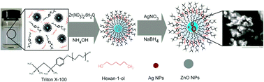
Water-in-oil microemulsions have been found to be good templates and suitable media for the synthesis of ZnO and ZnO@Ag nanoparticles offering themselves as ideal ‘nanoreactors’ for uniform fabrication of core@shell nanoparticles.


Semiconducting core@metal shell nanoparticles are one of the most fascinating materials of the modern age and the preparation, characterization and fabrication of materials of this variety has received an upsurge of interest. Such nanohybrid particles are expected to greatly contribute to the modern scientific world due, inter alia, to their ability to integrate multifunctional characteristics in one system. For instance, ZnO@Ag core@shell nanoparticles exhibit antibacterial activity1a and promising characteristics for photoelectrical devices1b and photoelectrochemical anode materials.1c

ZnO is a semiconductor with a wide band-gap and it possesses enhanced optical and electrical properties, electrochemical stability and high electron mobility at the nano-level. Silver on the other hand is considered to be a noble metal with remarkable catalytic activity, non-toxicity, cost-effectiveness and also shows antibacterial activity. Due to its excellent characteristics together with low-cost many researchers were prompted to prepare ZnO nanoparticles following a wide variety of methods. Direct synthesis of ZnO nanoparticles by basic hydrolysis of zinc nitrate in water-in-oil microemulsion (w/o)2a is a notable example. With a view to enhancing the properties of ZnO nanoparticles, Wang and co-workers reported the preparation of ZnO-based core@shell nanoparticles in reverse micelles where a SiO2 core was coated with a ZnO shell2b and ZnO based core@shell nanotubes and nanorods have been synthesized electrochemically.2c There have also been several attempts to fabricate ZnO both as the core and the shell, as in ZnO@Ag core@shell and Ag@ZnO core@shell nanoparticles. The crystalline mismatch between ZnO and silver places a stumbling block for the fabrication of ZnO@Ag or Ag@ZnO core@shell nanoparticles. ZnO in crystalline form is hexagonal cubic packed (HCP) whereas, silver is face centered cubic (FCC). The synthesis of ZnO microspheres coated with silver nanoparticles3a and ZnO nanorods coated with silver3b were reported earlier where attachment of silver on the surface of ZnO was described to be correlated with the presence of a selective and suitable surface.3a,b Pacholski et al. carried out site-specific deposition of silver nanoparticles onto ZnO nanorods by a photocatalytic wet chemical method where silver nanoparticles were located at one end of the ZnO nanorods due to the crystalline mismatch.3c This phenomenon is known as ‘rattle’. Noble silver nanoparticles were decorated with ZnO; but due to the difference in crystalline structure, a large number of silver nanoparticles remained in the final products, unattached since it was difficult to avoid self-nucleation and the core@shell nanostructure formed was dumbell-shaped rather than spherical.3d–f Despite several reports, a systematic approach to uniformly coat silver nanoparticles on ZnO in a controllable manner or vice versa remains a serious challenge.

The ‘water-in-oil’ (w/o) microemulsions can be thought of as true nanoreactors containing “water pools” and offer the potential to control the dimensions of nanoparticles.4 In this work, we report our approach towards preparation of heterostructured nanoparticles in w/o microemulsion and show evidence that uniform coating of silver can be successfully performed on ZnO nanoparticles and the properties may be systematically tuned by varying the thickness of the core and the shell. Direct synthesis of ZnO@Ag core@shell nanoparticles has been carried out for the first time using a double scheme microemulsion method and the limiting factor, that of crystalline mismatch, has been successfully overcome.

Triton X-100 and cyclohexane (Sigma Aldrich) were used as the surfactant and oil phase, respectively for preparation of w/o microemulsions. Hexanol (Sigma Aldrich) was used as a co-surfactant and eventually acted as an ‘anchor’ and hence played an important role in the stabilization of the microemulsion. 0.25 mol dm−3 of TX-100 solution was prepared in hexanol–cyclohexane and a molar ratio of 1[thin space (1/6-em)]:[thin space (1/6-em)]4 of TX-100–hexanol was maintained. Equal volumes of microemulsions were taken in two stoppered vials. 40 μL of 0.1 mol dm−3 Zn(NO3)2 (Riedel De Haenag Seelze Hannover) was incorporated in one microemulsion, and was mixed with another microemulsion with 80 μL of 1 mol dm−3 NH4OH (Merck) incorporated in it. The mixture was shaken until a transparent microemulsion was obtained. The resultant mixture was then kept overnight. The w/o microemulsion containing 0.42 mmol dm−3 of AgNO3 (Merck) with respect to the total microemulsion volume was then transferred to a vial containing ZnO nanoparticles synthesized in a w/o microemulsion followed by addition of NaBH4 incorporated in an equal volume of a microemulsion of TX-100. A dark brown solution formed, which faded with time and thus gave a reddish brown coloured solution. Scheme 1 shows the overall representation of the synthesis of the core@shell nanoparticles in w/o microemulsion.


image file: c4ra01046a-s1.tif
Scheme 1 Synthesis of ZnO@Ag core@shell nanoparticles synthesized in the water pool of w/o microemulsions.

The water to surfactant molar ratio (Wo) was maintained constant at 13.34. Under highly basic conditions, the [Zn(OH)4]2− ions that formed (due to basic hydrolysis of the zinc nitrate precursor) easily in the water pools of the w/o microemulsion were converted to ZnO since the coordination states of Zn2+ and O2− are very similar in both ZnO and [Zn(OH)4]2−.5 The in situ reduction of Ag(NO3)2 resulted in the production of silver which, via a nucleation process, deposited as a layer of silver on the ZnO nanoparticles.

FTIR spectrum was recorded over a range of 1000–400 cm−1 (cf. Fig. S1 ESI) to confirm the synthesis of ZnO nanoparticles. A band at around 555 cm−1 is assigned to Zn–O stretching and hence confirmed the presence of ZnO.6a The band appearing at 522 cm−1 can also be considered to be a characteristic peak of ZnO. However, there are split peaks at 522 cm−1 and 555 cm−1, which might be a result of the increasing polarity of the Zn–O bands and aggregation of nanoparticles.6b XRD measurement of the ZnO nanoparticles as well as the ZnO@Ag core@shell nanoparticles (centrifuged from microemulsions and calcined at 250 °C) was carried out (cf. Fig. S2(a) and (b) ESI). All the diffraction lines in the X-ray powder pattern are perfectly indexed as hexagonal cubic packed ZnO (cf. Fig. S2(a) ESI). The peaks show a single phase formation of ZnO nanoparticles with no impurities.2a Four additional, weak peaks are observed (cf. Fig. S2(b) ESI) which can be assigned to the (111), (200), (311) and (222) crystal planes of face-centered cubic (fcc) Ag crystallites.3a The peaks for bare ZnO and the ZnO of the core@shell nanoparticles are intense showing a higher degree of crystallinity and those of the silver shell are weak due to thin coating of silver. EDX spectra correspond to the total area shown in Fig. 1, which confirm the presence of the elements Zn, O and Zn, O and Ag in ZnO and the ZnO@Ag core@shell nanoparticles prepared from microemulsions with Wo = 13.34, respectively (cf. Fig. S3(a) and (b) ESI). When spotted on the dark portion, peaks are shown for Zn and O and when spotted on the brighter region, peaks for Ag appear along with Zn and O which suggests that silver nanoparticles are present along with ZnO (Fig. S3(b) ESI). The peak corresponding to carbon originated from the sample support as well as from carbon containing TX-100.


image file: c4ra01046a-f1.tif
Fig. 1 SEM image of ZnO@Ag core@shell nanoparticles synthesized at Wo = 13.34.

From the SEM image (model: JEOL JSM-6490LA) of the ZnO@Ag core@shell nanoparticles formed in w/o microemulsion with Wo = 13.34 it is clear that the core@shell nanoparticles formed are nearly spherical in shape (Fig. 1). Magnification of a selected part reveals two distinct regions; the inner dark part and a shiny portion surrounding the dark region. This indicates the formation of a very thin layer of metallic silver around the semiconducting ZnO core.

The absorbance spectra of ZnO, silver and ZnO@Ag core@shell nanoparticles synthesized at Wo = 13.34 were recorded (Fig. 2). The absorption maximum at 326 nm is attributed to the absorption of ZnO nanoparticles. The surface plasmon resonance (SPR) of ZnO@Ag core@shell nanoparticles is shifted to longer wavelength i.e. 420 nm (bathochromic shift) compared to the plasmon resonance of silver nanoparticles showing an absorption maximum at 408 nm. Interfacial coupling between silver nanoparticles may be the reason for the broadening and red shift of the surface plasmon absorption.7a


image file: c4ra01046a-f2.tif
Fig. 2 Absorbance spectra of ZnO, Ag and ZnO@Ag core@shell nanoparticles synthesized in a w/o microemulsion of TX-100–hexanol–cyclohexane–water at Wo = 13.34.

For coated nanoparticles, the plasmon peak position, λpeak2 is determined by:7b

image file: c4ra01046a-t1.tif
where s is the dielectric function of the shell layer, m is the dielectric function of the medium i.e. cyclohexane (the w/o microemulsion mostly comprises cyclohexane) and g is the volume fraction of the shell layer.

The refractive index and dielectric function is related by, ns = s1/2

image file: c4ra01046a-t2.tif
where bulk plasma wavelength, image file: c4ra01046a-t3.tif meff is the effective mass of free electron of the metal and N is the electron density of the metal.
nAg = 1.07 < ncyclohexane = 1.43248

Although the refractive index of silver is less than that of cyclohexane, calculation shows that λpeak is greater than λ, which explains the red shift.

If the particles are well separated (thick coating), the dipole–dipole coupling is fully suppressed and the plasmon band is located at nearly the same position as the individual metal particles. In this case, it can be presumed that a thin shell of silver has been formed. The ZnO nanoparticles synthesized in w/o microemulsion were kept overnight and the silver core was formed after one day. Therefore, the polydispersity of the ZnO nanoparticles might have increased thus showing such a broad peak for the core@shell nanoparticles formed which is near the SPR of silver nanoparticles. The work function of ZnO is more than that of silver (Fig. S4 ESI) and it is noticed that the Fermi level of Ag [Ef(Ag)] is more than that of ZnO [Ef(ZnO)] which results in the transfer of electrons from silver to ZnO until a new Fermi energy level (Ef) is formed in order to attain equilibration. The electron transfer from silver to ZnO enhances charge separation, which results in the deficiency of electrons on silver nanocrystals. Hence there is a shift in wavelength towards a higher value.8

DLS measurements of the reverse micelles in w/o microemulsion, and ZnO and ZnO@Ag core@shell in reverse micelles were carried out (cf. Fig. S5 ESI and Fig. 3 respectively). The size of the reverse micelle of the TX-100 microemulsion is 5 ± 1 nm which enables us to view them as nanoreactors (c.f. Fig. S5 ESI). The sizes of the ZnO nanoparticles are 43 ± 2 nm and those of the core@shell nanoparticles are 181 ± 8 nm (Fig. 3). The size and particle size distribution (PSD) have been found to increase significantly in the case of ZnO@Ag core@shell nanoparticles as compared to those of the ZnO nanoparticles. The size and PSD of the core@shell nanoparticles found by DLS measurement (181 ± 8 nm) is also supported by the size found from the field emission scanning electron microscope (FESEM) (model: JEOL 7600F) micrographs (159 ± 22 nm) (cf. Fig. S6(a) and (b)). Under different accelerating voltages (10 kV and 15 kV), FESEM micrographs reveal the sizes and PSDs of both the core (100 ± 16 nm) and shell (55 ± 15 nm). From the FESEM micrographs of a particular area, the sizes of the nanoparticles at a definite position are seen to range from 100 nm to 216 nm in which core size ranges from 58 nm to 173 nm and shell thickness ranges from 25 nm to 102 nm.


image file: c4ra01046a-f3.tif
Fig. 3 Size distribution of ZnO and ZnO@Ag core@shell nanoparticles synthesized in TX-100–hexanol–cyclohexane w/o microemulsion at Wo = 13.34.

Since the precursor salts of zinc and silver used are both water soluble, basic hydrolysis of Zn2+ and reduction of Ag+ occur in the hydrophilic water pool of the nanoreactors. Both ZnO and the silver nanoparticles remain confined in the hydrophilic core, which is said to contain water with special properties, such as lower micropolarity, altered nucleophilicity and viscosity to make it a medium different from ordinary water9a and water is said to exhibit major changes from bulk behaviour when Wo < 20.9b More energy is required for silver to undergo free nucleation. However, in these confined water pools of the reverse micelle, which has a ‘cage-like’ effect, the Gibbs energy required is lower for silver nanoparticles to undergo heterogeneous nucleation on the surface of the ZnO nanoparticles. The surfaces of the ZnO nanoparticles might have numerous defects and were thermodynamically unstable. Surface reconstruction would further decrease their energy and thus provide an active site for heterogenous nucleation and growth of silver nanoparticles.

It is interesting that even after the mismatch, silver nanoparticles are forced to form a layer around the ZnO nanoparticles and hence result in the formation of ZnO@Ag core@shell nanoparticles.

Conclusions

In conclusion, we have established a methodology for the synthesis of ZnO nanoparticles coated uniformly with thin silver nanoparticles in a w/o microemulsion of TX-100–hexanol–cyclohexane. Work is now underway to prepare ZnO@Ag core@shell nanoparticles with a wide variation in the thickness of the core and shell by systematic variation of the constituents and composition of the microemulsions and the concentration of the precursors. The w/o microemulsion system is cost effective and provides a very easy and convenient means to controllably synthesize core@shell nanoparticles with tunable optical, electrical, electronic and antimicrobial properties. This may open up a new route for preparing core@shell nanoparticles overcoming crystalline mismatch and could be used to systematically tune properties for task-specific applications.

References

  1. (a) S. Ghosh, V. S. Goudar, K. G. Padmalekha, S. V. Bhat, S. S. Indi and H. N. Vasan, RSC Adv., 2012, 2, 930 RSC ; (b) T. Wang, Z. Jiao, T. Chen, Y. Li, W. Ren, S. Lin, G. Lu, J. Ye and Y. Bi, Nanoscale, 2013, 5, 7552 RSC ; (c) Y. Wei, J. Kong, L. Yang, L. Ke, H. R. Tan, H. Liu, Y. Huang, X. W. Sun, X. Lu and H. Du, J. Mater. Chem. A, 2013, 1, 5045 RSC .
  2. (a) D. Sarkar, S. Tikku, V. Thapar, R. S. Srinivasa and K. C. Khilar, Colloids Surf., A, 2011, 381, 123 CrossRef CAS PubMed ; (b) J. Wang, T. Tsuzuki, L. Sun and X. Wang, ACS Appl. Mater. Interfaces, 2010, 2, 957 CrossRef CAS PubMed ; (c) Z. L. Wang, R. Guo, G. R. Li, L. X. Ding, Y. N. Ou and Y. X. Tong, RSC Adv., 2011, 1, 48 RSC .
  3. (a) F. Li, J. Wua, Q. Qin, Z. Li and X. Huang, Superlattices Microstruct., 2010, 47, 232 CrossRef CAS PubMed ; (b) F. Li, Y. Yuana, J. Luoa, Q. Qina, J. Wua, Z. Li and X. Huang, Appl. Surf. Sci., 2010, 256, 6076 CrossRef CAS PubMed ; (c) C. Pacholski, A. Kornowski and H. Weller, Angew. Chem., Int. Ed., 2004, 43, 4774 CrossRef CAS PubMed ; (d) G. Shan, L. Xu, G. Wang and Y. Liu, J. Phys. Chem. C, 2007, 111, 3290 CrossRef CAS ; (e) M. K. Lee, T. G. Kim, W. Kim and Y. M. Sung, J. Phys. Chem. C, 2008, 112, 10079 CrossRef CAS ; (f) R. Georgekutty, M. K. Seery and S. C. Pillai, J. Phys. Chem. C, 2008, 112, 13563 CrossRef CAS .
  4. (a) S. Hossain, U. K. Fatema, M. Y. A. Mollah, M. M. Rahman and M. A. B. H. Susan, J. Bangladesh Chem. Soc., 2012, 25, 71 CAS ; (b) S. Sultana, S. Saha, M. M. Islam, M. M. Rahman, M. Y. A. Mollah and M. A. B. H. Susan, J. Electrochem. Soc., 2013, 160, D524 CrossRef CAS PubMed ; (c) M. A. Haque, M. M. Rahman and M. A. B. H. Susan, J. Solution Chem., 2012, 41, 447 CrossRef CAS ; (d) T. I. Mredha, C. K. Roy, M. Y. A. M. Mollah and M. A. B. H. Susan, Electrochim. Acta, 2013, 97, 231 CrossRef PubMed ; (e) T. Afrin, S. N. Karabi, M. M. Rahman, M. Y. A. M. Mollah and M. A. B. H. Susan, J. Solution Chem., 2013, 42, 1488 CrossRef CAS ; (f) J. J. Keya, M. M. Islam, M. M. Rahman, M. Y. A. M. Rahman and M. A. B. H. Susan, J. Electroanal. Chem., 2014, 712, 161 CrossRef CAS PubMed .
  5. W. J. Li, E. W. Shi, W. Z. Zhong and Z. W. Yin, J. Cryst. Growth, 1999, 203, 186 CrossRef CAS .
  6. (a) N. Nasakumar and J. B. B. Rayappan, J. Appl. Sci., 2012, 12, 1758 CrossRef ; (b) G. Shan, L. Xu, G. Wang and Y. Liu, J. Phys. Chem. C, 2007, 111, 3290 CrossRef CAS .
  7. (a) G. Shan, S. Zheng, S. Chen, Y. Chen and Y. Liu, Colloids Surf., B, 2012, 94, 157 CrossRef CAS PubMed ; (b) A. C. Templeton, J. J. Pietron, R. W. Murray and P. Mulvaney, J. Phys. Chem. B, 2000, 104, 564 CrossRef CAS .
  8. G. Shan, L. Xu, G. Wang and Y. Liu, J. Phys. Chem. C, 2007, 111, 3290 CAS .
  9. (a) Reverse Micelles, ed. P. L. Luisi and B. E. Straube, Plenum, New York, 1984 Search PubMed ; (b) I. R. Piletic, D. E. Moilanen, D. B. Spry, N. E. Levinger and M. D. Fayer, J. Phys. Chem. A, 2006, 110, 4985 CrossRef CAS PubMed .

Footnote

Electronic supplementary information (ESI) available: FTIR, XRD and EDX spectra, energy band gap structure, DLS data, FESEM micrograph and XRD instrumentation. See DOI: 10.1039/c4ra01046a

This journal is © The Royal Society of Chemistry 2014
Click here to see how this site uses Cookies. View our privacy policy here.