DOI:
10.1039/C4RA00065J
(Paper)
RSC Adv., 2014,
4, 20247-20251
Gamma-irradiation induced direct fabrication of SERS-active Ag nanoparticles on glass substrates
Received
4th January 2014
, Accepted 6th March 2014
First published on 7th March 2014
Abstract
We have demonstrated here a facile gamma-irradiation induced direct fabrication of Ag nanoparticles on glass substrates for SERS applications. It has been found that the agents complexing with the Ag+ ions play a dominant role in enabling Ag particle growth directly on the glass substrates, whereas using bare AgNO3 solution only produced Ag particles in the solution but not on the glass substrate. Moreover, the complexing agent also decides the size and morphology of the Ag nanoparticles, where using ammonia leads to much larger Ag particles than when using ethylenediamine. The γ-ray dose can also influence the size of the Ag nanoparticles, and a higher dose usually results in larger Ag nanoparticles. The SERS performances of the as-fabricated Ag nanoparticles supported on glass substrates have been compared. The uniform Ag nanoparticles with smaller sizes prepared by using ethylenediamine as the complexing agent typically present superior SERS sensitivities. We believe that this facile and cost-effective gamma-irradiation induced fabrication of Ag nanoparticles will be of interest in SERS studies.
Introduction
The gamma-irradiation technique has been widely used in the synthesis of inorganic particles, taking advantage of the strong reductive nature of the produced hydrated electrons (eaq−).1–3 Submicrometer to micrometer sized single-crystalline Cu2O particles have been prepared by γ-irradiation under ambient conditions, with cetyltrimethyl ammonium bromide (CTAB) as a capping material or template.4 Copper oxide nanowires and cuprous oxide crystals have been synthesized through gamma-irradiation of aqueous CuCl2–NaOH–sodium dodecyl sulfate (SDS)–isopropyl alcohol solutions under ambient conditions.5 Ag nanocrystals with controlled morphologies can be facilely synthesized via gamma-irradiation of aqueous solutions containing AgNO3 salt and poly(vinyl pyrrolidone) (PVP) by adjusting the radiation dose.6 Torreggiani et al. fabricated Ag nanoparticles using γ-irradiation for the detection of fungicides based on surface enhanced Raman spectroscopy (SERS).7 In the past few years, we have also managed to prepare morphology-controlled magnetic metal nanostructures through the gamma-irradiation technique that showed improved microwave absorption properties.8–10
Among all of the metals, Ag has been regarded as the most promising material for use in SERS, and typically provides the highest enhancement factor (EF) of the adsorbed analytes.11–14 However, scattered Ag nanoparticles usually offer very limited enhancement of the Raman signals of target molecules, as SERS “hot spots” often reside in structures with sharp edges, intersections, and bifurcations.15–18 Therefore, various techniques have been developed to fabricate patterned SERS substrates with ordered nanostructures so as to reach higher EFs.18–21 In our previous works, we have managed to grow well-defined Ag nanostructures assembled on conducting polymer surfaces, which can be used as highly sensitive and cost-effective SERS substrates for chemical detection.22,23
Herein, we demonstrate a gamma-irradiation induced direct fabrication of Ag nanoparticles on glass substrates (Scheme 1). It has been found that agents complexing with the Ag+ ions play a dominant role in enabling silver particle growth directly on the glass substrates. The size and morphology of the Ag nanoparticles can be tuned by varying the dose of gamma-irradiation. The as-fabricated Ag nanoparticles supported on the glass substrates can be readily used as SERS substrates for chemical detection.
 |
| | Scheme 1 Schematic illustration of gamma-irradiation induced direct fabrication of Ag nanoparticles on glass substrates. | |
Experimental
Fabrication of silver nanoparticles
A glass slide was cut into 1 cm × 1 cm sized pieces. The glass pieces were thoroughly rinsed, first by base and acid treatment, and then by ultrasonication in ethanol for 30 min. In a typical experiment, 50 ml of a 0.05 M aqueous AgNO3 solution was mixed with aqueous ammonia or ethylenediamine to form a transparent solution. 5 ml of a radical scavenger, isopropanol or methanol, was then added to the complex solution under magnetic stirring. The solution was then transferred into a plastic bag, and three of the thoroughly cleaned glass pieces were put into the bag. The solution was then purged with nitrogen gas for 30 min to remove oxygen dissolved in the solution. Then, the plastic bag was sealed and placed in the field of a 60Co γ-ray source at a dose rate of 80 Gy min−1. After irradiation, the glass pieces were washed with distilled water and anhydrous ethanol several times each and then dried in a vacuum oven for 2 h at 50 °C. For comparison, experiments with a bare aqueous AgNO3 solution (without a complexing agent) were also carried out.
Characterization
Scanning electron microscopy (SEM) measurements were carried out on a FEI Inspect scanning microscope to study the morphology of the samples. The characteristics of the crystalline structure of the prepared samples were determined using an XRD-6000 X-ray diffractometer (Shimadzu) with a Cu Kα radiation source (λ = 1.5481 Å, 40.0 kV, 30.0 mA). The Ag nanoparticles supported on the glass substrates were optically characterized by using UV-visible transmission absorption spectroscopy (Varian Cary 300) over 340–800 nm. The SERS spectra were recorded on a Renishaw In Via micro-Raman spectroscopy system, using the TE air-cooled CCD array in a confocal Raman system (wavelength: 633 nm). The incident laser power was kept at 0.1 mW, and a total accumulation time of 10 s was employed.
Results and discussion
As has been shown,24 γ-irradiation of water leads to short-lived reducing species (hydrated electrons (eaq−) and hydrogen atoms (˙H)), a strong oxidizing species (hydroxyl radicals (˙OH)), and other molecular species (eqn (1)). eaq−, with a reduction potential of −2.77 eV, can theoretically reduce all metal ions except for alkali metal ions and alkaline earth metal ions (eqn (2)). However, in order to avoid metal ion oxidation by ˙OH, some alcohol (methanol, isopropanol, t-butanol, etc.) is usually added as a ˙OH radical scavenger (eqn (3)). Also, inert gas (nitrogen, argon) should be used to purge the solution to remove the dissolved oxygen so as to avoid the reaction of eaq− with O2.| |
H2O ![[radiolysis arrow - arrow with voltage kink]](https://www.rsc.org/images/entities/char_e116.gif) ˙H, eaq−, ˙OH, … ˙H, eaq−, ˙OH, …
| (1) |
| | |
CH3OH + ˙OH → ˙CH2OH + H2O
| (3) |
We found that the solutions in all experiments turned black after γ-irradiation, indicating the production of Ag particles. However, when bare aqueous AgNO3 solution was used, no Ag particle growth was witnessed on the glass pieces. Fig. 1 shows the SEM image of the Ag particles collected from the solution where bare aqueous AgNO3 solution was used. It can be seen that these Ag particles are very broad in size distribution. There are Ag bulks that are 1–3 μm in size, and Ag particles with sizes ranging from 100–500 nm. The diffraction peaks in the XRD pattern in Fig. 2 show that these particles have a face-centered-cubic (fcc) Ag structure, different from other Ag salts or impurities. Though Ag particles could be produced in the solution, it is disappointing that no Ag was found on the glass pieces. One possible reason is that when bare aqueous AgNO3 solution was used, the nucleation and growth of the Ag particles in the solution overwhelmed that on the glass surface. Therefore, our immediate thought was to control the growth rate of the Ag particles under the γ-irradiation and their affinity to the glass substrates.
 |
| | Fig. 1 SEM image of the Ag particles collected from the solution when bare aqueous AgNO3 solution was used. | |
 |
| | Fig. 2 XRD pattern of the Ag particles collected from the solution when bare aqueous AgNO3 solution was used. | |
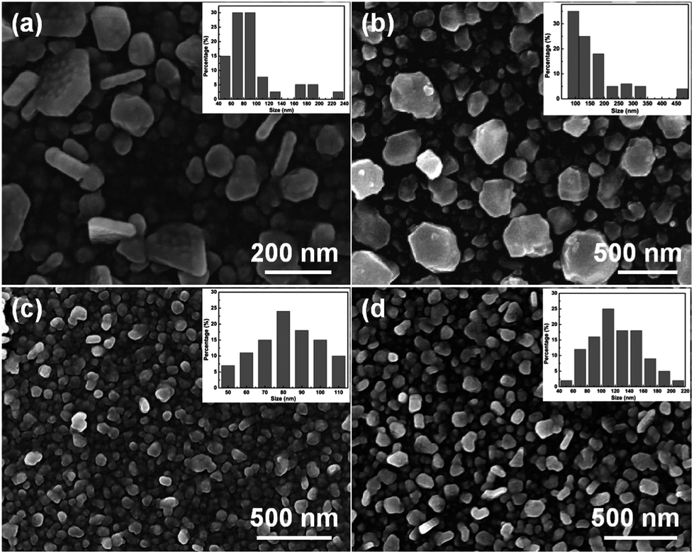
In order to reduce the growth rate of the Ag particles, we tried to use ammonia or ethylenediamine to form Ag+ ion complexes before the solution was put under the γ-irradiation. After γ-irradiation, one could still see that the solution color became dark, but the color was much lighter than that observed when using bare aqueous AgNO3 solution. Of note is that by using a Ag+ ion complex solution, we found that all three of the glass pieces were covered by a dense layer of Ag particles. Fig. 3 shows the SEM images of the Ag particles fabricated on the glass pieces using methanol as the radical scavenger with different agents complexed to the Ag+ ions. As can be seen in Fig. 3(a), when ammonia was used as the complexing agent, low doses of γ-rays led to Ag nanoparticles with sizes of 50–100 nm, and Ag nanosheets about 100–300 nm in size. With an increased γ-ray dose of 20 kGy, one can get Ag particles of larger sizes, as shown in Fig. 3(b). Besides Ag nanoparticles that are about 100 nm in size, one can also see big particles of about 200–500 nm. It is interesting to find that the complexing agent can dramatically change the size of the Ag nanoparticles on the glass substrates. When ethylenediamine was used as the complexing agent, Ag particles with a very narrow size distribution could be obtained, as can be seen in Fig. 3(c) and (d). A low γ-ray dose of 5 kGy resulted in Ag nanoparticles 80–100 nm in size, while a higher dose (20 kGy) led to Ag nanoparticles that were 100–150 nm in size. The above results indicate that there is a complexing agent effect on the final size and morphology of the Ag particles, which we believe is due to the wettability of the glass substrate by Ag+ ion complexes and the reduction rate of the Ag+ ion complexes by the γ-irradiation. Meanwhile, the γ-ray dose impacts the size of the Ag nanoparticles in such a way that larger sizes will be produced when using a higher dose.4,5
 |
| | Fig. 3 SEM images of the Ag nanoparticles fabricated on the glass pieces using methanol as the radical scavenger with different agents complexed to the Ag+ ions. (a) Ammonia, γ-ray dose: 5 kGy; (b) ammonia, γ-ray dose: 20 kGy; (c) ethylenediamine, γ-ray dose: 5 kGy; (d) ethylenediamine, γ-ray dose: 20 kGy. Insets show the size histograms of the Ag nanoparticles. | |
The solid-state UV-vis spectra of these Ag nanoparticles on the glass substrates can be well matched to their size characteristics (Fig. 4). With ammonia as the complexing agent, the mixture of 50–100 nm Ag nanoparticles and 100–300 nm Ag nanosheets (see Fig. 3(a)) produced by using a low dose of γ-rays presents a broad extinction peak centered at 510 nm (Fig. 4(a)); larger Ag particles produced by using a higher γ-ray dose display an even broader absorption feature (Fig. 4(b)). With ethylenediamine as the complexing agent, Ag nanoparticles of 80–100 nm in size produced using a γ-ray dose of 5 kGy show a sharp extinction peak at 445 nm (Fig. 4(c)), while a broader absorption feature with a peak at 455 nm can be seen for the Ag nanoparticles produced using a higher γ-ray dose that are 100–150 nm in size (Fig. 4(d)).
 |
| | Fig. 4 Solid-state UV-vis spectra of the as-fabricated Ag particles on the glass substrates. Spectra (a)–(d) are collected on the Ag particles shown in Fig. 3(a)–(d), respectively. | |
In order to test the SERS behavior of the as-fabricated Ag nanoparticles, we immersed the glass substrates in a 10−6 M 4-mercaptobenzoic acid (4-MBA) solution in ethanol for 10 min, and then rinsed the substrates in ethanol several times to remove surface residues. The Raman spectrum of 4-MBA is dominated by the ν8a (∼1590 cm−1) and ν12 (∼1080 cm−1) aromatic ring vibrations; other weak bands at ∼1150 and ∼1180 cm−1 are attributed to the C–H deformation modes.25 As can be seen from Fig. 5, the SERS performance was quite dependent on the size and morphology of the Ag particles. The Ag nanoparticles prepared using ethylenediamine as the complexing agent typically present much enhanced Raman signals of the 4-MBA molecules compared to those produced using ammonia as the complexing agent. It has been demonstrated that the gap between two closely adjacent metal nanoparticles usually has a much stronger electromagnetic field and can be a SERS “hot spot” for enhancing the Raman signal of a target analyte.26 Here, the Ag particles with sub-micrometer sizes prepared using ammonia as the complexing agent can only provide very limited SERS “hot spots”. Moreover, the surface area of these larger Ag particles should also be much smaller than that of the smaller, more uniform Ag nanoparticles, and thus fewer 4-MBA molecules can be adsorbed onto the surface of the sub-micrometer Ag particles. Therefore, we see SERS signals that are about 5 times less strong on the sub-micrometer Ag particles.
 |
| | Fig. 5 SERS spectra of 4-mercaptobenzoic acid (4-MBA) on the as-fabricated Ag particles. Spectra (a)–(d) are collected on the Ag particles shown in Fig. 3(a)–(d), respectively. | |
Since ethylenediamine is a better complexing agent for the direct fabrication of the Ag nanoparticles on the glass substrates, we used another radical scavenger, isopropanol, in the γ-irradiation induced preparation of Ag nanoparticles directly on the glass substrates. It is found that all three of the glass substrates are covered by dense Ag layers, and Fig. 6 shows the SEM images of the Ag nanoparticles resulting from different γ-ray doses. As can be seen in Fig. 6(a), a low dose of 5 kGy led to Ag nanoparticles with a very narrow size distribution (60–80 nm). An increased dose of 20 kGy resulted in 100–150 nm Ag nanoparticles that are embedded in an Ag layer consisting of 60–80 nm Ag nanoparticles. This result again verifies that ethylenediamine can be an efficient complexing agent to directly prepare uniform Ag nanoparticles on the glass substrates and that a larger dose of γ-rays will typically increase the size of the as-prepared Ag particles.
 |
| | Fig. 6 SEM images of the Ag nanoparticles fabricated on the glass pieces using isopropanol as the radical scavenger and ethylenediamine as the agent complexing with the Ag+ ions. (a) γ-ray dose: 5 kGy; (b) γ-ray dose: 20 kGy. Insets show the size histograms of the Ag nanoparticles. | |
The SERS performance of the Ag nanoparticles prepared by using isopropanol as the radical scavenger was compared with that of the Ag nanoparticles prepared using methanol as the radical scavenger, as shown in Fig. 7. It can be seen that with the radical scavenger isopropanol, SERS sensitivity of the larger Ag nanoparticles prepared using a dose of 20 kGy is about half that of the uniform 60–80 nm nanoparticles. However, the Ag nanoparticles prepared using methanol as the radical scavenger, as shown in Fig. 3(c), provide the best SERS performance. We think that this can be rationalized by the fact that although the Ag nanoparticles prepared using methanol as the radical scavenger are slightly larger in size than those shown in Fig. 6(a), they are somehow stacked into 3-dimensional structures, where edges and intersections of these nanoparticles may create more SERS “hot spots” for enhancing the Raman signal of the 4-MBA molecules.15 Nevertheless, all of the Ag nanoparticles shown in Fig. 3(c), (d) and 6(a) fabricated on the glass substrates are feasible for practical application in chemical detection based on the SERS technique.
 |
| | Fig. 7 SERS spectra of 4-mercaptobenzoic acid (4-MBA) on the as-fabricated Ag particles. Spectra (a)–(c) are collected on the Ag particles shown in Fig. 6(a), (b) and 3(c), respectively. | |
Conclusions
In summary, we have demonstrated a gamma-irradiation induced direct fabrication of Ag nanoparticles on glass substrates for chemical detection based on the SERS technique. It is found that using bare AgNO3 solution only produces Ag particles in the solution and not on the glass substrates. When ammonia or ethylenediamine is used to form Ag+ ion complexes, a layer of Ag nanoparticles can be deposited on the glass substrates, and ethylenediamine is more efficient in producing uniform Ag nanoparticles with smaller sizes. The dose of γ-rays can also impact on the size of the Ag nanoparticles, where a higher dosage typically leads to larger particles. These Ag nanoparticles fabricated on glass substrates can be used in chemical detection by the SERS technique. We believe these as-fabricated efficient and cost-effective SERS substrates are practically feasible in sensing applications.
Acknowledgements
We thank the support from the China Postdoctor Fund (2013M530149), Heilongjiang Postdoctor Fund (2013), NSFC (no. 21203045, 21101041, 51377048), Fundamental Research Funds for the Central Universities (grant no. HIT. NSRIF. 2010065 and 2011017, and HIT.BRETIII. 201223), and Scientific Research Foundation for Young Scientist of Harbin (2012RFQYG117, 2013RFQYG170).
Notes and references
- N. J. Withers, K. Sankar, B. A. Akins, T. A. Memon, T. Y. Gu, J. J. Gu, G. A. Smolyakov, M. R. Greenberg, T. J. Boyle and M. Osinski, Appl. Phys. Lett., 2008, 93, 173101 CrossRef PubMed.
- S. I. Bin Ahmad, M. S. B. Ahmad and S. Bin Radiman, Nanosci. Nanotechnol., 2009, 1136, 186–190 CAS.
- N. M. Huang, S. Radiman, H. N. Lim, P. S. Khiew, W. S. Chiu, T. K. Tan, A. Ahmad and H. Idris, Nanosci. Nanotechnol., 2009, 1136, 191–195 CAS.
- H. R. Liu, W. F. Miao, S. Yang, Z. M. Zhang and J. F. Chen, Cryst. Growth Des., 2009, 9, 1733–1740 CAS.
- Z. B. Hai, C. H. Zhu, J. L. Huang, H. R. Liu and J. F. Chen, Inorg. Chem., 2010, 49, 7217–7219 CrossRef CAS PubMed.
- Y. B. Zhao, A. H. Chen and S. Liang, J. Cryst. Growth, 2013, 372, 116–120 CrossRef CAS PubMed.
- A. Torreggiani, Z. Jurasekova, M. D'Angelantonio, M. Tamba, J. V. Garcia-Ramos and S. Sanchez-Cortes, Colloids Surf., A, 2009, 339, 60–67 CrossRef CAS PubMed.
- H. T. Zhao, X. J. Han, L. F. Zhang, G. Y. Wang, C. Wang, X. A. Li and P. Xu, Radiat. Phys. Chem., 2011, 80, 390–393 CrossRef CAS PubMed.
- H. T. Zhao, B. Zhang, J. S. Zhang, L. F. Zhang, X. J. Han, P. Xu and Y. Zhou, J. Phys. Chem. C, 2010, 114, 21214–21218 CAS.
- H. T. Zhao, X. J. Han, M. F. Han, L. F. Zhang and P. Xu, Mater. Sci. Eng., B, 2010, 167, 1–5 CrossRef CAS PubMed.
- M. T. Sun, Z. L. Zhang, H. R. Zheng and H. X. Xu, Sci. Rep., 2012, 2, 647 Search PubMed.
- M. Fan and A. G. Brolo, Phys. Chem. Chem. Phys., 2009, 11, 7381–7389 RSC.
- D. He, B. Hu, Q. F. Yao, K. Wang and S. H. Yu, ACS Nano, 2009, 3, 3993–4002 CrossRef CAS PubMed.
- M. T. Sun and H. X. Xu, Small, 2012, 8, 2777–2786 CrossRef CAS PubMed.
- B. Zhang, P. Xu, X. Xie, H. Wei, Z. Li, N. H. Mack, X. Han, H. Xu and H.-L. Wang, J. Mater. Chem., 2011, 21, 2495–2501 RSC.
- Y. Y. Xia and H. P. Xiao, J. Raman Spectrosc., 2012, 43, 469–473 CrossRef CAS.
- P. Xu, S. H. Jeon, N. H. Mack, S. K. Doorn, D. J. Williams, X. J. Han and H. L. Wang, Nanoscale, 2010, 2, 1436–1440 RSC.
- Y. Y. Xia, T. J. Li and J. Chen, Phys. Chem. Chem. Phys., 2013, 15, 11900–11903 RSC.
- C. L. Haynes, A. D. McFarland and R. P. Van Duyne, Anal. Chem., 2005, 77, 338a–346a CrossRef CAS.
- M. Yang, R. Alvarez-Puebla, H. S. Kim, P. Aldeanueva-Potel, L. M. Liz-Marzan and N. A. Kotov, Nano Lett., 2010, 10, 4013–4019 CrossRef CAS PubMed.
- M. Sun, Z. Zhang, P. Wang, Q. Li, F. Ma and H. Xu, Light: Sci. Appl., 2013, 2, e112 CrossRef CAS.
- P. Xu, B. Zhang, N. H. Mack, S. K. Doorn, X. J. Han and H. L. Wang, J. Mater. Chem., 2010, 20, 7222–7226 RSC.
- P. Xu, X. Han, B. Zhang, Y. Du and H.-L. Wang, Chem. Soc. Rev., 2014, 43, 1349–1360 RSC.
- J. L. Marignier, J. Belloni, M. O. Delcourt and J. P. Chevalier, Nature, 1985, 317, 344–345 CrossRef CAS.
- P. Xu, N. H. Mack, S. H. Jeon, S. K. Doorn, X. J. Han and H. L. Wang, Langmuir, 2010, 26, 8882–8886 CrossRef CAS PubMed.
- W. Y. Li, P. H. C. Camargo, X. M. Lu and Y. N. Xia, Nano Lett., 2009, 9, 485–490 CrossRef CAS PubMed.
|
| This journal is © The Royal Society of Chemistry 2014 |
Click here to see how this site uses Cookies. View our privacy policy here.