Mesoporous silica: a highly promising and compatible candidate for optical and biomedical applications

Sakthivel Gandhiab, Kavitha Thandavanb, Bong-Joon Kwona, Hyun-Joo Wooa, Soung Soo Yic, Ho Sueb Leea, Jung Hyun Jeongd, Kiwan Jang*a and Dong Soo Shine
aDepartment of Physics, Changwon National University, Changwon, Republic of Korea. E-mail: kwjang@changwon.ac.kr; Fax: +82-55-213-0263; Tel: +82-055-213-3427
bCentre for Nanotechnology & Advanced Biomaterials, SASTRA University, Thanjavur, India
cDepartment of Electronic Materials Engineering, Silla University, Busan, Republic of Korea
dDepartment of Physics, Pukyong National University, Busan, Republic of Korea
eDepartment of Chemistry, Changwon National University, Changwon, Republic of Korea

Received 9th December 2013 , Accepted 15th December 2013

First published on 16th December 2013


Abstract

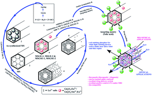
A simple post-co-precipitation strategy is adopted for the construction of a mesoporous silica framework in which the rare earth gadolinium oxide (Gd2O3:Eu3+& Gd2O3:Sm3+, Eu3+) is incorporated as an emissive probe for optical and bio-medical applications. The samples show good emission along with the required theranostic properties. In the blue region, a better red emissive behavior is observed in the case of Eu3+ doped sample while the Sm3+ and Eu3+ co-doped sample exhibit the same in the near ultra-violet region. Moreover, the structural and textural properties of mesoporous silica are preserved even in the presence of rare earth doped Gd2O3 and this can very well be utilized for theranostic applications. A clear confocal microscopy image shows a red spot, and thus its potential towards trackability is confirmed. In addition to this, other admirable properties are also associated with it namely, an excellent drug loading ability, bio-compatibility, easy surface modification by the targeting moiety and excellent longitudinal relaxivity (T1) under magnetic resonance imaging (MRI), which stands par for it to be utilized in the field of theranostics too. This material shows good luminescence properties along with the desired internal–external morphology, and a narrowly ordered porosity can validate the application of these materials in multi-faceted fields such as phosphors for LEDs, bio-imaging, theranostics etc.

Introduction

In the past two decades, an enormous amount of attention has been paid by researchers towards mesoporous silica material, due to its extraordinary properties such as high surface area, highly ordered narrow pore system and excellent hydrothermal stability.1,2 Among them, Santa Barbara Amorphous (SBA-15) material has attracted a lot of interest because of its extra ordinary features like the presence of micropores in the walls of mesopores and easy tunability of the porous system towards the required applications.2 The introduction of lanthanide (Ln3+) ions in the pores of mesoporous silica can offer a promising potential in the field of biomedical and phosphor research as well. Two well-known techniques are adopted for the synthesis of the material namely grafting (post-synthesis) or co-precipitation (pre-synthesis).3 A broad range of applications from optical to biomedical field utilize nanostructured materials owing to its inherent potency of giant impact and it will also offer a promising research in the future.

‘Energy conservation’ is the buzz word of the day where people rely on an efficient photochemical conversion system for day-to-day applications. Two factors namely durability and power savings are vital to furnish the need of the day, and these are associated with the white light emitting diodes (LEDs). LEDs are well known for their efficient luminescence and longer life time4 and hence it has drawn a pronounced attention. Though it is quite possible to obtain a full spectrum from LEDs, there is a need for suitable color conversion phosphors because of its associated critical factors namely varying brightness and life time at various emission wavelengths, and thus leads to different aging of different color LEDs etc.5 These cons associated with it have expanded the horizon of the phosphor field by the way of grabbing the attention of many researchers and manufacturers towards the making of different phosphors for a wide variety of applications. Earlier reports on LEDs confirm the direct relationship between the excitation wavelength and the emission efficiency,4 and hence it is very essential to prepare phosphors of high efficiency for the desired output because they absorb strongly in the blue or near ultra violet spectral region and emit with high intensity in the visible region.

A detailed scan of the literature shows that, a large number of reports are available on phosphors which emit light in the visible region of the spectrum. But there are only a few works on photoluminescent phosphors which can be excited efficiently by blue and near ultra violet LEDs are available. Hence the current hot field of researches focus on the development of host materials, so as to obtain phosphors of efficient excitation at the blue and near ultra violet region along with high intensity emission in the visible region. In the similar lines, various phosphors have been reported and among them, europium doped Gd2O3 phosphor is one of the important red emitting phosphors. Gd2O3, as such is a fluorescent material, but it shows a low quantum yield and hence it requires optimization by the way of doping with other rare-earth ions like Ln, Eu, Er, Tb, etc.

Many fabrication routes are adopted for the preparation of this material,6 but there remains some challenges to make it an excellent red emitting phosphor. Few of the notable challenges are the uniformity in the morphology of particles, non-agglomeration properties, ability to withstand high temperature & electron beam, narrow particle size distribution and the strong energy absorption during excitation in both the blue and near ultra violet region. These aforementioned challenges can be efficiently overcome by using a unique host material called mesoporous silica which provides all the required properties.

In the same pathway, Gd is an important contrast agent as it shortens the spin relaxation time for water protons inside the body which results in improved contrast to the magnetic resonance imaging (MRI).7 Due to the increasing scientific and clinical requirements, an indispensable need to upgrade the MRI contrast agents, fluorescence probes and drug delivery systems became important.8 Nowadays, the researchers are focusing more on the materials which can impart dual functions namely the simultaneous diagnosis and therapy (theranostic).9 This theranostic application requires the materials to be biocompatible and with high drug loading capability along with the tracking ability and thus it can be followed either by fluorescence imaging or by MR imaging. These requirements can be fulfilled by the mesoporous silica, which is incorporated with the Gd2O3:Eu3+ and thus the high surface area, ordered porosity along with more functionalities, resistivity and high stability.

Our research work, utilizes the post-co-condensation technique, for the synthesis of MSGE and MSGSE materials without compromising on its pore order and morphological factors. An extensive literature review confirms that, there are only a few reports available on the incorporation or doping of photoluminescence materials onto mesoporous silica10 and its utilization on bio-medical applications such as drug delivery, imaging, etc. Even though, there are reports available on the usage of Gd-derivatives such as Gd2O3, GdVO4, etc., alone for drug delivery and MRI,11 our work on the rare earths doped Gd2O3 incorporated mesoporous silica is the first of its kind for theranostic applications. Hence, our work emphasizes on the usage of mesoporous SBA-15 silica as a host reactor to prepare Gd2O3:Eu3+ (MSGE) and Gd2O3:Sm3+, Eu3+ (MSGSE). The advantages of using this mesoporous SBA-15 silica as host reactor are the ‘unidirectional distribution of size limited particles, increased stability towards high temperature & electron beam, narrow particle size distribution which ranges between 800 and 1000 nm along with the increased charge transfer property’. These properties may be due to the highly ordered narrow pore size distribution, more surface to volume ratio, presence of micropores, high hydrothermal stability and rigidity. In our present study, MSGE and MSGSE have been synthesized without compromising the structural and textural properties of mesoporous silica. The as-synthesized MSGE & MSGSE materials were calcined at 500 °C (MSGE-1 & MSGSE-1) & 1000 °C (MSGE-2 & MSGSE-2) respectively, and characterized using FT-IR, FE-SEM, FE-TEM, EDX, XRD and nitrogen isotherm. The utilization of these modified materials as phosphor for LED applications and as contrast agent for MRI applications have been demonstrated successfully. The in vivo cell viability and drug loading capability have also been studied systematically.

Results and discussion

Physico-chemical characterization of MS and its derivatives

Fig. 1 demonstrates the functional groups present in the mesoporous silica samples (a) and its derivatives MSGE-1 (b), MSGSE-1 (c), MSGE-2 (d) & MSGSE-2 (e) which were recorded using FT-IR. There occurs an intense band at 1080–1070 cm−1 & 910 cm−1 and a broad band observed at 3410–3300 cm−1 in MS (a), MSGE-1 (b) & MSGSE-1 (c) which corresponds to the –Si–O–Si– stretch, –Si–O–H bend & –O–H stretch respectively. There is no change observed in the functional moieties of MSGE-1 & MSGSE-1 which were treated at 500 °C along with Gd2O3:Eu3+ & Gd2O3:Sm3+, Eu3+ which confirms that the chemical structure of mesoporous silica is preserved in the samples. On the other hand, the spectra of MSGE-2 (d) & MSGSE-2 (e) showed very weak broad band of characteristic MS and this may be due to the distortion in the physical structures. The absorption band seen at ∼500 cm−1 in both the samples MSGE-2 (d) & MSGSE-2 (e) may be due to the stretching vibration of Gd–O.
image file: c3ra47414c-f1.tif
Fig. 1 FT-IR of MS (a), MSGE-1 (b), MSGSE-1 (c), MSGE-2 (d) & MSGSE-2 (e).

Fig. 2 shows the surface morphology of MS (a), MSGE-1 (b), MSGSE-1 (c), MSGE-2 (d) & MSGSE-2 (e) which were recorded by FE-SEM. All the samples show the characteristic rod like morphology of mesoporous SBA-15 silica. The introduction of Gd2O3:Eu3+ & Gd2O3:Sm3+, Eu3+ in the mesochannels did not affect the surface texture of mesoporous silica. The field emission transmission electron microscopic images of MS (a), MSGE-1 (b), MSGSE-1 (c), MSGE-2 (d) & MSGSE-2 (e) are shown in Fig. 3. The images reveal that the samples MS (a), MSGE-1 (b) & MSGSE-1 (c) are showing the ordered porous structure along with the hexagonally arranged pore system. The appearances of dark spots in all the samples other than MS are due to the presence of Gd2O3:Eu3+ & Gd2O3:Sm3+, Eu3+. The samples MSGE-1 (b) & MSGSE-1 (c) which were treated at 500 °C illustrate the ordered porous structure with the presence of inorganic oxides inside the pores. This confirms that the incorporation of inorganic oxides did not change the properties and physical structure of mesoporous silica which was also proved by FT-IR. But, the samples MSGE-2 (d) & MSGSE-2 (e) shows the distorted pores arrangement due to its high temperature treatment during synthesis.


image file: c3ra47414c-f2.tif
Fig. 2 FE-SEM images of MS (a), MSGE-1 (b), MSGSE-1 (c), MSGE-2 (d) & MSGSE-2 (e).

image file: c3ra47414c-f3.tif
Fig. 3 FE-TEM images of MS (a), MSGE-1 (b), MSGSE-1 (c), MSGE-2 (d) & MSGSE-2 (e).

Table 1 specifies the elemental composition present in the prepared mesoporous derivatives calculated using energy dispersive X-ray analysis technique (EDX) which was operated along with FE-TEM (Fig. S1). The result shows 28.13, 16.11 and 0.73% of Si, Gd & Eu respectively for the sample MSGE treated at 500 °C while it is 14.86 (Si), 29.43 (Gd) & 4.99% (Eu) for the sample treated at 1000 °C. The decrease in Si content and the increase in Gd & Eu content in MSGE-2 reveal that the pores had undergone distortion at high temperature (1000 °C). This results in the greater exposure of Gd & Eu which were inside the pores. Similar trend is also observed with the samples namely MSGSE-1 & MSGSE-2, in which the prior one shows 24.42 (Si), 8.9 (Gd), 1.24 (Sm) & 1.74% (Eu) content whereas the latter shows 5.12 (Si), 35.12 (Gd), 1.84 (Sm) & 3.92 (Eu).

Table 1 Elemental compositions of MSGE-1, MSGSE-1, MSGE-2 & MSGSE-2
Sample code Elemental compositiona (weight %)
Si Gd Sm Eu
a Others: Cu, O & C.
MSGE-1 28.13 16.11 0.73
MSGE-2 14.86 29.43 4.99
MSGSE-1 24.42 8.9 1.24 1.74
MSGSE-2 5.12 35.12 1.84 3.92


The nitrogen adsorption–desorption isotherm and BJH isotherm (dV/dD) of all the samples are shown in Fig. 4. All the samples except MSGE-2 & MSGSE-2 show the type IV adsorption–desorption isotherms (Fig. 4A(a–c)) with hysteresis, together with narrow pore size distribution (Fig. 4B(a–c)), indicating the capillary condensation of adsorbate inside the hexagonally arranged cylindrical channels of mesoporous material. From the result, it can be inferred that the introduction of Gd2O3:Eu3+ & Gd2O3:Sm3+, Eu3+ in MSGE-1 & MSGSE-1 samples does not cause any interruption in the basic physical structure of mesoporous silica. But, the samples treated at 1000 °C namely MSGE-2 & MSGSE-2 (Fig. 4A(d and e)) show the type V isotherm due to the interruption in the pore structure which results in the exposure of Gd2O3:Eu3+ & Gd2O3:Sm3+, Eu3+. The wide range of pore distribution is also noted in the BJH (Fig. 4B(d and e)) isotherm. The textural characteristics of the corresponding samples are summarized in Table 2. It can be observed that the samples MS, MSGE-1 & MSGSE-1 possess high BET surface area and pore volume along with the narrow pore size distribution which is in the range of 6.8–7.1 nm, indicating its potential application as a host in making highly dispersed uniform particles of Gd2O3:Eu3+ & Gd2O3:Sm3+, Eu3+. On the other hand, the decrease in pore volume is noted in MSGE-1 & MSGSE-1 while the surface area was found to be almost equal, and this may be attributed to the involvement of Gd2O3:Eu3+ & Gd2O3:Sm3+, Eu3+ inside the mesoporous channels, and not onto the surface. Further, the samples namely MSGE-2 & MSGSE-2 show a broad range of pores size distribution between 7 and 16 nm along with the steep decrease in the pore volume and surface area. This may be due to the distortion of pore order at the applied high temperature during preparation.


image file: c3ra47414c-f4.tif
Fig. 4 Nitrogen adsorption–desorption isotherm (A) and BJH (B) isotherm of MS (a), MSGE-1 (b), MSGSE-1 (c), MSGE-2 (d) & MSGSE-2 (e).
Table 2 Physical parameters of MS, MSGE-1, MSGSE-1, MSGE-2 & MSGSE-2 obtained from BET & BJH isothermsa
Sample code Physical parameters
SA (m2 g−1) PD (nm) PV (cm3 g−1)
a SA – surface area; PD – pore diameter; PV – pore volume.
MS 780 6.8 1.2328
MSGE-1 723 6.9 0.8442
MSGE-2 241 10.9 0.3150
MSGSE-1 708 7.1 0.7652
MSGSE-2 79 10.7 0.1221


Fig. 5 shows the XRD patterns of MS (a), MSGE-1 (b), MSGSE-1 (c), MSGE-2 (d) & MSGSE-2 (e) samples. The XRD pattern of host mesoporous silica (Fig. 5a) shows a broad peak between 18 and 30° which represents the characteristic diffraction peak of amorphous silica (JCPDS no. 29-0085). The same amorphous peak is also noted in the samples namely MSGE-1 & MSGSE-1 (Fig. 5b and c). However the FE-TEM and BET analysis confirm the presence of Gd2O3:Eu3+ & Gd2O3:Sm3+, Eu3+ inside the pores of amorphous mesoporous channels, while the characteristic peaks are absent in the XRD for both the samples which were treated at 500 °C. In the case of samples namely MSGE-2 & MSGSE-2 (Fig. 5d and e), there is disappearance of amorphous diffraction pattern, corresponding to mesoporous silica with increase in the crystalline nature of the sample. This may be because of the distortion of textural property by high thermal treatment (1000 °C) applied during preparation. Moreover, the MSGE-2 & MSGSE-2 samples are showing the characteristics diffraction peaks of cubic Gd2O3 at 2θ ∼ 20, 28, 33, 47 & 56 which correspond to the miller indices 211, 222, 400, 440 & 622 respectively. The values are in well agreement with the JCPDS no. 12-0797.


image file: c3ra47414c-f5.tif
Fig. 5 Powder XRD patterns of MS (a), MSGE-1 (b), MSGSE-1 (c), MSGE-2 (d) & MSGSE-2 (e).

Optical applications

To elucidate the photoluminescence behavior of all the samples synthesized, excitation was recorded initially at 615 nm (5D07F2) emission and it has been picturized as Fig. 6. The broad excitation band observed at ∼260 nm for all the samples may be correlated with the charge transfer state transition (CTS) from oxygen 2p state to 4f state of Eu3+ ion. On the other hand, it is noteworthy that the intensity of CTS transition is significantly weaker for all the samples. This may be due to the weak covalency between Eu3+ and O2− which results with the trivial energy transfer from O2− → Eu3+. But, there may be a possible energy transfer from oxygen 2p state to Sm3+ ion which may lead to further energy transformation to Eu3+ ion. The absorption bands obtained at ∼320, 365, 380 & 390 nm correspond to the higher energy level f–f transitions of Eu3+ ion. The sharp line-like absorption band obtained at 466 & 395 nm may be due to the 7F05DJ (J = 1, 2) and 7F05L6 transitions of Eu3+, but their intensities vary for the samples MSGE and MSGSE, which imply that those excitation lines may result from different sites of Eu3+ in the samples MSGE and MSGSE.
image file: c3ra47414c-f6.tif
Fig. 6 Photoluminescence excitation spectra of MSGE-1, MSGSE-1, MSGE-2 & MSGSE-2 monitored at 615 nm.

Fig. 7 shows the emission spectra of all the samples recorded at the blue region 466 nm (Fig. 7A) and near ultra violet region 395 nm (Fig. 7B). The spectrum obtained at 466 nm (blue region) for MSGE and MSGSE consists of a series of emission lines at ∼579, 586, 592, 597, 615, 623, 627 and 654 nm (Fig. 7A). These emission lines correspond to the transitions from 5D07FJ (J = 0, 1, 2, 3, 4) manifolds of Eu3+ in which the 5D07F1,3 is an allowed magnetic dipole transition, whereas the Eu3+ 5D07F2,4 is a forbidden electric dipole transition. The same trend of emission behavior was observed at 395 nm (NUV region) also. But, the sample MSGE showed very less intense emission at 395 nm when compared to MSGSE (Fig. 7B). This confirms that the introduction of Sm3+ ion in MSGSE plays a vital role for emitting intense red under the 395 nm near UV excitation. Moreover, the emission trends of MSGSE at 395 nm mimics the Eu3+ emission and this confirms that there is no energy transfer from Eu3+ → Sm3+. But, there is a possibility of energy transfer which can occur from 4G5/2 of Sm3+ to 5D0 of Eu3+ (4G5/25D0).12 There is also an increase in emission intensity for MSGE-2 & MSGSE-2 samples which were treated at 1000 °C, and this may be either due to distortion of silica frame work or more crystallization of Gd2O3:Eu3+ & Gd2O3:Sm3+, Eu3+. It is noted prominently that the remarkable excitation band at 466 nm is located around the emission wavelength of commercial blue light-emitting diodes (LEDs) (450–470 nm). This indicates that the samples MSGE-1, MSGSE-1, MSGE-2 & MSGSE-2 can act as a potential blue exciting phosphor. Moreover, the MSGSE-1 & MSGSE-2 samples in which the Sm3+ ions were introduced as co-dopant along with Gd2O3:Eu3+ can also be effectively excited at 395 nm which is more suitable for near ultraviolet (NUV) LEDs (350–420 nm). This refers that the strong and sharp excitation peak at 395 nm for MSGSE-1 & MSGSE-2 can also act as a potential NUV exciting phosphor. The color luminescence performances of materials were checked using CIE chromaticity coordinates which is demonstrated in Fig. 8 and the values are tabulated in Table 3. It can be noted that the CIE chromaticity coordinate values of all samples are closer to the standard national television system committee (NTSC) (x = 0.670, y = 0.330).


image file: c3ra47414c-f7.tif
Fig. 7 Photoluminescence emission spectra of MSGE-1, MSGSE-1, MSGE-2 & MSGSE-2 at 466 nm (A) & 395 nm (B) of excitation.

image file: c3ra47414c-f8.tif
Fig. 8 Chromaticity coordinates on CIE 1931 diagram. The marked places represent the color point of emission from MSGE (1) and MSGSE (2) under 395 nm and 466 nm excitation.
Table 3 CIE coordinate values of MSGE and MSGSE
Materials CIE coordinate values
x y
395 nm 466 nm 395 nm 466 nm
MSGE-1 0.638 0.640 0.361 0.359
MSGE-2 0.634 0.645 0.366 0.361
MSGSE-1 0.624 0.625 0.377 0.374
MSGSE-2 0.625 0.629 0.371 0.377


Theranostic applications

To utilize this material for theranostics, it should possess unique properties such as chemically modifiable outer surface to link target moieties, highly ordered surface & pore properties to use it as a drug carrier and fluorescent & magnetic properties to track this materials in confocal and molecular level. To satisfy the above properties and to put it in a nutshell, MSGE-1 was chosen as it shows good emissive behavior under NUV and blue region in fluorescence spectra along with the unique properties such as high surface area, well ordered pore structure and more functionalities. So, the surface has been modified with APTES initially and followed by FA. Folic acid can easily help the material to target the specific region as cancerous cell will have more folic acid receptors within it. Fig. 9 shows the FT-IR spectra of MSGE-1 (a), A-MSGE-1 (b) and FA-MSGE-1 (c). In the FT-IR spectra, apart from the bands observed at 1080–1070 cm−1, 910 cm−1 and a broad band located between 3410 and 3300 cm−1 for FA-MSGE-1 which arised due to mesoporous silica, there are also bands at 1639 cm−1 and 2950–2850 cm−1 which can be attributed to the amide carbonyl stretching and alkyl –C–H stretching respectively indicating the presence of folate in the sample. The band at 1395 cm−1 is due to the C–N stretch and the N–H bend appears at 1565 cm−1 in the spectrum for FA-MSGE-1 & A-MSGE-1 (Fig. 10b and c). In addition, a band at 1250 cm−1 in the spectrum appears due to –C–O–C– stretch.
image file: c3ra47414c-f9.tif
Fig. 9 FT-IR of MSGE-1 (a), A-MSGE-1 (b) & FA-MSGE-1 (c).

image file: c3ra47414c-f10.tif
Fig. 10 Cumulative drug loading capability of MS (a), MSGE-1 (b) & FA-MSGE-1 (c).

Drug loading

To evaluate the loading capacity of MS, MSGE-1 and FA-MSGE-1 towards drug carriers, DOX has been chosen as a model drug. DOX is water-soluble and also an anti-cancer drug. The drug loading was calculated as 147.2, 143.6 and 135.2 mg g−1 for MS, MSGE-1, FA-MSGE-1 respectively. Fig. 10 gives the percentage loading of drug in MS (a), MSGE-1 (b) & FA-MSGE-1 (c) which was analyzed using uv-visible spectroscopy. It clearly shows that the maximum amount of drug could be loaded in MS while it is lesser for other samples due to decrease in surface area and pore volume (Table 2). It is also observed that the loading of model drug showed a time dependant increase in all the samples up to 6 h after which it gets saturated. The maximum loading of 98.1% was achieved by MS while MSGE-1 (95.7%) showed a slight reduction due to decrease in surface area and pore volume. The introduction of target moiety in FA-MSGE-1 sample reduced the drug loading capacity to 90.1%.

Bio-imaging

To evaluate these hybrid materials (MSGE-1 and FA-MSGE-1) for bio-imaging capability, fluorescent and molecular imagings have been analyzed. Initially the fluorescent imaging of MSGE-1 & FA-MSGE-1 samples were monitored under confocal and displayed as Fig. 11. This shows a clear red emitting behavior under due to the presence of Eu3+ ions. This proves that the trackability of these samples under confocal can be used to follow the material and study the cell uptake.
image file: c3ra47414c-f11.tif
Fig. 11 Confocal laser scanning microscopy images of MSGE-1 (A) & FA-MSGSE-1 (B).

Molecular imaging

The potentiality of MSGE-1 & FA-MSGE-1 materials to be used as positive contrast agents for MRI is confirmed by T1-weighted MR images recorded at 4.7 T. Fig. 12 shows the MR images of conventional Gd2O3, MSGE-1 and FA-MSGE-1 at different concentrations which are dispersed in aqueous solution. The T1-weigted MR images of hybrid materials (MSGE-1 & FA-MSGE-1) are found to be having greater positive contrast when compared with the conventional Gd2O3. Moreover, the longitudinal (T1-weighted) images reveal a gradual increase in contrast with the increase in concentrations from 0.01 to 0.12 mg mL−1. This is reflected by the longitudinal relaxivities r1 values which are higher in the case of hybrid materials such as MSGE-1 and FA-MSGE-1 than for conventional Gd2O3 sample (Table 4). It is clearly noticeable that the presence of mesoporous silica with unique properties and the luminescent Eu3+ in MSGE-1, act as an excellent positive contrast agent. The Gd-based contrast agents along with large surface area provide the higher positive relaxivity (r1).11 Moreover, the introduction of FA in FA-MSGE-1 does not affect the positive contrast properties of the Gd3+ ions.
image file: c3ra47414c-f12.tif
Fig. 12 Magnetic resonance imaging properties: signal intensity (T1-weighted images) (A), T1 relaxation rate (B) of Gd2O3, MSGE-1 & FA-MSGE-1 at various concentrations and the mechanism associated for enhanced positive contrast by MSGE-1.
Table 4 T1 relaxivity values of Gd2O3, MSGE-1 & FA-MSGE-1a
Sample r1 (s−1 mg−1 mL) R
a r1 – longitudinal relaxation rate & R – regression coefficient.
Gd2O3 12.4 0.9794
MSGE-1 18.1 0.9949
FA-MSGE-1 16.9 0.9921


These observations bring us to the conclusions that the unique properties such as presence of well ordered mesoporous system (6.9 nm), large pore volume (0.8442 cm3 g−1), presence of micropores in the walls of mesopores and the high surface area (723 m2 g−1) of mesoporous silica help to absorb and circulate more water molecules to the Gd2O3 surface which is inside the pores and on the surface of mesoporous silica (Fig. 12C). These contacts between water and Gd2O3 in mesoporous silica make hydrogen bonding and increase the positive contrast. On the other hand, accommodation of more number of Gd2O3 in single particle of mesoporous silica, results in an increase in positive contrast. All these prove that MSGE-1 can be used as a potential T1 contrast agent in MR imaging.

In vivo cell viability

MTT assay was carried out to study the short-term cytotoxicity of the MS, MSGE-1 and FA-MSGE-1 samples on L929 fibroblast cells. From the Fig. 13 it can be clearly seen that all the samples show no obvious cytotoxicity against the fibroblast cells at a concentration of 25–200 μg mL−1 within 24 h. More than 90.3% cells remained alive when the concentration of all the samples reached the maximum of 200 μg mL−1. Even the presence of folic acid in FA-MSGE-1 does not influence the cytotoxicity of L929 cells. The absence of cytotoxicity in all dosages of MSGE-1 and FA-MSGE-1 even up to 24 h indicates its suitability for biomedical applications. On the whole, the mesoporous silica incorporated with Gd2O3:Eu3+ (MSGE-1) shows good diagnostic property under fluorescence & molecular imaging systems and also shows good therapeutic properties with its increased drug loading capacity. It also associates with it the specific targeting moiety on the surface. An absence of cytotoxicity is observed, which substantiates the use of this material for theranostic applications.
image file: c3ra47414c-f13.tif
Fig. 13 In vitro cell viability of L929 cells in the presence of MS, MSGE-1 & FA-MSGE-1 at specified concentrations upon exposure for 24 h.

Methods

Materials

Poly ethylene glycol–poly butylene glycol–poly ethylene glycol (P-123) tri block co-polymer, tetraethyl orthosilicate (TEOS), aminopropyl trimethoxysilane (APTES), gadolinium nitrate hexahydrate (Gd(NO3)3·7H2O), europium nitrate hexahydrate (Eu(NO3)3·7H2O), samarium nitrate hexahydrate (Sm(NO3)3·7H2O) and morpholino ethane sulphonic acid (MES) were purchased from sigma-Aldrich. Hydrochloric acid, ethanol, toluene and ammonium hydroxide were received from Daejung chemicals. All the above mentioned chemicals have been used as such, without any further purification.

Preparation of mesoporous silica

The mesoporous SBA-15 silica has been prepared according to our earlier report.13 Briefly, 2 g of P-123 was mixed with 35 g of deionized water, 5 g of glycerol and 35 g of 2 N HCl. It was then stirred at 40 °C for 4 hours to get a homogeneous solution. To it added the silica precursor (TEOS) drop wise and maintained at 40 °C with vigorous overnight stirring. The clear solution obtained was then subjected to aging process at 100 °C in static condition for 24 hours using an autoclave. The white precipitate obtained was filtered, washed with deionized water several times and dried overnight at 80 °C to get the as-synthesized material. Finally, the resultant as-synthesized material was calcined at 550 °C for 5 h to remove the organic templates and represented as ‘MS’.

Post-co-precipitation of Gd2O3:Eu3+ and Gd2O3:Sm3+, Eu3+ with MS

The incorporation of Gd2O3:Eu3+ and Gd2O3:Sm3+, Eu3+ to MS was achieved through post-co-precipitation technique in which the calcined mesoporous silica was treated with salts of inner transition elements. For preparing Gd2O3:Eu3+ incorporated MS (MSGE), 1.6 g of Gd(NO3)3 and 0.2 g of Eu(NO3)3 were dissolved in 10 mL of deionized water and stirred at room temperature for 30 minutes. To it added 500 mg of MS and left for overnight stirring. This solution was filtered and washed with water to remove the unbound inner transition element's salts. The precipitate was then dispersed in deionized water and sonicated for about 10 minutes after adding 5 mL of ammonium hydroxide (NH4OH) solution. In order to hydrolyze the inner transition elemental salts the prepared solution was then aged at 100 °C in an autoclave for 24 hours, with vigorous stirring. The white precipitate deposited in the solution was filtered, washed with water and dried at 100 °C to get the as-synthesized MSGE. The dried sample was bifurcated into two parts in which one was subjected to treatment at 500 °C (MSGE-1) and the other one at 1000 °C (MSGE-2) in a furnace at atmospheric air.

Along the same lines, the incorporation of Gd2O3:Sm3+, Eu3+ to MS (MSGSE) was carried out where the Sm(NO3)3 was also used in addition to Gd(NO3)3 and Eu(NO3)3. Thus the prepared samples were represented as MSGSE-1 & MSGSE-2 respectively.

Preparation of folic acid modified MSGE-1

In order to introduce the target moiety (folic acid) to MSGE-1 sample, the surface functionality has been modified with amine group and this substitution was done using APTES. Briefly, a toluenic dispersion of MSGE-1 (200 mg in 10 mL) was prepared by sonication and then APTES was added. The reaction mixture was kept under reflux condition at 80 °C for overnight and then filtered. Then it was washed with toluene, ethanol & water respectively and dried at 100 °C (A-MSGE-1). From the prepared A-MSGE-1, about 100 mg was weighed and added to the MES buffer of pH 5 and then added the MES solution of folic acid (1 mg mL−1) to the reaction mixture. The reaction mixture was stirred at room temperature for 30 min and the final product was isolated by centrifugation and marked as FA-MSGE-1.

Characterization

The presence of functional group in the MS and its modifications were analyzed using a Fourier transform infrared spectroscopy (FT-IR, Jasco-6300, USA). The analysis was performed between 4000 and 450 cm−1 averaging 10 scans for all the samples. The sample preparation was done by mixing the moisture-free sample with IR grade KBr and pelletized by applying a mechanical pressure on it and then the spectrum was recorded at room temperature.

The surface morphology was imaged using a thermal field emission scanning electron microscope (FE-SEM) (Tescan, MIRA IILMH, Brno, Czech Republic). The samples were affixed onto the carbon tape and coated with platinum to improve its conductivity. All the images were recorded at an applied voltage of 20 keV.

The internal morphology of the modified mesoporous silica samples were studied using a field emission transmission electron microscope (FE-TEM, JEM 2100F, JEOL, Japan). The samples were dispersed in 99.9% ethanol and drop casted on the carbon coated copper grid. It was allowed to dry and then the imaging was done. Energy dispersive X-ray analysis (EDX, model no. 6498, OXFORD, USA) was done to elucidate the elemental distribution in the modified materials.

Surface area, pore diameter and pore volume of modified mesoporous silica samples were derived from BET & BJH isotherms which was calculated using nitrogen adsorption–desorption isothermal analysis (Autosorb-1, Quantachrome, USA). The outgas temperature and time were maintained to be 350 °C and 8 h respectively.

X-ray diffraction studies (XRD, X'Pert Pro, PANalytical) were done to understand the transformation of crystallinity in the samples due to the change in temperature applied during synthesis. The samples were finely ground and mounted onto the polymeric sample holder. It was then analyzed between (2θ = ) 10 to 80° with the scan speed of 0.02° s−1.

The excitation and emission behavior of all the prepared samples were obtained using spectrofluorophotometer (FL, RF-5301PC, Shimadzu, Japan). The samples were finely ground and mounted on a polymer sample holder where the Xe lamp was used as light source.

Drug loading test

In a typical process for the loading of doxorubicin hydrochloride (DOX) on MS, MSGE-1 and FA-MSGE-1, about 20 mg of sample was weighed and added to the beaker containing 3 mg of DOX dispersed PBS solution (10 mL). The drug loading capability was analyzed by recording the absorbed radiation of supernatant liquid at 480 nm using uv-visible spectrophotometer at a constant interval of time. The drug loading efficiency was calculated as follows,
image file: c3ra47414c-t1.tif
where DLE, TC and CT represents the drug loading efficiency (in %), total concentration of drug (150 mg g−1) and concentration of drug in solution at time ‘t’ respectively. CT was calculated from the absorption intensity of supernatant liquid at 480 nm.

Confocal imaging

Bio-imaging of MSGE-1 and FA-MSGE-1 were achieved by confocal microscopy and magnetic resonance imaging (MRI) system. Initially, the confocal microscopic (Olympus, FV 1000) images were recorded by placing a drop of MSGE-1 and FA-MSGE-1 water dispersions on a micro slide.

Magnetic resonance imaging

T1 weighted MR images were recorded on a 4.7 T Bruker biospec instrument. Initially, different concentrations of Gd2O3, MSGE-1 and FA-MSGE-1 were dispersed individually in water and sonicated for 15 min. All the samples were placed carefully near the iso-centre of the magnet in MRI instrument. The T1 weighted images were recorded by varying TR between 54.6 and 10 000 ms while TE is maintained as a constant at 10.9 ms. After imaging, the intensities were measured by a manually drawn regions of interest (ROI). Relaxation rate r1 was calculated by mono-exponential curve fitting of the signal intensity vs. time (TE or TR) data using origin software.

In vitro cytotoxicity

The short time cell cytotoxicity of MS, MSGE-1 and FA-MSGE-1 were measured by the standard 3-(4,5-dimethylthiazol-2-yl)-2,5-diphenyltetrazolium bromide (MTT) assay. Initially, the L929 fibroblast cells were cultured in a 96 well plate using fresh culture medium for 24 h. varying concentrations of MS, MSGE-1 & FA-MSGE-1 were dispersed in PBS and sterilized under uv radiation. The cells were incubated for 24 h after the addition of all the samples. After incubation, MTT was added and cells were counted by reading the intensity at 570 nm using a microplate reader.

Conclusions

In summary, this work points out the successful post-co-precipitation strategy towards the synthesis of mesoporous silica along with the incorporation of Gd2O3:Eu3+ & Gd2O3:Sm3+, Eu3+ in the mesoporous frame work. The results reveal the retention of textural properties for the samples treated at 500 °C (MSGE-1 & MSGSE-1). The photo-luminescence spectroscopic results of all the samples showed improved red emission at 466 nm of excitation which is in the range of most of the blue LED's emitting wavelength. Moreover, the introduction of Sm3+ ions as co-dopant shows an improved red emission under 395 nm, and thus confirms its possible application as red component with near ultra violet LEDs. Meanwhile, the material MSGE-1 had been easily modified with target moiety and showed good drug loading capability along with the absence of cytotoxicity effect even with 200 μg mL−1 after 24 h. It also showed an excellent red emissive behavior under confocal and excellent positive contrast property (18.1 & 16.9 s−1 mg−1 mL) with MR imaging. Hence, it proves that the combination of photo-emitting Gd2O3:Eu3+ and the unique textural & structural properties of MSGE-1 & MSGSE-1 may expand the horizon for the researchers and industrialists to prepare these kinds of materials and utilize it as a potential multifunctional opto-electronic material for various applications.

Acknowledgements

This work was supported by the Priority Research Centers Program through the National Research Foundation of Korea funded by the Ministry of Education, Science and Technology (NRF-2010-0029634) and also this work was partially supported by the Korean government (NRF-2010-0023034).

Notes and references

  1. D. Ravichandran, R. Roy, W. B. White and S. Erdei, J. Mater. Res., 1997, 12, 819 CrossRef CAS; P. Schmidt-Winkel, W. W. Lukens, D. Y. Zhao, P. D. Yang, B. F. Chmelka and G. D. Stucky, J. Am. Chem. Soc., 1999, 121, 254 CrossRef; Y. Xia and R. Mokaya, Microporous Mesoporous Mater., 2005, 86, 231 CrossRef PubMed; J. Y. Ying, C. P. Mehnert and M. S. Wong, Angew. Chem., Int. Ed., 1999, 38, 56 CrossRef; S. Gandhi, S. Sethuraman and U. M. Krishnan, Dalton Trans., 2012, 41, 12530 RSC.
  2. D. Zhao, J. Feng, Q. Huo, N. Melosh, G. H. Fredrickson, B. F. Chmelka and G. D. Stucky, Science, 1998, 279, 548 CrossRef CAS; D. Zhao, Q. Huo, J. Feng, B. F. Chmelka and G. D. Stucky, J. Am. Chem. Soc., 1998, 120, 6024 CrossRef.
  3. D. Zhang, D. Tang, X. Wang, Z. Qiao, Y. Li, Y. Liu and Q. Huo, Dalton Trans., 2011, 40, 9313 RSC; P. Yang, Z. Quan, Z. Hou, C. Li, X. Kang, Z. Cheng and J. Lin, Biomaterials, 2009, 30, 4786 CrossRef CAS PubMed.
  4. E. F. Schubert and J. K. Kim, Science, 2005, 308, 1274 CrossRef CAS PubMed; J. Kido, M. Kimura and K. Nagai, Science, 1995, 267, 1332 CrossRef PubMed; S. Tasch, E. J. W. List, O. Ekstrom, W. Graupner, G. Leising, P. Schlichting, U. Rohr, Y. Geerts, U. Scherf and K. Mullen, Appl. Phys. Lett., 1997, 71, 2883 CrossRef PubMed; S. Pimputkar, J. S. Speck, S. P. DenBaars and S. Nakamura, Nat. Photonics, 2009, 3, 180 CrossRef; N. Kimura, K. Sakuma, S. Hirafune, K. Asano, N. Hirosaki and R. J. Xie, Appl. Phys. Lett., 2007, 90, 51109 CrossRef PubMed.
  5. A. Kitai, Luminescent materials and applications, John Wiley & sons, Ltd., 2008 Search PubMed.
  6. M. F. Yan, T. C. D. Huo and H. C. Ling, J. Electrochem. Soc., 1987, 134, 493 CrossRef CAS PubMed; S. Erdei, R. Roy, G. Harshe, S. Juwhari, H. D. Agrawal, F. W. Ainger and W. B. White, Mater. Res. Bull., 1995, 30, 745 CrossRef; L. E. Shea, J. McKittrick, O. A. Lopez and E. Sluzky, J. Am. Ceram. Soc., 1996, 79, 3257 CrossRef; Y. Gao, Q. Zhao, Q. Fang and Z. Xu, Dalton Trans., 2013, 42, 11082 RSC; Y. Song, Y. Huang, L. Zhang, Y. Zheng, N. Guo and H. You, RSC Adv., 2012, 2, 4777 RSC; S. Gai, P. Yang, D. Wang, C. Li, N. Niu, F. He and X. Lia, CrystEngComm, 2011, 13, 5480 RSC.
  7. V. P. Matthews, K. S. Caldemeyer, M. J. Lowe, S. L. Greenspan, D. M. Weber and J. L. Ulmer, Radiology, 1999, 211, 257 CrossRef CAS PubMed; P. Sharma, S. C. Brown, G. Walter, S. Santra, E. Scott, H. Ichikawa, Y. Fukumori and B. M. Moudgil, Adv. Powder Technol., 2007, 18, 663 CrossRef PubMed.
  8. L. M. Manus, D. J. Mastarone, E. A. Waters, X. Q. Zhang, E. A. Schultz Sikma, K. W. MacRenaris, D. Ho and T. J. Meade, Nano Lett., 2010, 10, 4848 CrossRef CAS PubMed; J. C. Knight, P. G. Edwards and S. J. Paisey, RSC Adv., 2011, 1, 1415 RSC; J. L. Bridot, A. C. Faure, S. Laurent, C. Rivière, C. Billotey, B. Hiba, M. Janier, V. Josserand, J. L. Coll, L. Vander Elst, R. Muller, S. Roux, P. Perriat and O. Tillement, J. Am. Chem. Soc., 2007, 129, 5076 CrossRef PubMed; C. Louis, R. Bazzi, C. A. Marquette, J. L. Bridot, S. Roux, G. Ledoux, B. Mercier, L. Blum, P. Perriat and O. Tillement, Chem. Mater., 2005, 17, 1673 CrossRef.
  9. J. L. Vivero-Escoto, R. C. Huxford-Phillips and W. Lin, Chem. Soc. Rev., 2012, 41, 2673 RSC; P. Singh, K. Joshi, D. Guin and A. A. Prabhune, RSC Adv., 2013, 3, 22319 RSC; S. Gandhi, S. Venkatesh, U. Sharma, N. R. Jagannathan, S. Sethuraman and U. M. Krishnan, J. Mater. Chem., 2011, 21, 15698 RSC; Z. Wang, G. Niu and X. Chen, Pharm. Res., 2013 DOI:10.1007/s11095-013-1103-7; H. He, A. David, B. Chertok, A. Cole, K. Lee, J. Zhang, J. Wang, Y. Huang and V. C. Yang, Pharm. Res., 2013, 30, 2445 CrossRef CAS PubMed.
  10. J. Sauer, F. Marlow, B. Spliethoff and F. Schuth, Chem. Mater., 2002, 14, 217 CrossRef CAS; C. Y. Peng, H. J. Zhang, J. B. Yu, Q. G. Meng, L. S. Fu and H. R. Li, J. Phys. Chem. B, 2005, 109, 15278 CrossRef PubMed; L. N. Sun, H. J. Zhang, C. Y. Peng, J. B. Yu and Q. G. Meng, J. Phys. Chem. B, 2006, 110, 7249 CrossRef PubMed.
  11. F. H. Zheng, W. X. Wang and P. P. Yang, Optoelectron. Adv. Mater. Rapid Commun., 2011, 5, 596 Search PubMed; X. Li, M. Yu, Z. Y. Hou, G. G. Li, P. A. Ma, W. X. Wang, Z. Y. Cheng and J. Lin, J. Solid State Chem., 2011, 184, 141 CrossRef CAS PubMed; Y. H. Zheng, H. P. You, G. Jia, K. Liu, Y. H. Song, M. Yang and H. J. Zhang, Cryst. Growth Des., 2009, 9, 5101 Search PubMed; M. Gu, Q. Lin, S. P. Mao, D. L. Mao and C. K. Chang, Cryst. Growth Des., 2008, 8, 1422 Search PubMed; X. Kang, D. Yang, P. Ma, Y. Dai, M. Shang, D. Geng, Z. Cheng and J. Lin, Langmuir, 2013, 29, 1286 CrossRef PubMed.
  12. J. Zhang, L. Wanga, Y. Jin, X. Zhang, Z. Hao and X. Wang, J. Lumin., 2011, 131, 429 CrossRef CAS PubMed.
  13. S. Gandhi, S. Sethuraman and U. M. Krishnan, J. Porous Mater., 2011, 18, 329 CrossRef CAS.

Footnote

Electronic supplementary information (ESI) available. See DOI: 10.1039/c3ra47414c

This journal is © The Royal Society of Chemistry 2014
Click here to see how this site uses Cookies. View our privacy policy here.