Biomimetic and bioinspired synthesis of titania and titania-based materials

Zhenwei Tong ae, Yanjun Jiang b, Dong Yang ade, Jiafu Shi ace, Shaohua Zhangae, Chuang Liua and Zhongyi Jiang *ae
aKey Laboratory for Green Technology, School of Chemical Engineering and Technology, Tianjin University, Tianjin 300072, China. E-mail: zhyjiang@tju.edu.cn; Fax: +86 22 2350 0086; Tel: +86 22 2740 6646
bSchool of Chemical Engineering and Technology, HeBei University of Technology, Tianjin 300130, China
cSchool of Environmental Science and Engineering, Tianjin University, Tianjin 300072, China
dKey Laboratory of Systems Bioengineering of Ministry of Education, School of Chemical Engineering and Technology, Tianjin University, Tianjin 300072, China
eCollaborative Innovation Center of Chemical Science and Engineering (Tianjin), Tianjin 300072, China

Received 5th December 2013 , Accepted 11th February 2014

First published on 19th February 2014


Abstract

Titania, one of the most important metal oxides, is extremely useful in a broad range of applications due to its unique physical/chemical properties as well as good biocompatibility. Inspired by the exquisite biomineralization process in nature, biomimetic and bioinspired methods which utilize biomolecules and their mimics as the inducers have been evolved as a novel protocol for facile, efficient synthesis of titania and titania-based materials. Compared to the most commonly employed sol–gel, hydrothermal methods, the biomimetic and bioinspired methods are often conducted under much milder conditions (aqueous solution, neutral or near neutral pH, ambient temperature and pressure). In this review, the representative advances have been highlighted for the biomimetic mineralization of titania and titania-based materials over the past decades. According to the types of inducers, the biomimetic and bioinspired synthesis of titania with noncrystalline and crystalline phase via proteins, designed peptides and their mimics have been reviewed. Moreover, the synthesis of titania-based materials through this biomimetic and bioinspired strategy coupled with self-assembly or bioadhesion has also been presented. This review may shed light on the exploration of a green, controllable platform for synthesis of diverse kinds of inorganic materials.
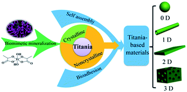

image file: c3ra47336h-p1.tif

Zhenwei Tong

Zhenwei Tong received her bachelor's degree in chemical engineering and technology from Sichuan University in 2012. She is pursuing a Ph.D. under Prof. Zhongyi Jiang at School of Chemical Engineering and Technology, Tianjin University. She is currently a Ph.D. student whose research interests include hierarchically structured materials, biocatalysis & photocatalysis.

image file: c3ra47336h-p2.tif

Yanjun Jiang

Yanjun Jiang obtained a Ph.D. in Biochemical Engineering at the Tianjin University in 2009 under the tutelage of Prof. Zhongyi Jiang. He started working at School of Chemical Engineering and Technology, HeBei University of Technology, where he is currently an associate professor in biochemical engineering. His research interests include biocatalysis, biomass and bioenergy. He has coauthored more than 40 SCI papers, with a total citation time of over 300 and hindex of 10.

image file: c3ra47336h-p3.tif

Dong Yang

Dong Yang obtained a Ph.D. at the Northeast Forestry University in 2001 under the guidance of Prof. Lijia An. After a stint with Prof. Limin Qi as a Postdoctoral at Peking University, he joined the faculty of Tianjin University in 2004, where he is currently an associate professor in biochemical engineering. His research interests encompass functional nanomaterials, membranes and membrane processes. Till now, he has coauthored more than 40 SCI papers, with a total citation time of over 700 and h-index of 17.

image file: c3ra47336h-p4.tif

Jiafu Shi

Jiafu Shi obtained a Ph.D. in Chemical Engineering from Tianjin University in 2013 under the tutelage of Prof. Zhongyi Jiang. After graduation, he joined the faculty of Tianjin University, and started working at the School of Environmental Science and Engineering. His research interests include biocatalysis and hierarchically structured materials, which are related to environment.

image file: c3ra47336h-p5.tif

Zhongyi Jiang

Zhongyi Jiang is a professor at School of Chemical Engineering and Technology of Tianjin University. He obtained a Ph.D. degree from Tianjin University in 1994. He was a visiting scholar of University of Minnesota with prof. Edward Cussler in 1997 and California Institute of Technology with Prof. David Tirrell in 2009. He is the winner of National Science Fund for Distinguished Young Scholars in China. His research interest includes biomimetic and bioinspired membranes and membrane processes, biocatalysis, photocatalysis. Till now, he has coauthored over 220 SCI papers, the total citation times is over 3500.


1 Introduction

Titania is considered as one of the most fascinating inorganic materials mainly due to its excellent physical and chemical properties, such as nontoxicity, high chemical and thermal stability and photosensitivity. Multimillion tons of powdered titania materials are produced each year by the flame oxidation of purified titanium tetrachloride,1,2 and applied in the fields including photocatalysis, coating, solar energy conversion, etc. The exhibited performance of titania materials is strongly dependent on their structural characters, i.e. crystal phase, particle size and surface structure,3–5 which is closely related to the synthetic method. Thus, the synthesis of titania materials has received increasing attention in recent years. Till now, a variety of methods have been developed for the synthesis of titania materials, such as anodization method,6,7 sol–gel method,8 hydrothermal method,9,10 chemical vapor deposition.11 However, these methods usually involve harsh or toxic conditions including high temperature and pressure, organic solvents or strong acid–base catalysts, which severely restrict the wide application (especially bio-related applications, such as biosensor, biocatalysis and drug delivery) of titania materials. To address these problems, it is essential to explore green, facile and controllable approaches to synthesize high-performance titania materials.

In nature, living organisms can synthesize diverse inorganic minerals as the form of skeletons, shells, teeth, etc., through biomineralization process. This intracellular biomineralization process exhibits a high level of time and spatial control over the formation of biominerals, which combine complex morphology over several hierarchy levels with superior materials properties. Surprisingly, almost all biomineralization process in nature takes place at ambient temperature, near neutral pH in the aqueous solution.12–15 Based on the insight into molecular mechanism of biomineralization, biomimetic and bioinspired strategies to synthesize inorganic materials extracellularly were developed tens of years ago.15,16 In 1998, silicateins, isolated from the sponge Tethya aurantia, were verified to have the ability to catalyze the formation of silica through the so-called biosilification process.17,18 Subsequently, Morse et al. found that silicateins from siliceous sponges could also induce the formation of amorphous/nanocrystalline titania materials from water soluble titanium precursors.19 This pioneering work opened the avenue for preparing non-natural metal oxides by the biomimetic and bioinspired strategy in vitro.17,20 Roughly speaking, biomimetic synthesis means the exploratory research with the focus on the molecular understanding of structure–function relations without dealing with the complexity of the biological archetypes; in comparison, bioinspired synthesis means developmental researches which adopt basic principles from biology without obvious resemblance to the biological archetypes. In a way, bioinspiration builds a delicate connection between fundamental science and applied engineering.21

In this review, the recent developments and advances in the biomimetic and bioinspired synthesis of titania and titania-based materials will be highlighted. Firstly, we will give a detail illustration about the formation of crystalline and non-crystalline titania by using biomimetic and bioinspired mineralization. Then, the synthesis and characterization of series hierarchically structured titania-based materials (nanoparticle, nanowire or nanotube, film, 3D porous monolith, etc.) will be highlighted thoroughly by combining biomimetic and bioinspired mineralization with other approaches (e.g., self-assembly, bioadhesion, etc.). Finally, the major challenge and opportunity for future research related to the biomimetic and bioinspired preparation of titania and titania-based materials will be discussed. Hopefully, this review will be of some assistance to the synthesis of diverse kinds of inorganic materials with desirable performances through the biomimetic and bioinspired strategies (Fig. 1).


image file: c3ra47336h-f1.tif
Fig. 1 Outline of this review: biomimetic and bioinspired synthesis of titania and titania-based materials.

2 Biomimetic synthesis of titania and related chemistry

Since the first report of amorphous titania synthesized by biomimetic mineralization,19 a variety of titania and titania-based materials (Fig. 2) have been synthesized.20,22,24,26,29,31 Up to date, numerous cationic molecules including peptides, proteins, polyamines, amino acids, etc.22,24,32,33 (Table 1) have been adopted as the inducers and play crucial roles for the “bottom-up” synthesis of titania in vitro.14 These inducers can act as catalyst and/or template (structure-directing agent) to control the morphology, crystal phase, and orientation of titania, leading to two typical types of titania with noncrystalline and crystalline phase.25 In addition, several studies have recently demonstrated that titanium can be metabolically inserted into the patterned biosilica of the diatom,34,35 which presented a novel method using living organisms to controllably fabricate TiO2 materials nanostructures by a bottom-up self-assembly process.
image file: c3ra47336h-f2.tif
Fig. 2 Selected examples of structures and morphologies of titania that could be produced using biomimetic chemistry. Particles formation using (A) R5 peptide,22 (B) poly-L-lysine,23 (C) lysozyme.24 Use of (D) rSliC25 to produce titania microspheres with pores inside. Nanofibers and nanonetwork formed using (E) silicatein19 and (F) (LK)8-PEG70.26 Titania coating produced using (G) protamine,27 (H) amyloid peptide,28 and (I) b-PEI.29 Hollow titania spheres using (J) protamine.30 All the images have been reproduced with permission from the respective publishers and the original copyrights remain with the respective publishers. The original references are cited in this legend and detailed references can be found at the end of this article.
Table 1 Summary of the bioinspired and biomimetic mineralization of titania in vitro discussed in main text. The table contains the inducer, the morphology, the crystallization of titania and the relevant references
Inducer classification Inducer Titania morphology Titania crystallization Ref.
Proteins and peptides Silicatein Titania-coated filaments Amorphous and nanocrystalline 19
Recombinant silicatein Layered morphology Amorphous 36
Lysozyme Spherical particles 10–50 nm Amorphous 24
Protamine Spherical particles ∼50 nm Amorphous 30
Viral capsids Irregular particles ∼20 nm Amorphous and β-titania 29
Glucose oxidase Spherical particles ∼30 nm Rutile 71
Catalase Spherical particles ∼30 nm Anatase 71
R5peptide (H2N-SSKKSGSYSG- SKGSKRRIL-COOH) Spherical particles 30–70 nm Amorphous 22
QPYLFATDSLIK, GHTHYHAVRTQT Spherical particles ∼4 nm Amorphous 52
Poly(L-lysine) Spherical particles 80–200 nm Amorphous 23
(LK)8-PEG70 3D network with nanofibers Amorphous 26
Polyamines Poly(2-(dimethyl-amino)ethyl methacrylate) Film with thickness ∼300 nm Amorphous 53
Poly(allylamine hydrochloride) Hollow spheres ∼10 nm Anatase and rutile 76
Small molecules Arginine, lysine, glycine Spherical particles 30–350 nm Amorphous 33 and 63
Spermidine Irregular particles 100–80 nm Amorphous 32
Spermine Irregular particles 50–300 nm Amorphous 32


2.1 Synthesis of noncrystalline titania

2.1.1 Proteins and peptides as the inducers. As shown above, silicateins, the proteins isolated from a marine sponge, were able to catalyze in vitro formation of titania from a water-soluble titanium precursor under ambient conditions.19 Specifically, titania could be synthesized and coated on the silicatein filaments through incubating silicatein (24 h at 20 °C) in an aqueous solution of titanium bis(ammonium lactato)-dihydroxide (Ti-BALDH, a typical water-soluble titanium precursor). The as-obtained titania coating was composed of nanocrystalline anatase near the silicatein filament and amorphous titania in regions away from the filament/inorganic interface (Fig. 3).19 The formation process of titania included thermally induced crystal grain growth and phase transformation from anatase to rutile, which were accomplished more easily than those of titania produced from the same precursor via alkali catalysis or thermal pyrolysis. Besides, in comparison with the acid–base-catalytic process, silicatein-mediated process can yield a product with much smoother surface, suggesting that the silicatein filaments played at least two roles: (1) catalyzing the hydrolysis and polycondensation of Ti-BALDH to form titania and (2) templating assembly of titania to generate nanoaggregates or nanocrystals.19,20 This pioneering work promoted the subsequent development of the biomimetic synthesis of titania.
image file: c3ra47336h-f3.tif
Fig. 3 Silicatein-catalyzed synthesis of titania. (A) SEM micrograph of silicatein filaments isolated from sponge spicules. (B) SEM micrograph of titania encrusted silicatein filaments following exposure to a neutral pH aqueous solution containing Ti-BALDH. (C) TEM and electron diffraction (as inset) characterization of the titania coating composed of both amorphous and crystalline titania on a silicatein filament.19 Reproduced from ref. 19 with permission.

Thair and co-workers demonstrated that the surface-bound silicatein immobilized on self-assembled polymer layers could catalyze the formation of laminated titania. During this process, a reactive ester polymer, poly(acetoxime methacrylate), was designed to react with primary amines for immobilizing silicatein onto surfaces. The recombinant protein with a short affinity sequence of histidines (histidine-tag) was capable of binding metal ion complexes, such as nitrilotriacetic acid (NTA).36 By using this process, a layer-like appearance of titania nanoparticles with diameters of 50–60 nm can be obtained, which indicated that the surface-bound silicatein might act initially as a catalyst and template to generate small nuclei,36,37 leading to the formation of final titania products with smooth surface.

Similar to silicatein, silaffin (another protein-type inducer) may also have the titania-forming capability, owing to the cooperative effects between the titanium precursor and mineral-interacting protein domains. In order to verify this hypothesis, selected regions of silaffin genes were expressed to form the recombinant silaffins rSilC.38 As expected, rSilC exhibited the ability to induce the formation of titania. Kharlampieva et al.38 reported that rSilC tethered on the surface of polyelectrolyte multilayers could create a monolayer composed of individual 4 nm titania nanoparticles after the addition of Ti-BALDH (Fig. 4). The surface-tethered silaffin molecules were aggregated into nanoscale domains, which then served as the templates for the formation of titania nanoparticles, and prevented their further aggregation. Meanwhile, this process can be repeated to build up thick, multifunctional nanoparticle composites. To mimic the titania-forming abilities of silicateins, exogenous proteins, such as lysozyme,24,39 viral capsids40 and protamine,41,42 have also been exploited as inducers for synthesizing titania materials. Unlike silicateins, the relatively low price, facile purification, and easy availability ensured these inducers as the promising titania-precipitating reagents in practical applications.


image file: c3ra47336h-f4.tif
Fig. 4 (A) Silaffin rSilC, adsorbed on (PAH/PAS)20 surface initiated the nucleation and growth of 4 nm titania nanoparticles with the addition of Ti-BALDH in a dispersed manner at 24 °C. (B) TEM image of titania nanoparticles grown on rSilC-tethered polyelectrolyte surfaces.38 Reproduced from ref. 38 with permission.

Hen egg lysozyme, a low-cost cationic polypeptide (pI = 10.5), which comprises several biomineralization-mediating groups including nucleophilic and hydrogen-bonding acceptor groups, can also be utilized as an efficient inducer.43 Recent progress on adopting lysozyme as inducer during the biomineralization of silica and calcium carbonate inspired the exploitation of utilizing lysozyme for the biomimetic synthesis of titania.24,44 Luckarift and co-workers reported that lysozyme could induce the formation of lysozyme/titania composite materials. More specifically, lysozyme catalyzed the rapid precipitation of titania when added to a solution of either potassium hexafluorotitanate (PHF–Ti) or Ti-BALDH at ambient conditions. Interestingly, the titania formed from lysozyme-induced mineralization of Ti-BALDH exhibits an open, highly interconnected network of rather fine nanoparticles (10–50 nm),24 which differed conspicuously from that produced from PHF–Ti precursors (Fig. 5).24 The results of thermal gravimetric analysis (TGA) and the increase of specific surface area after pyrolysis demonstrated that lysozyme was entrapped in the lysozyme/titania precipitates. This process may have great potential in enzyme immobilization owing to the following two aspects: the physical encapsulation can protect the enzymes from denaturation, whereas the co-immobilized lysozyme can offer the protection from microbial degradation.


image file: c3ra47336h-f5.tif
Fig. 5 SEM and TEM micrographs of titania nanoparticles formed by precipitation with lysozyme. (a) SEM and (b) TEM micrographs of titania from lysozyme and PHF–Ti; (c) SEM and (d) TEM micrographs of titania from lysozyme and Ti-BALDH. Insets: selected-area electron diffraction (SAED) patterns of precipitates.24 Reproduced from ref. 24 with permission.

Subsequently, Jiang's group first proposed that protamine may be another ideal candidate to induce and template the precipitation of titania in vitro.42 Protamine, a globular protein consisting of 32 amino acid residues, contains 20–22 cationic arginine residues,45 which endows it a very high pI value (12.0–12.4) with outstanding ability and unique application in the biomimetic synthesis of titania materials.42 When protamine was added to the Ti-BALDH solution, a white precipitate formed within several minutes at room temperature, validating the catalytic function of protamine in the formation of titania. In general, the weight of titania increased as the pH value of the reaction solution increased.41 However, when the pH value was higher than 6.0, only minor changes were observed. It was inferred that the positive charge on the surface of protamine was insufficient in the acidic solution, which weakened its electrostatic interaction with the titanium precursor.41 The protamine-induced titania precipitates were amorphous, and exhibited fused-particle morphology of 50 nm in size, which was more regular than that synthesized by using ammonium hydroxide as catalyst. In view of the superior stability of titania/protamine nanoparticle composites, an enzyme was encapsulated in titania particles during the protamine-induced biomimetic mineralization process. When the negatively charged enzyme immersed into the positively charged protamine solution, the guanidyl groups of protamine were prone to be attracted to enzyme through electrostatic interaction, aggregates were thus formed.30 The obtained enzyme-containing titania particles displayed excellent mechanical and catalytic properties.30

In some cases, a peptide sequence, which is much shorter than proteins, has also been employed as inducer for in vitro catalyzing hydrolysis and condensation of titanium precursors under mild conditions. Typically, R5 peptide (a bioinspired analogue derived from the silaffin protein in Cylindrotheca fusiformis) is the first peptide inducer that was used to catalyze formation of titania.22 Specifically, Pender et al.46 firstly found that R5 peptide could mediate the formation of carbon nanotube/titania composite materials. Then, in 2006, Wright and co-workers23 reported more specific investigations about the synthesis of titania nanoparticles utilizing R5 peptide as inducer. In Wright's report, R5 peptide was able to induce the formation of titania within several seconds and leaded to a relatively high yield, which was primarily due to its distinctly high arginine and lysine content. This finding also implied that the self-assembled peptide structure was vital for the production of titania in a manner similar to the R5-mediated silica.23

Insight into the molecular mechanism of R5 peptide in the synthesis of titania, some artificial peptides such as poly(L-lysine) (PLL) and P1R5, etc. have also been introduced to produce titania nanoparticles under mild conditions. For example, PLL can promote the hydrolysis and condensation of Ti-BALDH to form TiO2 nanoparticles. The interaction between PLL and TiO2 modulated the phase transformation of TiO2 from anatase to rutile, resulting in lower transition temperatures than that of other biogenic TiO2.23 Peptides that displayed on the surface of biological systems (such as bacteria, yeast and bacteriophage) have also been utilized for the biomimetic synthesis of inorganic materials.47,48 For example, Pender designed a multifunctional fusion peptide P1R5 that could induce the precipitation of titania from water-soluble precursors on the surface of the nanotubes.46 During this process, the R5 peptide domain was in charge of precipitating titania, whereas the P1 dodecapeptide identified from a combinatorial phage peptide display library was responsible for coating and suspending single-wall carbon nanotubes. Then, the work conducted by Sandhage and coworkers revealed that the 12-mer peptides enriched in basic residues (arginine, lysine, histidine) were particularly effective for inducing the synthesis of Ti–O-bearing precipitates.49 In their study, four peptide sequences were synthesized to precipitate titania, including Ti-1 (RKKRTKNPTHKLGGGW), Ti-2 (MRMIRRFPSSLKGGGW), Ti-3 (KSLSRHDHIHHHGGGW), and Ti-4 (TQHLSHPRYATKGGGW), which contained different number of basic amino acids (lysine (K), arginine (R), and histidine (H)). Once the peptide solution was added to a Ti-BALDH phosphate/citrate buffer solution (pH 6.3), a white precipitate appeared.50 The titania precipitation activities of the peptides increased with the number of positive charges carried by the peptide at pH 6.3. Generally, the designed peptide sequences of Ti-1 precipitated the largest amount of titania, which was due to this peptide possessing the suitable proportions of basic residues (Table 2). These findings confirm the hypothesis that the positive charges are essential to promote the interaction of the peptide with negatively charged titanium precursors. Although the enrichment of K, R, H in artificial peptides would confer them increased titania precipitation activity, the resultant amount of titania did not simply increase with the number of positive charges in the peptides. In detail, the net charge number of lysine and arginine were similar, but the peptide sequence individually composed of lysine residues performed better mineralization-inducing ability than the sequence composed of arginine residues. Furthermore, to understand the role of histidine in the biomimetic synthesis of titania, a peptide containing two serine–histidine pairs was investigated.51 Interestingly, this peptide showed significantly high precipitation activity despite low positive charge.

Table 2 Titania precipitation activities of peptidesa
Peptide (purified) pI H K + R Number of positive charges g titania/mmol peptide
a pI = isoelectric point. H = number of histidine residues. K + R = combined number of lysine and arginine residues. The ranges indicated for the titania precipitation activities and yields refer to one standard deviation. Reproduced from ref. 50 with permission.
dTi-1(RKK) 12.8 0 12 12 3.7 ± 0.2
Ti-1 12.4 1 6 6.3 3.1 ± 0.1
dTi-1(H/R) 12.6 0 7 7 2.8 ± 0.1
Ti-3 10.1 5 2 3.7 2.3 ± 0.1
Ti-2 12.3 0 4 4 1.7 ± 0.1
Ti-4 10.0 2 2 2.7 0.6 ± 0.1
Ti-9 11.0 3 1 2 0.2 ± 0.0
Ti-8 7.1 0 2 2 0.0 ± 0.0
Ti-18 7.8 1 0 0.3 0.0 ± 0.0


In an effort to better clarify the relationship between precipitation activity and structure-directing function, researchers designed a series of peptides to explore the functionality of different amino acids in initiating and precipitating titania materials. Specifically, the (leucine-lysine)8-PEG70 ((LK)8-PEG70) peptide, which was composed of hydrophilic and hydrophobic amino acid residues, was rationally designed to mineralize titania with a morphology of three-dimensional (3D) nanonetwork.26 The artificial peptide could self-assemble into nanofiber via the interaction between the hydrophobic leucine side chains, which acted as the catalyst and template to induce the nucleation of titania and control the morphology of titania materials. According to this work, it was clearly demonstrated that the peptide could manipulate the structure-directing function and precipitation activity, simultaneously. Furthermore, a similar viewpoint was put forward by Puddu and co-workers. They reported that two un-conventional artificial peptides (Ti.1 (QPYLFATDSLIK) and Ti.2 (GHTHYHAVRTQT)) can efficiently promote the formation of titania sols with 4 nm nanoparticles.52 The high affinity and specific binding between the titania surface and the two peptides were considered as the driving force for trigging the capping process, which could effectively stabilize the titania particles and inhibiting their further growth. To further understand the mineralization process, the conversion of titanium precursor as a function of its concentration was investigated both in phosphate buffer and water using Ti.1 as a peptide inducer.52 When the initial peptide was fixed at 0.28 mmol L−1, the conversion was remarkably enhanced with the increase of precursor concentration. Moreover, the Ti.1 peptide had a superior precipitation capacity in phosphate buffer than in water.

2.1.2 Polyamines and polysaccharides as the inducers. Besides proteins and peptides, the polyamines (such as poly(2-(dimethyl-amino)ethyl methacrylate) (PDMAEMA), poly(allylamine hydrochloride) (PAH), amine-terminated dendrimers) and cationic polysaccharides (such as xanthan) may also have the capability to induce the mineralization of titania due to their high content of net positive charges. Choi, Yang and their colleagues53 reported the formation of biological titania thin films under ambient conditions by using PDMAEMA (a synthetic counterpart of silaffins) as the inducer.53,54 To the best of our knowledge, this was the first example of biomimetic synthesis of titania thin films at neutral pH in vitro by using tertiary and quaternary amine-containing polymers as inducers. Previous study has indicated that not only silicateins,55 but also its special variants were designed and used for catalyzing inorganics mineralization. For example, silicatein α exhibited the ability of catalytic conversion of silicon precursors.56 Based on this finding, Hire57 synthesized a cationic block copolymer, poly(hydroxylated isoprene-b-2-vinylpyridine), which can successfully induce the hydrolysis and condensation of titanium iso-propoxide to form titania at neutral pH. The functional group of this block copolymers mimicked serine and histidine residues of silicatein α, which were required for biomineralization. The results of this report indicated that a synthetic biomimetic polymer could act as a catalyst for the production of highly condensed titania. Notably, the catalytic activity of this synthetic polyamine during the titania mineralization process was much higher than that of conventional acid/alkane catalytic sol–gel process. Meanwhile, as the hydrolysis step is separate from the condensation step, the rate of each step can be individually controlled, and the morphology of the titania can also be controlled.57 Additionally, linear polyethylenimines (PEIs) have excellent ability to self-organize into one-dimensional nanostructure in aqueous solution, and these nanofiber aggregates could be used as template and catalyst for the biomimetic formation of hybrid nanofiber-based titania under ambient conditions on the silicon substrates. Compared to the titania powders formed in solution, the interface-mediated titania maintained anatase phase in majority even at 900 °C, which indicated that this kind of titania exhibited dramatically improved thermostability.58

Polysaccharides, the abundant naturally-occurring biomacromolecules, are kinds of biocompatible, low-cost and versatile polymeric materials. With the remarkable progress in investigating the role of polysaccharides in biomineralization, polysaccharides were finally introduced to mineralize the titania formation.59 As is well known, xanthan as a typical polysaccharide usually contains hydroxyl and carboxyl groups, which may bind to the titanium precursor, and facilitate the cross-linking between polysaccharides and titanium precursors through hydrogen bonding.60 After contacting with water, the introduced precursor would instantly hydrolyze and condense. Unlike traditional inducers, polysaccharide in the synthetic reactions only served as a template other than a catalyst. By changing the concentration of precursor, xanthan, and water, it was possible to manipulate the morphology of titania ranging from fibrillar, particulate, to plate-like structures.59

2.1.3 Small molecules as the inducers. As described above, proteins, peptides and polyamines with high percentage of positively-charged amino acid residues (e.g., arginine and lysine) or amine functionalities are effective in the precipitation of titania materials. To extend the biomimetic mineralization process, amino acids or small amine molecules served as inducers were also utilized for the preparation of titania materials. For example, several amino acids, such as aspartic acid (Asp), glutamic acid (Glu), and serine (Ser), have been introduced in an aqueous solution of titanium precursor for the biomimetic synthesis of titania. It was found that the precipitation amount and crystallization of titania were affected by the pH value of solution. Pure anatase could be synthesized at a wide range of pH values in the presence of Glu, Asp, or Ser, while the same product was precipitated with histidine (His) only at high or low pH (1 or 6).61 Under all synthetic conditions, the anatase particles were lower than 8 nm in size, which were much smaller than those prepared by a hydrothermal treatment (20–100 nm).62 Moreover, the side chain property of amino acids was of particular importance in regard to the regulation of the structure and morphology of titania.

In order to investigate the effect of the amino acid number on the titania precipitation, a series of oligo(L-lysine)s (OLLs) were chosen as model molecules, and tested for the biomimetic mineralization of titanium precursor.63 Experimental results showed that significant enhanced precipitation could be achieved once the number of lysine in OLLs was high up to three. Besides, OLLs with less than eight sequenced lysines failed to control the morphology of titania, suggesting that longer peptides were required to guide the shape of titania products. Specific activities of K5 (Lys-Lys-Lys-Lys-Lys), and K8 were higher than those of R5 peptide (SSKKSGSYSGSKGSKRRIL) and its derivatives, but much lower than that of poly(L-lysine) (containing ∼430 lysine per single chain).23,63 Additionally, the specific activity of R5 peptide was lower than K4 although it had three lysine (KK and K), and consecutive lysine and arginine components (KRR) inside. This suggested that the number of consecutive lysine, other than the total number of lysine in a peptide, critically influenced the precipitation capability.

Alternatively, biomimetic techniques to produce titania films by using amino acid inducers have been developed.64–66 The presence of biological species allowed a higher deposition rate and a better control over the structure and morphology of the coating. For example, Durupthy et al.67 demonstrated that the addition of specific amino acids, such as histidine or glutamic acid, could acquire much smoother and denser films. The thickness of the deposited films increases with decreasing the concentration of the applied amino acids. For the highest amino acid concentration (R = 0.25), the resulting films were thinner than 50 nm. Moreover, due to the different chemical functions of their side chains, the amino acid inducers significantly influenced the deposition process and the roughness of the films. The solid–amino acid interface played a key role in the film deposition on the parent substrate. Hopefully, this deposition process can be applied to fabricate titania coatings on the irregular organic and inorganic substrates and there-by offering new opportunities for the fabrication of next-generation devices, for example, biocompatible implants or self-cleaning objects.67

Besides amino acids, small amine molecules32 (e.g., spermidine (SPDN) and spermine (SPN)) also exhibited the ability to induce the mineralization of titanium precursors to titania materials. Similar to amino acid inducers, at least three amine functionalities were required in small amine molecules for rapid mineralization reaction. With four amine functionalities, SPN were more complex and could condense titanium atoms much easier than SPDN.32 Additionally, once the mineralization process was completed, small amine molecules remained in solution, which could only act as the catalyst. This phenomenon is different from the R5 peptides and PLL induced process (the peptide and PLL may be encapsulated in the as-formed nanoparticles).

2.1.4 Mechanism of biomimetic mineralization of titania. Insight into the molecular mechanisms underlying titania mineralization has attracted considerable research attentions on purifying and characterizing natural proteins and other molecule inducers, which can enable the biomimetic and bioinpsired process for titania synthesis. The mechanism of biomimetic synthesis of titania was first proposed in Sumerel's work. The hydrogen bonding between the hydroxyl groups of a specific serine–histidine pair in silicatein increased the nucleophilicity of the serine oxygen. Thus, an attack on the titanium in Ti-BALDH facilitated to form a transitory Ti–O bond with the protein, finally initiating the hydrolysis of Ti-BALDH at neutral pH.19 Subsequently, a deeper understanding of the peptide-enabled titania synthesis was illustrated: the hydroxyl group in serine attacked the titanium atom in Ti-BALDH acting as a nucleophile, and a pair of electrons on the histidine nitrogen accepted the hydrogen from the serine hydroxyl group.67 Nucleophilic attack on the titanium atom displaced ammonium lactate, and a transitory peptide–Ti-BALDH intermediate, which would be stabilized through a donor bond from the imidazole nitrogen atom, formed by releasing ammonium lactate. Furthermore, the amine-rich peptide template was also demonstrated to act as a general acid–base catalyst to promote the hydrolysis of the negatively charged precursor Ti-BALDH through a co-effect of electrostatic and hydrogen bonding interactions.23 And the metal complexation induced by the amines of inducers brings two titanium atoms in close proximity, which would facilitate the condensation process.32

On the basis of previous finding, a mechanism of biomimetic mineralization of Ti-BALDH induced by protamine was also proposed by Jiang and coworkers. As shown in Scheme 1, globular protamine molecules are positively charged at pH 7.0, which can absorb and concentrate the negatively charged Ti-BALDH.41 Hydrogen bonding between Ti–OH of the titanium precursor and C[double bond, length as m-dash]NH of the protamine induced the nucleophilic substitution of a Ti–O oxygen atom on another adjacent titanium atom, and the polycondensation reaction occurred subsequently. Meanwhile, the complexation of titanium with protamine molecules offered some additional contribution to the acceleration of the polycondensation,32 and then aggregation with other adjacent titania particles through the condensation reaction between the surface hydroxyl groups, ultimately leading to the formation of titania nanoparticles.41


image file: c3ra47336h-s1.tif
Scheme 1 Tentative mechanism of titania formation induced by protamine molecules.41 Reproduced from ref. 41 with permission.

2.2 Synthesis of crystalline titania (anatase, rutile or brookite)

The above summarized reports indicate that titania prepared through biomimetic mineralization is mainly amorphous or partially crystallized. Calcination of the resultant titania can acquire fully crystalline phase, which is similar to that of conventional methods. It is known that the crystallization of titania is closely relevant to its technological applications, therefore synthesizing crystalline titania through biomimetic mineralization at ambient temperature will be a promising development in the future research.

In 2006, an exciting discovery was achieved by Kröger and co-workers: the recombinant silaffin rSilC could induce the formation of rutile titania under ambient conditions and neutral pH, which opened up new possibility to produce functional hybrid materials for optical applications. The presence of oriented rutile microcrystals in the rSilC–titania precipitate was confirmed by the XRD analysis. Typically, when rSilC was added, the Ti-BALDH solution instantly became turbid, and precipitates were formed after only a few minutes. The rSilC-induced rutile titania microspheres contained two types of structures.25 One type consisted of aggregates of spherical amorphous titania particles (Fig. 6). The other type consisted of large microspheres and revealed a hierarchical architecture.25 These microspheres were composed of abundant rutile crystals, in which numerous rectangular pores could be observed. The transformation process from partially amorphous titania aggregation into highly organized rutile microspheres usually requires the reorganization of the condensed TiO6 octahedral,25 which can only be achieved at high temperatures or under extremely acidic conditions in conventional methods.68–70 In the rSilC-induced mineralization process, the numerous lysine residues of rSilC can donate and accept protons, and the highly repetitive spacing of functional groups in rSilC may lower the activation energy for the alignment of TiO6 octahedral into a rutile lattice. These results indicated that the distribution of positive charges, the amino acid sequence/composition, and a combination of these factors were all crucial to the transformation process and the formation of rutile.25 The ability of rSliC precipitating rutile titania at ambient temperature was unprecedented, and opened up new perspectives to produce functional hybrid materials. This significant finding was crucial in promoting the penetration of biological methods in the field of materials science.


image file: c3ra47336h-f6.tif
Fig. 6 SEM micrographs of hierarchically structured rutile titania microspheres generated through the dehydration of rSilC titania. Arrow heads in (D) highlight elongated rectangular pores in the cross section of a rutile microsphere. Scale bars correspond to 20 μm in (A), 5 μm in (B), 1 μm in (C), and 500 nm in (D).25 Reproduced from ref. 25 with permission.

Subsequently, Xia71 reported three examples of synthesizing titania nanocrystallites through enzyme-induced (lysozyme, catalase, and glucose oxidase) mineralization. Rutile, anatase, and mixed phases of monoclinic β-titania were obtained by the unique combination of enzyme and titanium precursor followed by aging for three to four weeks at room temperature (Fig. 7). For the first case (that is lysozyme induced mineralization of titania), previous literature had proven that lysozyme-stabilized titania was amorphous,24 whereas, in Xia's report, mixed phases of monoclinic β-titania and anatase were observed in the XRD pattern of sample after three weeks of aging, indicating a slow phase transformation from amorphous to partially crystallized anatase phase and β-titania.71 For the second case (that is catalase induced mineralization of titania), the tentative mechanisms were proposed that catalase acted as an acid–base catalyst promoting the hydrolysis of precursors and guiding the rearrangement of the TiO6 octahedra into a crystal lattice. This result agreed well with the reported transformation from anatase to rutile through a “dissolution–recrystallization” mechanism, which had only been favored in strong acid media (pH <1) at high temperatures.72,73 For the third case (that is glucose oxidase induced mineralization of titania), glucose oxidase might provide nucleation sites and lower the activation energy of titania nucleation. The precipitated titania possessed pore size less than 2 nm, which was in accordance with the results revealed by the TEM images (Fig. 7C).71 One plausible mechanism was that in the process of the slow phase transformation from the amorphous state (less denser phase) to the crystalline state (denser phase), pores were left behind.71


image file: c3ra47336h-f7.tif
Fig. 7 TEM image of sample 1–3: crystalline titania precipitate mediated by (A) glucose oxidase, (B) and (C) catalase, (D) lysozyme. The scale bar corresponds to 50 nm.71 Reproduced from ref. 71 with permission.

Shortly afterwards, it was found that the polysaccharide could also be applied for the biomimetic synthesis of crystalline titania. Chen and co-workers reported a novel biomimetic synthetic route based on the glucan polymers-mediated formation of titania using titanium sulfate as the precursor at room temperature. Compared with the structure of proteins, the glucan polymers have the similar chain structure but different charged groups,74 which afforded the possibility to investigate the contributions from the chemical groups. Chitosan, cellulose and sodium alginate were also demonstrated to catalyze the hydrolysis and polycondensation of the titanium precursor to form titania with different shapes and polymorphs.75 Particularly, the phase transformation from anatase to rutile was observed in the chitosan-mediated titania process. The mechanism of precipitation and pores formation were similar to the above process induced by enzymes. The phase transformation was distinctly dependent on the protonation level of the amino groups in chitosan. More specifically, the protonated amino groups of chitosan might act as a ‘proton reservoir’,75 donating protons to impact on the surface protonation of titania, thus facilitating the well-known ‘dissolution–reprecipitation’ process.

Additionally, poly(allylamine hydrochloride) (PAH) and poly(diallyldimethylammonium chloride) (PDDA) were used to induce the biomimetic mineralization of titania under mild conditions in Chen's another work. Hollow spheres with mixed phase of anatase/rutile or anatase/β-titania were obtained.76 The results revealed that the protonation level of amino groups in PAH might facilitate the formation of hollow structure as well as the phase transformation from anatase to rutile using Ti(SO4)2 as precursor. In detail, the protonated amino groups of PAH in the composites might serve as “acid–base” catalyst, which could release protons to direct the rearrangement of TiO6 octahedra into anatase lattice. PDDA, a long chain quaternary ammonium polymer, induced formation of a hollow structure with a mixture of anatase and TiO2-β in the Ti-BALDH solution. However, when using Ti(SO4)2 as precursor, the phase transformation can not occur and the hollow structure could not be obtained, which may be related to the hydrophilic/hydrophobic performance in different solutions. Besides, the different charged groups of polymers affected the size, shape and polymorph of the acquired titania particles strongly.76 This work may ultimately offer novel approach to tailoring polymorphs, size, shape, surface, and other physicochemical properties of titania.

3. Synthesis of hierarchically structured titania-based materials using biomimetic and bioinspired tools

After in-depth understanding of the biomimetic mineralization of titania, synthesis of hierarchically structured titania-based materials can be realized by using the combination of biomimetic mineralization with other approaches such as self-assembly and bioadhesion processes.

3.1 Synthesis of titania-based materials through synergy between self-assembly and biomimetic mineralization

In the last decade, organic–inorganic composite materials have drawn much attention due to their combined and unique properties.28,77 Inspired by the delicate structure of cell, a new method was proposed to prepare titania-based composite materials utilizing the combination of layer-by-layer (LbL) self-assembly technique with biomimetic mineralization. In this review, we focus on two main classes of templates that have been employed to prepare titania-based composite materials: hard templates and soft templates.
3.1.1 Preparation of titania-based composite materials by using hard templates. The deep-going understanding of the mechanisms of biomineralization has opened up the bioinspired approaches for the synthesis of functional organic–inorganic hybrid materials under mild reaction conditions. Recently, the method through the combination of LbL self-assembly with bioinspired mineralization has been utilized for preparing titania-based materials with hierarchical architectures. Some hard templates, for example CaCO3,30,78 SiO2,79 Al2O3 (ref. 80) etc., were used in these processes (Scheme 2).79 Jiang and coworkers used the porous CaCO3 microparticles as sacrificial templates for the synthesis of protamine–titania hybrid microcapsules. The LbL assembly started with deposition of protamine layer onto the surface of CaCO3 microparticles. The protamine layer with positive charge can absorb the titanium precursor (Ti-BALDH) with negative charge, and then induce the formation of titania layer. The negative charged titania layer could allow the next cycle of protamine deposition. By repeating the above steps, the multilayer with defect-free structure could be formed. After dissolving the CaCO3 microparticles, protamine–titania hybrid microcapsules were obtained.30 These capsules exhibited desirable encapsulation efficiency for yeast alcohol dehydrogenase (YADH) and the immobilized YADH showed improved recycling stability.30 By using a double templating strategy, hybrid double membrane microcapsules (HDMMCs) with mitochondria-like structure was constructed through synergy between biomimetic mineralization and LbL self-assembly. The organic inner membrane was acquired via LbL self-assembly of oxidized alginate (o-alginate) and protamine on the CaCO3 template. The silica layer was then formed onto the inner membrane through the biomimetic silicification process. The titania outer membrane was acquired via biomimetic titanification process. After removing the CaCO3 and silica subsequently, the HDMMCs with microscale lumen and nanoscale intermembrane space were obtained. Based on the HDMMCs, a multi-enzyme system containing formate dehydrogenase (FateDH, enzyme 1) and formaldehyde dehydrogenase (FaldDH, enzyme 2) was constructed for converting CO2 into formaldehyde. The activity, selectivity and recycling stability of HDMMCs-immobilized multi-enzyme system were significantly improved compared to the free form of the enzymes.
image file: c3ra47336h-s2.tif
Scheme 2 Protamine-induced layer-by-layer (LbL) deposition of titania on hard templates.79 Reproduced from ref. 79 with permission.

Kröger and coworkers79 fabricated the biocatalytic nanoscale coatings through the combination of LbL self-assembly with biomimetic mineralization process by using silica spheres as hard template. As shown in Scheme 2, protamine modified glucose oxidase (GOx-PA) was used as the inducer. During protamine-enabled LbL mineralization on silica spheres, GOx-PA was entrapped into nanoscale layers (∼5–7 nm thickness) of Ti–O and Si–O.79 The resultant hybrid materials exhibited between 20 and 100% of the catalytic activity of free GOx, which depended on the layer location of GOx and the type of mineral (silica or titania). This approach can be extended to a wide variety of surfaces and functional biomolecules, and the enzyme-containing layers are interesting for numerous applications such as enzyme-based biofuel cells and biosensors.

This approach was also extended to prepare freestanding microscale titania with morphologies and structures inherited from complex-shaped, three-dimensional (3-D) biosilica templates (diatom frustules) and alumina templates.80,81 The arginine/lysine-rich protamine was utilized for the sequential deposition and buildup of a conformal and continuous titania coating on complex 3-D diatom frustule and alumina templates. After calcination, the conformal and continuous nature of the resulting anatase titania was obtained. With selective removal of the biosilica or alumina template, freestanding titania structures that with the 3-D diatom frustule shape or titania nanotubes were obtained, respectively (Fig. 8).80,81 The intricate and fascinate structure of the titania materials controlled by this approach can be utilized in a number of chemical, optical, photochemical and biochemical devices. Additionally, this approach can also be used to fabricate other inorganic/organic composites materials with desired shape/dimensions and morphological features.82,83


image file: c3ra47336h-f8.tif
Fig. 8 Secondary electron images of protein-enabled titania coating of diatom frustules (A) and (B) a complete and a partial titania-coated Aulacoseira frustule, after 12 cycles of protamine/Ti-BALDH exposure. Scale bars: 2 μm.81 SEM of top-down (C) and cross-sectional (D) views of through-hole anodic aluminum oxide templates that were covered with a protamine/Ti–O-bearing composite coating via exposure to 8 protamine/Ti-BALDH deposition cycles.80 (C) and (D) Reproduced from ref. 80 with permission; (A) and (B) Reproduced from ref. 81 with permission.

It was reported that the polyelectrolyte LbL films could be used as structured soft matrix to control recombinant silaffin (rSilC) adsorption and direct titania nanoparticle nucleation, growth, and organization on the tailored surfaces. The titania nanoparticles obtained by this method were composed of 4 nm titania cores (mixed amorphous and anatase phases) surrounded by 1 nm protein shells.38 In contrast to previous studies, this method was a facile way to synthesize hybrid surfaces with dispersed titania nanoparticles at high density. Chen and cowokers fabricated a protamine/titania composite layer on nickel foam through the combination of LbL assembly and biomimetic mineralization. However, unlike the negatively charged surface of diatom frustules, silica spheres, alumina, and CaCO3 particles, the surface of nickel foam was often inert and hydrophobic, making it difficult to adsorb protamine. To overcome this problem, the nickel foam was modified with a hydrophilic polydopamine (PDA) layer.27 Thus, protamine could be successfully attached on the PDA layer. Subsequently, the protamine-coated nickel foams were treated with titanium precursor to create a titania-containing hybrid layer. The layer, composed of amorphous carbon, titania, and anatase titania nanoparticles, was formed upon organic pyrolysis under a reducing atmosphere. The obtained titania/anatase/carbon-coated nickel foam was used as a binder-free electrode in lithium–ion batteries and exhibited high reversible capacity and fast charge–discharge properties. This report indicated that the bioinspired approach can be developed into a general pathway to synthesize titania-based coatings for a wide range of applications.27

3.1.2 Preparation of titania-based composite materials by using soft templates. Proteins and peptides were designed to be folded in desired conformations, and these 3D building blocks may yield supramolecular structures through self-assembly process, which had been successfully utilized as soft templates to produce a variety of self-assembled structured inorganic materials under relatively mild conditions.84 Katsuya Kato and coworkers prepared the titania by using (leucinelysine)8-PEG70 ((LK)8-PEG70) as the soft reactive template. In a basic environment, (LK)8-PEG70 peptide can spontaneously self-assemble to 3D nanofiber network (Scheme 3). The well-controlled peptide secondary structure present in (LK)8-PEG70 as an organic template rendered the desired nanofibrous TiO2. Such a nanonetwork simultaneously controlled the morphology and crystal phase of mineralized titania. It was speculated that the amino group of the lysine residue captured the titanium complex, confining the titanium-atom position on the lysine side chain in the earliest stage; biomimetic mineralization of titania then started at the lysine side chain. It was thought that the ion-capturing functional group's position was important and affected the subsequent growth of the mineralized titania.26 More interesting, this method could be used to prepare the rutile phase of titania at low temperature. When it was used in photocatalytic test, the high activity could be achieved by the synergetic effect of surface area, mixed-crystal phases, and elemental doping after sintered at 700 °C.26
image file: c3ra47336h-s3.tif
Scheme 3 (a) Hierarchical self-assembly of β-sheet peptide into a 3D nanofiber network; (b) titania mineralization occurs at the surface of the amino group side of the peptide nanonetwork.26 Reproduced from ref. 26 with permission.

Additionally, self-assembled amyloid-like peptides have emerged as a unique class of materials with potential applications as soft templates for nanotube and nanowire growth, and 1-D nanostructure organization,85 which rendered suitable soft templates to direct the mineralization process with their inherent ability to self-assemble into fibrillar nanostructures and their functional group.28,86 In the work reported by Acar et al., an amyloid-like peptide (ALP) was designed and used as template to fabricate 1-D titania nanofibers. The self-assembly of ALP molecules was achieved by a specially designed short peptide sequence, Ac-KFFAAK-Am, which formed sheet like hydrogen-bonded structures.28 The functional groups of lysine residues on the side chains of ALP served as nucleation sites for the successive deposition of titanium precursor. By using this self-assembly nanofibers as organic templates, 1-D titania with high surface area was obtained.28 This new approach could yield wide opportunities to produce high-aspect-ratio inorganic nanostructures with high surface areas, which endowed the ALP-templated process with vast potential in the fields of catalysis and electronic materials. Similarly, β-lactoglobulin amyloid protein with self-assembled fibrillar nanostructures was also employed to direct the synthesis of titania-based hybrid nanowires.86 Protein fibrils acted as soft templates to generate closely packed titania nanoparticles on the surface of the fibrils, resulting in the titania-coated amyloid hybrid nanowires. Importantly, the titania nanoparticles were sintered uniformly, thus producing a rather homogeneous layer with the thickness of 20–25 nm on the surface of the protein fibrils.86 The possible mechanism for the titania synthesis was similar with the report by Kharlampieva et al.38 The obtained amyloid–titania hybrid nanowires can serve in heterojunction photovoltaic devices. To demonstrate this, Mezzenga and coworkers prepared a photovoltaic active layer by spin coating the blended mixture of polythiophene-coated fibrils and amyloid–titania hybrid nanowires. Compared to photovoltaic solar cells based on fully organic homologue active layers, these photovoltaic devices exhibited enhanced fill factor, increased photovoltaic current density and much higher power conversion efficiency. In addition, cell-assemblies with different shapes were also used as templates in the synthesis of hierarchical macro–mesoporous titania with tunable macroporous morphology and this material exhibited enhanced photocatalytic performance.87 Such aforementioned model studies indicated that these titania-based materials with hierarchical nanostructures can be used in catalysis and heterojunction hybrid solar cells. Overall, by using hard or soft templates, through the synergy of self-assembly and biomimetic mineralization, hierarchical titania-based materials can be fabricated, which may be an interesting way to produce bulk amounts of titania materials for various industrial and technological applications.

3.2 Synthesis of titania-based materials through synergy between bioadhesion and biomimetic mineralization

Mussels are promiscuous fouling organisms and can attach to virtually all types of surfaces, ranging from natural inorganic and organic materials to synthetic materials. Inspired by the structure of adhesive proteins in mussels, dopamine, a small molecule was discovered by Messersmith et al.88,89 Dopamine can form thin, surface-adherent films onto a wide range of materials through self-polymerization. By combining of this so-called bioadhesion process with biomimetic mineralization, Zhang et al.90 developed a green and efficient approach to prepare titania-based hybrid microcapsules. More specifically, the hybrid microcapsules were fabricated by sequential deposition of titania and polydopamine layers on the surface of CaCO3 microparticles, followed by dissolving of CaCO3 microparticles (Fig. 9). The titania layer (biomineral layer) was formed through biomimetic mineralization of Ti-BALDH induced by protamine. The polydopamine layer (PDA, bioadhesive layer) was formed through spontaneous polymerization of dopamine. The quaternary ammonium groups and carboxyl groups in PDA layer can endow abundant reaction sites on the surface of hybrid microcapsules. The thickness and porosity of the biomineral layer and bioadhesive layer can be flexibly tuned by altering the concentration of titanium precursor/dopamine, temperature and pH of the aqueous solution.90
image file: c3ra47336h-f9.tif
Fig. 9 Schematic representation of the formation process of the PTi-PDA (PSi-PDA) microcapsule.90 Reproduced from ref. 90 with permission.

By combining of the bioadhesion and biomimetic mineralization process, size-controlled and surface-functionalized titania nanoparticles (TiNPs) were also prepared by Shi et al.33 The initial formation and subsequent growth of TiNPs were induced by arginine via biomimetic mineralization, while this process can be terminated by pre-polymerized dopa (oligodopa).33 By adjusting the addition time of oligodopa, the particle size of TiNPs could be facilely tailored from ca. 30 nm to 350 nm (Fig. 10); meanwhile, the surface of TiNPs could be functionalized by oligodopa. In addition, the layer of oligodopa can anchor proteins through amine–catechol adduct formation.27,33 Thus, the TiNPs were used to construct spatially separated multienzyme system for the conversion of carbon dioxide to formaldehyde. Specifically, the first enzyme was entrapped accompanying the formation of TiNPs through arginine induced biomimetic mineralization. After in situ surface functionalization of TiNPs with oligodopa, the second enzyme was conjugated on the TiNPs through amine–catechol adduct reaction between oligodopa and the second enzyme. Compared to co-immobilized and free multienzyme system, the spatially separated multienzyme system exhibited significantly enhanced activity and selectivity.91 This approach which combines bioadhesion and biomimetic mineralization may evolve as a generic platform for preparation of titania-based materials for a variety of applications.


image file: c3ra47336h-f10.tif
Fig. 10 Control over of arginine concentration and termination time on the morphology and size of surface-functionalized TiNPs. SEM images (bar: 500 nm) of surface-functionalized TiNPs prepared at different arginine concentrations (up to down 0.1, 0.2, 0.3, 0.5 M) and termination time (left to right 0.5, 1.0, 2.0, 5.0, 10.0 min).33 Reproduced from ref. 33 with permission.

4 Conclusions and prospective

Bioinspired/biomimetic approach has become a burgeoning branch of materials chemistry, which opens a new and green avenue to synthesis inorganic materials. Till now, a wide variety of (bio)molecules have been explored for fabricating titania and titania-based materials with outstanding and unique properties through powerful biomimetic and bioinspired tools. The mechanisms regulating the generation of titania and the interactions between inducers and titanium precursor have been extensively elucidated.

Albeit the considerable progress, many issues related to the bioinspired/biomimetic synthesis of titania materials remain to be addressed: (1) although many inducers have been validated, more biomolecules and synthetic molecules should be explored as versatile catalysts and templates in the biomimetic mineralization process, thus building titania materials with complex 2D or 3D structures; (2) to further harness the nature's ability for titania formation in vitro, continued efforts in fundamental research of the biomimetic titanification process and mechanisms are definitely required. In particular, methods that can monitor the process in real time and analyze the process at molecular, organelle, cellular, tissue, organ, and organism levels need to be further developed to better understand and fully exploit the biomimetic titanification process; (3) the structure and crystallization of titania materials directly govern their application performances, thus more efforts should be dedicated to gather more information on the relationships among inducers' structure and composition, the structure and crystallization of titania materials; (4) although some multifunctional hybrid materials have been produced by the combination of biomimetic titaniafication with LbL self-assembly and/or bioadhesion, more novel methods and approaches that of combining biomimetic titaniafication with other techniques should be always pursued to obtain materials with controllable hierarchical structures, compositions and novel properties.

To our pleasure, in recent years, a huge amount of knowledge has been accumulated with respect to the relevant biology, chemistry, physics and engineering of biomimetic titania and titania-based materials. Biomimetic and bioinspired synthesis of titania materials has grown up as a generic and potent platform technology with many inherent and potential advantages. Doubtlessly, with the rational combination of exploratory biomimetics and developmental bioinspiration, massive production of titania materials with energy saving, environmental friendliness, better control attributes could be achieved in the near future.

Acknowledgements

The authors thank the financial support from the National Science Fund for Distinguished Young Scholars (21125627), National Basic Research Program of China (2009CB724705).

References

  1. R. H. West, M. S. Celnik, O. R. Inderwildi, M. Kraft, G. J. Beran and W. H. Green, Ind. Eng. Chem. Res., 2007, 46, 6147–6156 CrossRef CAS.
  2. J. C. Deberry, M. Robinson, M. D. Pomponi, A. J. Beach, Y. Xiong and K. Akhtar, U.S. Patent. No. 6387347, 2002.
  3. J. Zhu, J. Yang, Z.-F. Bian, J. Ren, Y.-M. Liu, Y. Cao, H.-X. Li, H.-Y. He and K.-N. Fan, Appl. Catal., B, 2007, 76, 82–91 CrossRef CAS PubMed.
  4. U. Diebold, Surf. Sci. Rep., 2003, 48, 53–229 CrossRef CAS.
  5. E. Pouget, E. Dujardin, A. Cavalier, A. Moreac, C. Valéry, V. Marchi-Artzner, T. Weiss, A. Renault, M. Paternostre and F. Artzner, Nat. Mater., 2007, 6, 434–439 CrossRef CAS PubMed.
  6. J. M. Macak, M. Zlamal, J. Krysa and P. Schmuki, Small, 2007, 3, 300–304 CrossRef CAS PubMed.
  7. J. M. Macak, H. Tsuchiya and P. Schmuki, Angew. Chem., Int. Ed., 2005, 44, 2100–2102 CrossRef CAS PubMed.
  8. A. C. Pierre and G. M. Pajonk, Chem. Rev., 2002, 102, 4243–4266 CrossRef CAS PubMed.
  9. C.-C. Wang and J. Y. Ying, Chem. Mater., 1999, 11, 3113–3120 CrossRef CAS.
  10. X. G. Han, Q. Kuang, M. S. Jin, Z. X. Xie and L. S. Zheng, J. Am. Chem. Soc., 2009, 131, 3152–3153 CrossRef CAS PubMed.
  11. J.-J. Wu and C.-C. Yu, J. Phys. Chem. B, 2004, 108, 3377–3379 CrossRef CAS.
  12. E. Bäuerlein, Biomineralization: Progress in biology, molecular biology and application, Wiley-VCH Verlag GmbH & Co. KGaA, Weinheim, Germany, 2004 Search PubMed.
  13. S. Mann, Oxford University Press, Oxford, 2002.
  14. K. H. Sandhage, M. B. Dickerson and R. R. Naik, Chem. Rev., 2008, 108, 4935–4978 CrossRef PubMed.
  15. A.-W. Xu, Y. Ma and H. Cölfen, J. Mater. Chem., 2007, 17, 415–449 RSC.
  16. E. Dujardin and S. Mann, Adv. Mater., 2002, 14, 775 CrossRef CAS.
  17. K. Shimizu, J. Cha, G. D. Stucky and D. E. Morse, Proc. Natl. Acad. Sci. U. S. A., 1998, 95, 6234–6238 CrossRef CAS.
  18. J. N. Cha, K. Shimizu, Y. Zhou, S. C. Christiansen, B. F. Chmelka, G. D. Stucky and D. E. Morse, Proc. Natl. Acad. Sci. U. S. A., 1999, 96, 361–365 CrossRef CAS.
  19. J. L. Sumerel, W. Yang, D. Kisailus, J. C. Weaver, J. H. Choi and D. E. Morse, Chem. Mater., 2003, 15, 4804–4809 CrossRef CAS.
  20. K. M. Roth, Y. Zhou, W. Yang and D. E. Morse, J. Am. Chem. Soc., 2005, 127, 325–330 CrossRef CAS PubMed.
  21. V. I. Vullev, J. Phys. Chem. Lett., 2011, 2, 503–508 CrossRef CAS.
  22. K. E. Cole, A. N. Ortiz, M. A. Schoonen and A. M. Valentine, Chem. Mater., 2006, 18, 4592–4599 CrossRef CAS.
  23. S. L. Sewell and D. W. Wright, Chem. Mater., 2006, 18, 3108–3113 CrossRef CAS.
  24. H. R. Luckarift, M. B. Dickerson, K. H. Sandhage and J. C. Spain, Small, 2006, 2, 640–643 CrossRef CAS PubMed.
  25. N. Kröger, M. B. Dickerson, G. Ahmad, Y. Cai, M. S. Haluska, K. H. Sandhage, N. Poulsen and V. C. Sheppard, Angew. Chem., 2006, 118, 7397–7401 CrossRef.
  26. T. Nonoyama, T. Kinoshita, M. Higuchi, K. Nagata, M. Tanaka, K. Sato and K. Kato, J. Am. Chem. Soc., 2012, 134, 8841–8847 CrossRef CAS PubMed.
  27. X. Wang, Y. Yan, B. Hao and G. Chen, ACS Appl. Mater. Interfaces, 2013, 5, 3631–3637 CAS.
  28. H. Acar, R. Garifullin and M. O. Guler, Langmuir, 2011, 27, 1079–1084 CrossRef CAS PubMed.
  29. B. Hao, Y. Yan, X. Wang and G. Chen, ACS Appl. Mater. Interfaces, 2013, 5, 6285–6291 CAS.
  30. Y. Jiang, D. Yang, L. Zhang, Q. Sun, X. Sun, J. Li and Z. Jiang, Adv. Funct. Mater., 2009, 19, 150–156 CrossRef CAS.
  31. R. Andre, M. N. Tahir, F. Natalio and W. Tremel, FEBS J., 2012, 279, 1737–1749 CrossRef CAS PubMed.
  32. K. E. Cole and A. M. Valentine, Biomacromolecules, 2007, 8, 1641–1647 CrossRef CAS PubMed.
  33. J. Shi, D. Yang, Z. Jiang, Y. Jiang, Y. Liang, Y. Zhu, X. Wang and H. Wang, J. Nanopart. Res., 2012, 14, 1–13 Search PubMed.
  34. C. Jeffryes, T. Gutu, J. Jiao and G. L. Rorrer, ACS Nano, 2008, 2, 2103–2112 CrossRef CAS PubMed.
  35. Y. Lang, F. del Monte, B. J. Rodriguez, P. Dockery, D. P. Finn and A. Pandit, Sci. Rep., 2013, 3, 3205 Search PubMed.
  36. M. N. Tahir, P. Theato, W. E. Muller, H. C. Schroder, A. Borejko, S. Faiss, A. Janshoff, J. Huth and W. Tremel, Chem. Commun., 2005, 5533–5535 RSC.
  37. M. N. Tahir, M. Eberhardt, H. A. Therese, U. Kolb, P. Theato, W. E. Muller, H. C. Schroder and W. Tremel, Angew. Chem., Int. Ed., 2006, 45, 4803–4809 CrossRef CAS PubMed.
  38. E. Kharlampieva, T. Tsukruk, J. M. Slocik, H. Ko, N. Poulsen, R. R. Naik, N. Kröger and V. V. Tsukruk, Adv. Mater., 2008, 20, 3274–3279 CrossRef CAS.
  39. C. Liu, D. Yang, Y. Jiao, Y. Tian, Y. Wang and Z. Jiang, ACS Appl. Mater. Interfaces, 2013, 5, 3824–3832 CAS.
  40. M. T. Klem, M. Young and T. Douglas, J. Mater. Chem., 2008, 18, 3821–3823 RSC.
  41. Y. Jiang, D. Yang, L. Zhang, L. Li, Q. Sun, Y. Zhang, J. Li and Z. Jiang, Dalton Trans., 2008, 4165–4171 RSC.
  42. Y. Zhang, H. Wu, J. Li, L. Li, Y. Jiang, Y. Jiang and Z. Jiang, Chem. Mater., 2007, 20, 1041–1048 CrossRef.
  43. Y. Jiang, D. Yang, L. Zhang, Y. Jiang, Y. Zhang, J. Li and Z. Jiang, Ind. Eng. Chem. Res., 2008, 47, 1876–1882 CrossRef CAS.
  44. T. Shiomi, T. Tsunoda, A. Kawai, F. Mizukami and K. Sakaguchi, Chem. Mater., 2007, 19, 4486–4493 CrossRef CAS.
  45. E. Raukas and R. H. Mikelsaar, BioEssays, 1999, 21, 440–448 CrossRef CAS.
  46. M. J. Pender, L. A. Sowards, J. D. Hartgerink, M. O. Stone and R. R. Naik, Nano Lett., 2006, 6, 40–44 CrossRef CAS PubMed.
  47. M. Sarikaya, C. Tamerler, A. K.-Y. Jen, K. Schulten and F. Baneyx, Nat. Mater., 2003, 2, 577–585 CrossRef CAS PubMed.
  48. J. M. Galloway and S. S. Staniland, J. Mater. Chem., 2012, 22, 12423–12434 RSC.
  49. Y. Fang, N. Poulsen, M. B. Dickerson, Y. Cai, S. E. Jones, R. R. Naik, N. Kröger and K. H. Sandhage, J. Mater. Chem., 2008, 18, 3871 RSC.
  50. M. B. Dickerson, S. E. Jones, Y. Cai, G. Ahmad, R. R. Naik, N. Kröger and K. H. Sandhage, Chem. Mater., 2008, 20, 1578–1584 CrossRef CAS.
  51. C.-X. Zhao, L. Yu and A. P. J. Middelberg, RSC Adv., 2012, 2, 1292 RSC.
  52. V. Puddu, J. M. Slocik, R. R. Naik and C. C. Perry, Langmuir, 2013, 29, 9464–9472 CrossRef CAS PubMed.
  53. S. H. Yang, K. Kang and I. S. Choi, Chem. – Asian J., 2008, 3, 2097–2104 CrossRef CAS PubMed.
  54. D. J. Kim, K.-B. Lee, Y. S. Chi, W.-J. Kim, H.-j. Paik and I. S. Choi, Langmuir, 2004, 20, 7904–7906 CrossRef CAS PubMed.
  55. L. A. Bawazer, M. Izumi, D. Kolodin, J. R. Neilson, B. Schwenzer and D. E. Morse, Proc. Natl. Acad. Sci. U. S. A., 2012, 109, E1705–E1714 CrossRef CAS PubMed.
  56. R. L. Brutchey and D. E. Morse, Chem. Rev., 2008, 108, 4915–4934 CrossRef CAS PubMed.
  57. C. C. Hire, H. C. Genuino, S. L. Suib and D. H. Adamson, Chem. Mater., 2013, 25, 2056–2063 CrossRef CAS.
  58. J.-J. Yuan and R.-H. Jin, Langmuir, 2010, 26, 4212–4218 CrossRef CAS PubMed.
  59. Y. Shchipunov and I. Postnova, Colloids Surf., B, 2009, 74, 172–177 CrossRef CAS PubMed.
  60. J. L. Arias and M. a. S. Fernández, Chem. Rev., 2008, 108, 4475–4482 CrossRef CAS PubMed.
  61. O. Durupthy, J. Bill and F. Aldinger, Cryst. Growth Des., 2007, 7, 2696–2704 CAS.
  62. K. Kanie and T. Sugimoto, Chem. Commun., 2004, 1584–1585 RSC.
  63. S. Ahn, S. Park and S.-Y. Lee, J. Cryst. Growth, 2011, 335, 100–105 CrossRef CAS PubMed.
  64. K. D. Anderson, K. Marczewski, S. Singamaneni, J. M. Slocik, R. Jakubiak, R. R. Naik, T. J. Bunning and V. V. Tsukruk, ACS Appl. Mater. Interfaces, 2010, 2, 2269–2281 CAS.
  65. P. Gerstel, P. Lipowsky, O. Durupthy, R. C. Hoffmann, P. Bellina, J. Bill and F. Aldinger, J. Ceram. Soc. Jpn., 2006, 114, 911–917 CrossRef CAS.
  66. P. Gerstel, R. C. Hoffmann, P. Lipowsky, L. P. H. Jeurgens, J. Bill and F. Aldinger, Chem. Mater., 2005, 18, 179–186 CrossRef.
  67. O. Durupthy, L. P. H. Jeurgens and J. Bill, ACS Appl. Mater. Interfaces, 2011, 3, 1624–1632 CAS.
  68. M. Wu, J. Long, A. Huang, Y. Luo, S. Feng and R. Xu, Langmuir, 1999, 15, 8822–8825 CrossRef CAS.
  69. D. Zhang, L. Qi, J. Ma and H. Cheng, J. Mater. Chem., 2002, 12, 3677–3680 RSC.
  70. S. Yang, Y. Liu, Y. Guo, J. Zhao, H. Xu and Z. Wang, Mater. Chem. Phys., 2003, 77, 501–506 CrossRef CAS.
  71. G. Chen, M. Li, F. Li, S. Sun and D. Xia, Adv. Mater., 2010, 22, 1258–1262 CrossRef CAS PubMed.
  72. S. Aruna, S. Tirosh and A. Zaban, J. Mater. Chem., 2000, 10, 2388–2391 RSC.
  73. J. Ovenstone and K. Yanagisawa, Chem. Mater., 1999, 11, 2770–2774 CrossRef CAS.
  74. S. R. Hall, Biotemplating: complex structures from natural materials, Imperial College Press, 2009 Search PubMed.
  75. Y. Yan, B. Hao and G. Chen, J. Mater. Chem., 2011, 21, 10755–10760 RSC.
  76. G. Chen, Y. Yan, B. Hao and X. Wang, Dalton Trans., 2013, 12179–12184 Search PubMed.
  77. J. Li, Z. Jiang, H. Wu, L. Zhang, L. Long and Y. Jiang, Soft Matter, 2010, 6, 542–550 RSC.
  78. Y. Jiang, Q. Sun, L. Zhang and Z. Jiang, J. Mater. Chem., 2009, 19, 9068–9074 RSC.
  79. N. R. Haase, S. Shian, K. H. Sandhage and N. Kröger, Adv. Funct. Mater., 2011, 21, 4243–4251 CrossRef CAS.
  80. J. D. Berrigan, T.-S. Kang, Y. Cai, J. R. Deneault, M. F. Durstock and K. H. Sandhage, Adv. Funct. Mater., 2011, 21, 1693–1700 CrossRef CAS.
  81. Y. Fang, Q. Wu, M. B. Dickerson, Y. Cai, S. Shian, J. D. Berrigan, N. Poulsen, N. Kröger and K. H. Sandhage, Chem. Mater., 2009, 21, 5704–5710 CrossRef CAS.
  82. K. H. Sandhage, Google Patents, 7067104 B2, 2006.
  83. K. H. Sandhage, Google Patents, 7204971 B2, 2007.
  84. X. Liu and S. K. Mallapragada, On Biomimetics: Bioinspired synthesis of organic/inorganic nanocomposite materials mediated by biomolecules, InTech, Ames, 2011.
  85. I. Cherny and E. Gazit, Angew. Chem., Int. Ed., 2008, 47, 4062–4069 CrossRef CAS PubMed.
  86. S. Bolisetty, J. Adamcik, J. Heier and R. Mezzenga, Adv. Funct. Mater., 2012, 22, 3424–3428 CrossRef CAS.
  87. X. Sun, C. Zheng, M. Qiao, J. Yan, X. Wang and N. Guan, Chem. Commun., 2009, 4750–4752 RSC.
  88. H. Lee, S. M. Dellatore, W. M. Miller and P. B. Messersmith, Science, 2007, 318, 426–430 CrossRef CAS PubMed.
  89. S. H. Ku, J. Ryu, S. K. Hong, H. Lee and C. B. Park, Biomaterials, 2010, 31, 2535–2541 CrossRef CAS PubMed.
  90. L. Zhang, J. Shi, Z. Jiang, Y. Jiang, R. Meng, Y. Zhu, Y. Liang and Y. Zheng, ACS Appl. Mater. Interfaces, 2011, 3, 597–605 CAS.
  91. J. Shi, X. Wang, Z. Jiang, Y. Liang, Y. Zhu and C. Zhang, Bioresour. Technol., 2012, 118, 359–366 CrossRef CAS PubMed.

Footnote

Zhenwei Tong and Yanjun Jiang contributed equally.

This journal is © The Royal Society of Chemistry 2014