DOI:
10.1039/C3RA46818F
(Paper)
RSC Adv., 2014,
4, 11831-11839
Synthesis and photocatalytic activity of a bentonite/g-C3N4 composite
Received
19th November 2013
, Accepted 3rd February 2014
First published on 3rd February 2014
Abstract
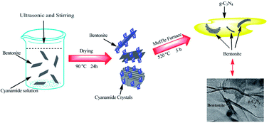
Novel bentonite/g-C3N4 composite photocatalysts were synthesized through a conventional calcination method and systematically characterized by thermogravimetric analysis (TG), powder X-ray diffraction (XRD), transmission electron microscopy (TEM), Fourier transform infrared spectroscopy (FT-IR), UV-vis diffuse reflection spectroscopy (DRS), X-ray photoelectron spectroscopy (XPS), photoluminescence (PL) and photocurrent–time measurement (PT). The optimal sample bentonite/g-C3N4 (5.4%) composite shows an enhanced photocurrent value (about 8 times as high as that of g-C3N4) under visible light irradiation and high efficiency for the degradation of methylene blue (the photoreaction kinetics constant value is about 2.5 times that of g-C3N4) under visible light. Results show the improved photoactivity is mainly attributed to the electrostatic interaction between g-C3N4 and negatively charged bentonite, this leads to the efficient migration of the photogenerated electrons and holes of g-C3N4. This study reports an inexpensive and environmentally friendly photocatalyst for pollution degradation and prospective photoelectric materials.
1 Introduction
Due to the incoming energy and environmental problems, photocatalysis is an attractive but very challenging process to convert solar energy into chemical energy. One of the central tasks to be solved before its practical application is to develop highly efficient, sustainable, visible-light-responsive photocatalytic materials.
Recently, Wang et al. reported that a metal-free polymeric photocatalyst, graphitic carbon nitride (g-C3N4), showed a good photocatalytic performance for hydrogen or oxygen production via water splitting under visible light irradiation.1 The metal free g-C3N4 photocatalyst possesses very high thermal and chemical stability as well as interesting electronic properties, which make it valuable for photocatalytic applications. However, the photocatalytic efficiency of bare g-C3N4 is limited due to the high recombination rate of photogenerated electron hole pairs.2,3 To resolve this problem, efforts have been made to improve the photocatalytic performance of g-C3N4, for example, by controlling shape and size,4,5 introducing heteroatoms (e.g. Au,6 Ag,7 Zn,8 B–F,9 S,10 P2 and CNT11) and coupling with other semiconductors (e.g. ZnO,12 WO3,13,14 BiPO4,15 YVO4,16 BiOBr,17 AgX18 and TaON19). Therefore, it is significant to search for efficient materials to modify g-C3N4.
Being cheap, abundant, non-toxic and environmental friendly, bentonite clay represents an attractive substrate for immobilization of variety of photocatalysts. The basic structural unit of bentonite is composed of two tetrahedrally coordinated sheets of silicon ions surrounding a sandwiched and octahedrally coordinated sheet of aluminum ions. The isomorphous substitution of Al3+ for Si4+ in the tetrahedral layer and Mg2+ or Zn2+ for Al3+ in the octahedral layer results in a net negative charge on the clay surface.20,21 Compared with other clays, it has excellent adsorption properties and possesses sorption sites available within its interlayer space as well as on the outer surface and edges.20 The above factors make bentonite a good adsorbent for the removal of dye in aqueous solutions.22–24 Furthermore, studies have confirmed that bentonite is a good matrix for synthesizing composite photocatalysts with higher photocatalytic activity, such as ZnO/bentonite,25 TiO2/bentonite,26 MoS2/bentonite,27 BiVO4/bentonite,28 etc. These composites all showed more reactivities towards the degradation of organic pollutants due to proper dispersion of nanosized photocatalyst in a solid supports thus generating distinct reaction sites. Moreover, all as-prepared bentonite-composites show much higher SBET values and improved adsorption capacity, which led to better photocatalytic efficiency compared to bare nanoparticles and less amount of photocatalyst required for the degradation of organic pollutants. As a result, combining semiconductor photocatalysts with bentonite may increase the photocatalytic efficiency by extending the visible light absorption and increasing the charge separation. However, the photocatalytic effect of bentonite combining with g-C3N4 has not been discussed, and the photocatalytic mechanism needed to be further investigated.
It is well known that both g-C3N4 and bentonite clay are layered materials,29,30 so the interface of the two layered compounds may be tight, which allow the prompt migration of light-induced charge, thus resulting in an efficient photocatalytic reaction.31 Further more, the composite structure of bentonite/g-C3N4 with a large surface area can be prepared readily thus generating more distinct reactions,25 which is beneficial to the increasing of photocatalytic activity. Remarkably, both bentonite and g-C3N4 do not contain heavy metal ion and all elements are rich in the soil. Above factors are favorable for bentonite/g-C3N4 as a potential “green” photocatalyst in photodegradation pollutants application.
Herein, a facile calcination route was proposed to synthesize the novel visible-light-responsive bentonite/g-C3N4 composite. In this work the combining process of bentonite with g-C3N4 was studied. The photodegradation experiments were performed on methylene blue (MB) and the possible mechanisms of enhancement of photocatalytic activity were also investigated.
2 Experimental
2.1 Preparation of photocatalysts
All starting regents were purchased from sinopharm company sources and used without further purification. Distilled water was used throughout.
Fig. 1 shows schematic illustration of the fabrication of bentonite/g-C3N4 composites. Synthesis of bentonite/g-C3N4: first, in order to obtain well dispersed bentonite sheets, three different amounts of bentonite (0.05 g, 0.10 g, 0.15 g) was dispersed in 50 mL distilled water with 30 minutes continuous ultrasonic at 80 °C. Subsequently 3 g dicyanamide was added to the bentonite solutions respectively with stirring at 25 °C overnight. Second, the mixture solution was dried at 90 °C for 24 h, during this process part of the bentonite sheets maybe aggregate and accompany with the precipitation of dicyanamide crystals. The resulting dried mixtures were heated at a rate of 20 °C min−1 to reach a temperature to 520 °C and kept at this temperature for another 5 h in a muffle furnace with a semiclosed system. The obtained products are grayish yellow, which were obtained from three reactant systems (0.05 g bentonite + 3 g dicyanamide, 0.10 g bentonite + 3 g dicyanamide, 0.15 g bentonite + 3 g dicyanamide). According to thermogravimetric analysis (TG) results, the weight contents of bentonite in bentonite/g-C3N4 composites were estimated to be 2.2%, 5.4% and 13.2%, respectively. As a result, the bentonite/g-C3N4 composites were named as bentonite/g-C3N4 (2.2%), bentonite/g-C3N4 (5.4%) and bentonite/g-C3N4 (13.2%).
 |
| | Fig. 1 Schematic illustration of the fabrication of bentonite/g-C3N4 composites. | |
Bulk g-C3N4 was prepared as follows: 5 g dicyanamide was directly heated to 520 °C at a rate of 20 °C min−1 and then kept at this temperature for another 4 h.
Based on the TG result of bentonite/g-C3N4 (5.4%) composite, bentonite and g-C3N4 mechanical mixture were prepared by mixing bentonite (0.1 g) with g-C3N4 (1.76 g), which was denoted as bentonite/g-C3N4 mixture.
2.2 Characterization of photocatalysts
The crystalline phases of bentonite/g-C3N4 composites were analyzed by XRD using Bruker D8 diffractometer with Cu Kα radiation (λ = 1.5418 Å) within the range of 2θ = 5–80°. The morphologies and structure of the obtained samples were examined by TEM (H-600-II, Hitachi). FT-IR spectra of samples were recorded on a Nicolet Avatar-370 spectrometer at room temperature. Ultraviolet visible (UV-vis) diffuse reflection spectra were measured using a UV-vis spectrophotometer (Shimadzu UV-2450, Japan) in the range of 200 nm to 800 nm. BaSO4 was used as the reflectance standard material. XPS analysis was performed on an ESCA Lab MKII X-ray photo-electron spectrometer using the Mg Kα radiation. TG analysis was done on STA-449C Jupiter (NETZSCH Corporation, Germany). The experiment temperature ranged from 25 °C to 790 °C at a constant heating rate of 10 °C min−1 under air condition. The nitrogen adsorption–desorption isotherms at 77 K were investigated using a TriStar II 3020 surface area and porosity analyzer (Micromeritics Instrument Corporation, USA). PL spectra of the catalyst were measured on the QuantaMaster & TimeMaster Spectrofluorometer, QuantaMaster™ 40 (Photon Technology International, Inc.). The samples were excited at wavelength of 325 nm to measure the emission spectrum.
2.3 Photocurrent–time measurement
Photocurrent measurements were performed on an electro-chemical analyzer (CHI660D, CHI Shanghai, Inc.) in a standard three-electrode configuration with a Pt wire as the counter electrode and Ag/AgCl (in saturated KCl) as a reference electrode. Irradiation proceeded with a Xe arc lamp. Na2SO4 (0.1 M) aqueous solution was used as the electrolyte. The typical working electrode was prepared as follows: the 5 mg ground sample was mixed with 1 mL distilled water solution to make slurry. The slurry was then dispersed onto an ITO glass electrode with 1 cm × 1 cm area and the electrode was dried at 50 °C for 8 h.
2.4 Photocatalytic activity
The photocatalytic activity of the samples was evaluated by the degradation of MB dye under visible light irradiation. An aqueous solution of MB (100 mL, 20 mg L−1) was placed in a glass, and 100 mg photocatalyst was added. Photocatalytic activity of the sample was evaluated under a 300 W Xe lamp with a 400 nm cutoff filter. Prior to irradiation, the suspensions were magnetically stirred in the dark for about 60 min to ensure the establishment of an adsorption–desorption equilibrium between the photocatalysts and MB dye. At certain time intervals, 3 mL aliquots were sampled and centrifuged to remove the photocatalyst particles. Then the filtrates were analyzed by recording variations of the absorption band maximum (663 nm) in the UV-vis spectra of MB by using an UV-vis spectrophotometer (UV-2450 Shimadzu). The photocatalytic degradation efficiency was obtained by the concentration decrease of MB.
3 Results and discussion
3.1 XRD analysis
Fig. 2 shows the powder XRD patterns of bentonite, g-C3N4 and various bentonite/g-C3N4 composites. As can be seen from Fig. 2A, for g-C3N4, two pronounced peaks can be indexed to (100) and (002) diffraction planes of the graphite-like carbon nitride, this corresponds to the interlayer stacking and the characteristic interlayer stacking peak of aromatic systems, respectively.1,32 For the bentonite clay, the peaks marked by (M) are the characteristic of the montmorillonite in the bentonite sample. The other peaks correspond to the impurities of feldspar, quartz and Fe(Ca)CO3.33 In the case of bentonite/g-C3N4 composites, crystalline bentonite is not obviously observed in the bentonite/g-C3N4 photocatalysts with low bentonite loadings. However, in the enlarged Fig. 2B from 25° to 30°, the as-prepared bentonite/g-C3N4 composites with higher bentonite loadings (>5.4%) exhibited a crystalline bentonite peak at 26.66°, the peak intensity increased with the increase of bentonite loading. This result suggested that the bentonite/g-C3N4 composites are constituted by g-C3N4 and bentonite.
 |
| | Fig. 2 (A) XRD pattern of bentonite, g-C3N4 and bentonite/g-C3N4 composite, (B) enlarged XRD pattern of bentonite, g-C3N4 and bentonite/g-C3N4 composites from 25° to 30°. | |
3.2 BET analysis
In order to investigate the surface property changing of the bentonite/g-C3N4 composites after introducing bentonite into g-C3N4, the SBET dates were obtained. The SBET of the bentonite/g-C3N4 (2.2%), bentonite/g-C3N4 (5.4%) and bentonite/g-C3N4 (13.2%) composites are 18.3 m2 g−1, 19.6 m2 g−1 and 24.1 m2 g−1, respectively, which are much higher than that of pure g-C3N4 (5.9 m2 g−1). In view of the large surface area bentonite, the SBET increases with the increasing weight content of bentonite. Similarly, in BiVO4/bentonite28 and TiO2/bentonite34 system, after BiVO4 and TiO2 were loaded on the bentonite clay, increased SBET of the composites were obtained. This fact implies that the introduction of bentonite is favorable for the formation of high specific surface areas bentonite/g-C3N4 composite.
3.3 TEM analysis
Fig. 3 shows the size and morphology of g-C3N4, bentonite and bentonite/g-C3N4 composites. It can be seen that g-C3N4 sample has thick layered structures (Fig. 3A) and bentonite also has sheet like morphology (Fig. 3B). In contrast to g-C3N4, it can be clearly observed that bentonite/g-C3N4 (2.2%) and (5.4%) composites are composed of thin and curved layered structure (Fig. 3C–F). In the enlarged TEM (Fig. 3D), the bentonite layers can be observed clearly, suggesting bentonite layers combining well with g-C3N4 layers. After introducing more bentonite into g-C3N4, the bentonite/g-C3N4 (13.2%) composite can be observed with abundant fluffy sheets (Fig. 3G and H) and parts of the sample shows dark grey color, which may be due to some aggregating products. The fluffy sheets are in accordance with the enlarged surface area in BET analysis.
 |
| | Fig. 3 TEM images of (A) g-C3N4, (B) bentonite, (C and D) bentonite/g-C3N4 (2.2%) composite, (E and F) bentonite/g-C3N4 (5.4%) composite and (G and H) bentonite/g-C3N4 (13.2%) composite. | |
3.4 TG analysis
Fig. 4 shows the TG curves of the bentonite/g-C3N4 composites. It can be observed that bentonite shows slight weight loss from 25 °C to 790 °C, which is associated with residual water and dehydroxylation of the aluminosilicate.35 As for bentonite/g-C3N4 composites, they have no obvious difference in the thermal stability. The initial decomposition temperature of bentonite/g-C3N4 composites is about at 530 °C, which is lower than that of g-C3N4 at 550 °C. The reason may be attributed to the existence of fluffy sheets of g-C3N4 in the composites (Fig. 3C–H). However, the complete decomposition temperature of bentonite/g-C3N4 composites is about at 730 °C, which is higher than that of g-C3N4 at 670 °C. The reason is due to the presence of bentonite nanolayers, which can act as barriers maximizing the heat insulation.36,37
 |
| | Fig. 4 TG curves for heating bentonite, g-C3N4 and bentonite/g-C3N4 composites from 25 °C to 790 °C at a heating rate of 10 °C min−1. | |
3.5 XPS analysis
XPS measurements were performed to determine the valence states of various species. Fig. 5A shows the survey scan XPS spectra of g-C3N4 and the bentonite/g-C3N4 (5.4%) composite. The result indicates the presence of Al, Si, C, N and O in the bentonite/g-C3N4 (5.4%) composite. High resolution spectra of C 1s and N 1s are shown in Fig. 5B and C. The C 1s peak positions in g-C3N4 are at 288.1 eV and 284.5 eV, respectively. The binding energy at 288.1 eV is assigned to a C–N–C coordination.38 The other C 1s peak at 284.5 eV is attributed to the adventitious carbon on the surface of g-C3N4.39 Fig. 5C shows the N 1s binding energy of g-C3N4 at 398.7 eV, which is assigned to sp2-hybridized nitrogen (C
N–C).38,40 In contrast to g-C3N4, the C 1s and N 1s binding energies of bentonite/g-C3N4 (5.4%) composite show minor negative shifts to 288.0 eV and 398.6 eV, respectively. This result suggests that there exists some interaction between bentonite and g-C3N4. Therefore, with the combination of the XRD, TEM and XPS investigation, the results confirmed that there were both bentonite and g-C3N4 species in the composite.
 |
| | Fig. 5 XPS spectra of g-C3N4 and bentonite/g-C3N4 (5.4%) composite: (A) survey spectrum, (B) C 1s and (C) N 1s. | |
3.6 FT-IR analysis
Fig. 6 shows the FT-IR spectra of bentonite, g-C3N4, and bentonite/g-C3N4 composites. As can be seen in Fig. 6, several bands in the 1200–1650 cm−1 regions (1241, 1322, 1405, 1561 and 1642 cm−1) are associated with the typical stretching modes of CN heterocycles.41 The peaks at 1642 cm−1 and 1561 cm−1 belonged to the C
N stretching vibrations and s-triazine ring modes,42,43 while the peaks at 1241 cm−1, 1322 cm−1 and 1405 cm−1 to the aromatic C–N strechings.3,43 For bentonite sample, the band at 794 cm−1 confirms the presence of quartz in the sample. The band at 691 cm−1 corresponds to the deformation modes of the Si–O bond.33 The bands at 522 cm−1 and 1044 cm−1 correspond to Al–O–Si bending vibrations and Si–O bending vibrations,33,44 respectively. In the case of bentonite/g-C3N4 composites, the bands of g-C3N4 can be observed clearly, whereas the typical bands of bentonite can not be obviously observed, which is attributed to the low content of bentonite and existence of g-C3N4.
 |
| | Fig. 6 FT-IR spectra of bentonite, g-C3N4, and bentonite/g-C3N4 composites. | |
3.7 DRS analysis
Fig. 7 shows the UV-vis DRS spectra of pure g-C3N4 and bentonite/g-C3N4 composites. In Fig. 7A, the pure g-C3N4 has an absorption edge at about 460 nm, which can be assigned to the intrinsic band gap of g-C3N4 (2.70 eV).1 The absorption edges of bentonite/g-C3N4 composites shift significantly to longer wavelengths compared with the pure g-C3N4, indicating that there may be some interaction between bentonite and g-C3N4. In previous BET and TEM analysis, the bentonite/g-C3N4 composites show enlarged surface area and much fluffy layer structure. In view of the enlarged surface area and the partly imperfect polymer-like structure of g-C3N4,45 the g-C3N4 in the as-obtained bentonite/g-C3N4 composites might have more defects compared with the pure g-C3N4, which leads to the enhanced light harvesting in the visible light range.46,47
 |
| | Fig. 7 (A) UV-vis spectra of g-C3N4 and bentonite/g-C3N4 samples, (B) plot of (αhν)1/2 versus energy (hν) for the band gap energy of g-C3N4 and bentonite/g-C3N4 composites. | |
The band gap energy (Eg) can be estimated from the intercept of tangents to the plots of (αhν)1/2 vs. photon energy. As shown in Fig. 7B, the calculated Eg value of pure g-C3N4 is 2.70 eV, in the case of bentonite/g-C3N4 composites, the Eg values of bentonite/g-C3N4 (2.2%, 5.4%, 13.2%) composites are 2.63 eV, 2.59 eV and 2.55 eV respectively, which are lower than that of pure g-C3N4. Therefore, the visible light responses of bentonite/g-C3N4 composites are significantly improved.
3.8 Photocatalytic activity
Fig. 8A shows the photocatalytic activity of g-C3N4 and bentonite/g-C3N4 samples for the degradation of MB (20 mg L−1) under visible light. When there is no photocatalyst involved with visible light irradiation, the organic contaminant MB is very low, indicating that MB self-degradation was negligible. In the presence of various bentonite/g-C3N4 composites the MB undergoes pronounced decomposition under visible light irradiation. Significantly, the bentonite/g-C3N4 (5.4%) induced 90% MB degradation within 3 h under visible light irradiation, which is much higher than that of g-C3N4 (62%) and bentonite/g-C3N4 mixture (66%). Fig. 8B shows the corresponding first-order kinetics plot by the equation of ln(C0/C) = kt, where C0 and C are the MB concentrations in solution at times 0 and t, respectively, and k is the apparent first-order rate constant. It can be seen from Fig. 8B, the k values of pure g-C3N4, bentonite/g-C3N4 mixture and bentonite/g-C3N4 (2.2%, 5.4%, 13.2%) composites were 0.30 h−1, 0.34 h−1, 0.39 h−1, 0.74 h−1 and 0.67 h−1 respectively. In the bentonite/g-C3N4 composite system, the rate constant of the bentonite/g-C3N4 (5.4%) photocatalyst is up to 2.5 times as high as that of the pure g-C3N4. However, further increasing the proportion of bentonite, the degradation rate shows a slight decrease though it remains higher than that of g-C3N4. The decrease in the activity of samples with a heavy loading of bentonite is likely due to the shading effect, which can seriously block the absorption of the incident light by g-C3N4, In MoS2/mpg-CN system, the excess doping of MoS2 led to the agglomeration of MoS2 nanoparticles on the surface of mpg-CN, which could decrease the catalytic activity.31 Therefore, excess loading of bentonite could prevent g-C3N4 from absorbing the visible light and then lead to a deceasing of photocatalytic activity.
 |
| | Fig. 8 (A) Photocatalytic degradation efficiency of MB by g-C3N4 and bentonite/g-C3N4 composites, (B) kinetic fit for the degradation of MB with g-C3N4 and bentonite/g-C3N4 composites, (C) adsorption activity and rate constants of (a) g-C3N4, (b) bentonite/g-C3N4 mixture, (c) bentonite/g-C3N4 (2.2%), (d) bentonite/g-C3N4 (5.4%) and (e) bentonite/g-C3N4 (13.2%) composite, (D) absorption spectral changes of MB under visible light: bentonite/g-C3N4 (5.4%) as photocatalyst. | |
 |
| | Fig. 9 Photoluminescence (PL) spectra of g-C3N4 and bentonite/g-C3N4 composites. | |
Because adsorption plays an important role in most photocatalytic reactions, the comparison of the adsorption capacity and rate constants of all catalysts during the degradation process was investigated (Fig. 8C). Compared with pure g-C3N4, bentonite/g-C3N4 mixture has the superior adsorption capacity; almost 56% of MB molecules were adsorbed on the mixture sample, which is much higher than that of g-C3N4 (20%). However, rate constant of the bentonite/g-C3N4 mixture (0.34 h−1) only increased a little compared with that of g-C3N4 (0.30 h−1).
The adsorption capacity of bentonite/g-C3N4 composites is not superior to that of bentonite/g-C3N4 mixture, whereas the rate constants are much higher than that of bentonite/g-C3N4 mixture. Remarkably, bentonite/g-C3N4 (5.4%) composite shows the optimal rate constant (0.74 h−1), which is around 2.2 times that of the bentonite/g-C3N4 mixture (0.34 h−1). From these data, it is obvious that the high degradation activity of bentonite/g-C3N4 composites is not mainly attributed to the adsorption capability any more. The enhanced photocatalytic activity can be ascribed to the significant differences in the interface of the samples, as the hybrid composites show a closely contacted interface (shown in Fig. 3), whereas only a diffuse interface can be formed after a simple mechanically mixing. Fig. 8D shows temporal evolution of the spectral changes during photocatalytic degradation of MB over bentonite/g-C3N4 (5.4%) composite. It can be seen that with the increase of irradiation time in bentonite/g-C3N4 (5.4%) composite aqueous suspension, the intensity of the absorption peak at 663 nm decreased drastically within 3 h. The bentonite/g-C3N4 (5.4%) composite shows high photocatalytic activity under the experimental condition.
3.9 PL analysis
In the previous studies, PL analysis was used to reveal the efficiency of charge carrier trapping, transfer, and separation and to investigate the fate of photogenerated electrons and holes in semiconductors, because the PL emission results from the recombination of free charge carriers.48,49 Herein, we present the PL measurement for pure g-C3N4 and the bentonite/g-C3N4 composites. As can be seen in Fig. 9, for the pure g-C3N4, one main emission peak appears at about 460 nm, which is attributed to the inner band gap of g-C3N4 (2.70 eV).1 After introducing bentonite into g-C3N4, the intensities of the PL signal for the bentonite/g-C3N4 composites are lower than that of g-C3N4, indicating that the composites have a lower recombination rate of electrons and holes under visible-light irradiation.
 |
| | Fig. 10 Photocurrent profiles of bentonite, g-C3N4 and bentonite/g-C3N4 (5.4%) composite. | |
3.10 Photocurrent–time analysis
The transient photocurrent responses of g-C3N4 and g-C3N4/bentonite (5.4%) composite electrodes were recorded under visible light irradiation. Fig. 10 shows the I–t curves for the aforementioned samples with two on–off cycles of intermittent irradiation. The photocurrent value of bentonite is very small and can be ignored. The photocurrent value of g-C3N4 and g-C3N4/bentonite (5.4%) composite rapidly decreases to zero as soon as the irradiation of light turned off and the photocurrent come back to a constant value when the light is on again, which was reproducible. Significantly, the photocurrent of the g-C3N4/bentonite (5.4%) electrode was about 8 times as high as that of the pure g-C3N4 electrode. The enhancement of photocurrent of g-C3N4/bentonite (5.4%) composite indicated there was not only a strong interaction between bentonite and g-C3N4 but also more efficient carrier separation at the g-C3N4 and bentonite interfaces.12,48
 |
| | Fig. 11 Proposed mechanism for the photodegradation of MB on bentonite/g-C3N4 (5.4%) composite. | |
3.11 Mechanism of enhancement of photocatalytic activity
As discussed above, four main reasons for the increase in the photocatalytic efficiency of coupled g-C3N4/bentonite composites are: (i) the absorption edges of bentonite/g-C3N4 composites shift significantly to longer wavelengths compared with the pure g-C3N4, which indicates the composites can be excited by more visible light photons; (ii) the enlarged BET surface of bentonite/g-C3N4 composites, the increased surface area offers more surface active sites for adsorption and photocatalytic reaction;50,51 (iii) the improved adsorptive activity of bentonite/g-C3N4 composites compared with that of g-C3N4, the improved adsorptivity for the MB molecules could accelerate the photodegradation reaction;51,52 (iv) the electrostatic interaction, the negatively charged bentonite can promote the immigration of electrons and holes, thus suppresses the charge recombination. The mechanism for the enhanced photocatalytic activity is illustrated in Fig. 11. As can be seen in Fig. 11, although bentonite has an overall neutral charge, it has excess negative charges on its crystal lattice due to the isomorphous substitution. This kind of negative charges is compensated for by exchangeable cations.23 When bentonite is placed in water, the exchangeable cations, such as sodium ions, diffuse away from the solid surface, leaving the bentonite surface with negative charges.23,53 Considering the fact that bentonite is known as a very good electrical insulator,54 bentonite can not be excited and only g-C3N4 can be activated, the electron–holes of g-C3N4 have no opportunity to migrate to bentonite and should still present on g-C3N4 surface. The excited electrons and holes of g-C3N4 should be driven to migrate efficiently due to electrostatic interaction between g-C3N4 and the negatively charged bentonite. Thus, the charge recombination could be easily suppressed, leaving more charge carriers and enhancing the photocatalytic activity. Similarly, in other insulator–semiconductor system, such as h-BN/TiO2, owing to the electrostatic interaction, the negatively charged h-BN can promote the immigration of h+ from the bulk of TiO2 to its surface and consequently improves the photocatalytic activity.55 Driven by the calcinations process, the layers of bentonite and g-C3N4 are splitted into many fluffy and thin sheets and a tight contact of bentonite with g-C3N4 can be achieved (shown in Fig. 3), which can strengthen the electrostatic interaction between them.
Therefore, the high photoactivity of bentonite/g-C3N4 composite arises from the synergistic effects between bentonite and g-C3N4 interfaces. Further study is necessary to clarify the mechanism and the synthesis route can be applied to other bentonite/semiconductor photocatalysts.
4 Conclusions
A novel bentonite/g-C3N4 composite was successfully synthesized via a calcination method. XPS, DRS, PL and photocurrent–time analyses reveal that the obtained bentonite/g-C3N4 composites are a hybridization photocatalyst. Bentonite/g-C3N4 composites exhibited higher photocatalytic activity than the pure g-C3N4 in the degradation of MB (20 mg L−1). The bentonite/g-C3N4 (5.4%) composite induced 90% MB degradation within 3 h under visible light irradiation, which is much higher than that of pure g-C3N4 (62%). The significant enhancement in the photocatalytic performance of bentonite/g-C3N4 composites is ascribed not only to its high surface area, adsorptivity and enhanced absorptivity in visible light but also to the electrostatic interaction between g-C3N4 and negatively charged bentonite, this can promote the efficient migration of the photogenerated electrons and holes of g-C3N4 and consequently improves the photocatalytic activity.
Acknowledgements
The authors appreciate the financial support of this work from the National Nature Science Foundation of China (no. 21177050, 21007021, 21076099 and 21175061) Society Development Fund of Zhenjiang (SH2012020, SH2011011), Postdoctoral Foundation of China (2012M521014) and Foundation of Jiangsu University (04JDG048).
References
- X. Wang, K. Maeda, A. Thomas, K. Takanabe, G. Xin, J. M. Carlsson, K. Domen and M. Antonietti, Nat. Mater., 2009, 8, 76–80 CrossRef CAS PubMed.
- Y. Zhang, T. Mori, J. Ye and M. Antonietti, J. Am. Chem. Soc., 2010, 132, 6294–6295 CrossRef CAS PubMed.
- J. X. Sun, Y. P. Yuan, L. G. Qiu, X. Jiang, A. J. Xie, Y. H. Shen and J. F. Zhu, Dalton Trans., 2012, 41, 6756–6763 RSC.
- X. Bai, L. Wang, R. Zong and Y. Zhu, J. Phys. Chem. C, 2013, 117, 9952–9961 CAS.
- F. Goettmann, A. Fischer, M. Antonietti and A. Thomas, Angew. Chem., Int. Ed., 2006, 45, 4467–4471 CrossRef CAS PubMed.
- J. A. Singh, S. H. Overbury, N. J. Dudney, M. Li and G. M. Veith, ACS Catal., 2012, 2, 1138–1146 CrossRef CAS.
- L. Ge, C. Han, J. Liu and Y. Li, Appl. Catal., A, 2011, 409–410, 215–222 CrossRef CAS PubMed.
- B. Yue, Q. Y. Li, H. Iwai, T. Kako and J. H. Ye, Sci. Technol. Adv. Mater., 2011, 12, 034401 CrossRef.
- Y. Wang, J. Zhang, X. Wang, M. Antonietti and H. Li, Angew. Chem., Int. Ed., 2010, 49, 3356–3359 CrossRef CAS PubMed.
- J. S. Zhang, J. H. Sun, K. Maeda, K. Domen, P. Liu, M. Antonietti, X. Z. Fu and X. C. Wang, Energy Environ. Sci., 2011, 4, 675–678 CAS.
- Y. Xu, H. Xu, L. Wang, J. Yan, H. Li, Y. Song, L. Huang and G. Cai, Dalton Trans., 2013, 42, 7604–7613 RSC.
- Y. J. Wang, R. Shi, J. Lin and Y. F. Zhu, Energy Environ. Sci., 2011, 4, 2922–2929 CAS.
- L. Y. Huang, H. Xu, Y. P. Li, H. M. Li, X. N. Cheng, J. X. Xia, Y. G. Xu and G. B. Cai, Dalton Trans., 2013, 42, 8606–8616 RSC.
- Y. Zang, L. Li, Y. Zuo, H. Lin, G. Li and X. Guan, RSC Adv., 2013, 3, 13646–13650 RSC.
- C. S. Pan, J. Xu, Y. J. Wang, D. Li and Y. F. Zhu, Adv. Funct. Mater., 2012, 22, 1518–1524 CrossRef CAS.
- J. Cai, Y. He, X. Wang, L. Zhang, L. Dong, H. Lin, L. Zhao, X. Yi, W. Weng and H. Wan, RSC Adv., 2013, 3, 20862–20868 RSC.
- J. Di, J. Xia, S. Yin, H. Xu, M. He, H. Li, L. Xu and Y. Jiang, RSC Adv., 2013, 3, 19624–19631 RSC.
- H. Xu, J. Yan, Y. Xu, Y. Song, H. Li, J. Xia, C. Huang and H. Wan, Appl. Catal., B, 2013, 129, 182–193 CrossRef CAS PubMed.
- S. C. Yan, S. B. Lv, Z. S. Li and Z. G. Zou, Dalton Trans., 2010, 39, 1488–1491 RSC.
- E. Eren and B. Afsin, J. Hazard. Mater., 2008, 151, 682–691 CrossRef CAS PubMed.
- P. F. Luckham and S. Rossi, Adv. Colloid Interface Sci., 1999, 82, 43–92 CrossRef CAS.
- A. S. Özcan, B. Erdem and A. Özcan, Colloids Surf., A, 2005, 266, 73–81 CrossRef PubMed.
- Q. H. Hu, S. Z. Qiao, F. Haghseresht, M. A. Wilson and G. Q. Lu, Ind. Eng. Chem. Res., 2005, 45, 733–738 CrossRef.
- A. Özcan, Ç. Ömeroğlu, Y. Erdoğan and A. S. Özcan, J. Hazard. Mater., 2007, 140, 173–179 CrossRef PubMed.
- S. Meshram, R. Limaye, S. Ghodke, S. Nigam, S. Sonawane and R. Chikate, Chem. Engin. J., 2011, 172, 1008–1015 CrossRef CAS PubMed.
- Z. Sun, Y. Chen, Q. Ke, Y. Yang and J. Yuan, J. Photochem. Photobiol., A, 2002, 149, 169–174 CrossRef CAS.
- K. H. Hu, D. F. Zhao and J. S. Liu, Trans. Nonferrous Met. Soc. China, 2012, 22, 2484–2490 CrossRef CAS.
- J. G. Qu, N. N. Li, B. J. Liu and J. X. He, Mater. Sci. Semicond. Process., 2013, 16, 99–105 CrossRef CAS PubMed.
- Y. Wang, X. Wang and M. Antonietti, Angew. Chem., Int. Ed., 2012, 51, 68–89 CrossRef CAS PubMed.
- P. C. Joshi, M. F. Aldersley, J. W. Delano and J. P. Ferris, J. Am. Chem. Soc., 2009, 131, 13369–13374 CrossRef CAS PubMed.
- Y. Hou, A. B. Laursen, J. Zhang, G. Zhang, Y. Zhu, X. Wang, S. Dahl and I. Chorkendorff, Angew. Chem., Int. Ed., 2013, 52, 3621–3625 CrossRef CAS PubMed.
- S. C. Yan, Z. S. Li and Z. G. Zou, Langmuir, 2009, 25, 10397–10401 CrossRef CAS PubMed.
- S. Yang, J. Li, Y. Lu, Y. Chen and X. Wang, Appl. Radiat. Isot., 2009, 67, 1600–1608 CrossRef CAS PubMed.
- J. Li, Suyoulema, W. Wang and Sarina, Solid State Sci., 2009, 11, 2037–2043 CrossRef CAS PubMed.
- F. G. Ramos Filho, T. J. A. Mélo, M. S. Rabello and S. M. L. Silva, Polym. Degrad. Stab., 2005, 89, 383–392 CrossRef CAS PubMed.
- A. A. Sapalidis, F. K. Katsaros, T. A. Steriotis and N. K. Kanellopoulos, J. Appl. Polym. Sci., 2012, 123, 1812–1821 CrossRef CAS.
- K. E. Strawhecker and E. Manias, Chem. Mater., 2000, 12, 2943–2949 CrossRef CAS.
- S. C. Yan, Z. S. Li and Z. G. Zou, Langmuir, 2010, 26, 3894–3901 CrossRef CAS PubMed.
- P. Niu, G. Liu and H. M. Cheng, J. Phys. Chem. C, 2012, 116, 11013–11018 CAS.
- X. F. Li, J. Zhang, L. H. Shen, Y. M. Ma, W. W. Lei, Q. L. Cui and G. T. Zou, Appl. Phys. A: Mater. Sci. Process., 2009, 94, 387–392 CrossRef CAS.
- F. Dong, L. Wu, Y. Sun, M. Fu, Z. Wu and S. C. Lee, J. Mater. Chem., 2011, 21, 15171–15174 RSC.
- Y. J. Wang, X. J. Bai, C. S. Pan, J. He and Y. F. Zhu, J. Mater. Chem., 2012, 22, 11568–11573 RSC.
- J. L. Zimmerman, R. Williams, V. N. Khabashesku and J. L. Margrave, Nano Lett., 2001, 1, 731–734 CrossRef CAS.
- H. Zaitan, D. Bianchi, O. Achak and T. Chafik, J. Hazard. Mater., 2008, 153, 852–859 CrossRef CAS PubMed.
- Y. Zhang, A. Thomas, M. Antonietti and X. Wang, J. Am. Chem. Soc., 2008, 131, 50–51 CrossRef PubMed.
- X. Wang, K. Maeda, X. Chen, K. Takanabe, K. Domen, Y. Hou, X. Fu and M. Antonietti, J. Am. Chem. Soc., 2009, 131, 1680–1681 CrossRef CAS PubMed.
- J. Sun, J. Zhang, M. Zhang, M. Antonietti, X. Fu and X. Wang, Nat. Commun., 2012, 1139 CrossRef.
- Q. Xiang, J. Yu and M. Jaroniec, J. Phys. Chem. C, 2011, 115, 7355–7363 CAS.
- Y. M. He, J. Cai, T. T. Li, Y. Wu, H. J. Lin, L. H. Zhao and M. F. Luo, Chem. Eng. J., 2013, 215, 721–730 CrossRef PubMed.
- Y. Zheng, J. Liu, J. Liang, M. Jaroniec and S. Z. Qiao, Energy Environ. Sci., 2012, 5, 6717–6731 CAS.
- J. Xu, Y. Wang and Y. Zhu, Langmuir, 2013, 29, 10566–10572 CrossRef CAS PubMed.
- H. Zhang, X. Lv, Y. Li, Y. Wang and J. Li, ACS Nano, 2009, 4, 380–386 CrossRef PubMed.
- M. G. Neumann, F. Gessner, C. C. Schmitt and R. Sartori, J. Colloid Interface Sci., 2002, 255, 254–259 CrossRef CAS.
- Y. Fujishiro, S. Uchida and T. Sato, Int. J. Inorg. Mater., 1999, 1, 67–72 CrossRef CAS.
- X. Fu, Y. Hu, Y. Yang, W. Liu and S. Chen, J. Hazard. Mater., 2013, 244–245, 102–110 CrossRef CAS PubMed.
|
| This journal is © The Royal Society of Chemistry 2014 |
Click here to see how this site uses Cookies. View our privacy policy here.