Bio-inspired green surface functionalization of PMMA for multifunctional capacitors

Vijay Kumar Thakura, Danny Vennerbergb, Samy A. Madboulybc and Michael R. Kessler*a
aSchool of Mechanical and Materials Engineering, Washington State University, Pullman, WA, USA. E-mail: MichaelR.Kessler@wsu.edu
bDepartment of Materials Science and Engineering, Iowa State University, Ames, IA, USA
cCairo University, Faculty of Science, Chemistry Department, Giza, Egypt

Received 12th November 2013 , Accepted 24th December 2013

First published on 3rd January 2014


Abstract

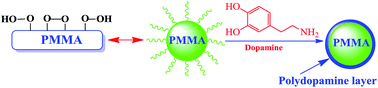
Poly(methyl methacrylate) (PMMA)-based dielectric materials are of prime interest for many electronic and power devices. However, the low dielectric constant and hydrophobic nature of PMMA limit its success in many applications. This work presents an environmentally friendly approach to surface-functionalize PMMA, exploring the use of dopamine (DOPA), to develop multifunctional polymers with enhanced dielectric properties. The reaction of dopamine with plasma pre-activated PMMA was carried out in an aqueous medium without using any toxic chemicals. The functionalized PMMA films exhibited enhanced dielectric properties compared to pristine PMMA films. As an example, below 100 Hz, the dielectric constant of functionalized PMMA films increased by 70% compared to pristine PMMA films. Interestingly, functionalization changed the storage modulus and the Tg of PMMA. The enhanced dielectric properties of the functionalized PMMA films may originate from the excellent intrinsic properties of DOPA, which was effectively functionalized on the surface of PMMA.


Introduction

Recently, dielectric materials have attracted considerable interest as high performance capacitors owing to their possible use in applications that efficiently store and control electrical energy in many modern electronics and electric systems.1–3 In particular, the continuing miniaturization in high energy density capacitors and with simultaneous increase in energy density sets ever higher requirements for advanced dielectric materials in terms of reliability, performance, and processing techniques.4–6 An ideal dielectric material should have a high dielectric constant and exhibit low dielectric loss and high breakdown strength in order to be considered for energy storage and other applications such as pulsed lasers, electromagnetic generation, and particle accelerators.7–11 Traditional ceramic-based dielectric materials exhibit high dielectric constants, but they suffer from high dielectric loss and low breakdown strength.12–15 Furthermore, it is difficult to process ceramics into desirable final products, especially in mobile electronic devices, hybrid electric vehicles, stationary power systems, and pulse power applications.16–19 Thus, there is a growing need for easily processable and low-cost dielectric materials for electronics and electrical power systems that can store sufficient amounts of energy and deliver it nearly instantaneously when required.20–22 Compared to traditional ceramics, polymers are easy to process and mechanically robust, with high breakdown strength and low dielectric loss.23–25 These unique characteristics of polymers encouraged the development of new dielectric polymers that are capable of storing high-density electric energy while exhibiting improved mechanical properties and thermal stability.26–28

Among various polymers, poly(methyl methacrylate) (PMMA) has been widely investigated for a variety of applications, including several in the electronics industry.28–30 PMMA exhibits excellent material properties such as exceptional mechanical strength, hardness, high rigidity, transparency, and good insulation properties.31,32 However, some inherent properties of PMMA, such as hydrophobicity and relatively low dielectric constant compared to ceramics/fluoropolymers and other materials, restrict its applications in high-energy density capacitors. The hydrophobic surface of PMMA prevents adhesion to other substances, limiting its spectrum of applications. Some efforts have been made recently to incorporate high dielectric constant materials, such as inorganic ceramic particles/CNTs/metal particles, into PMMA to obtain high energy density capacitors.18 However, the resulting composite materials still fell short of expectations because of the significant difference in permittivities of the two components. The inhomogeneity between the two phases also led to low energy density.18–21 Therefore, the development of new polymer systems with enhanced dielectric properties and with suitable surface-functionalization groups is of prime importance for various practical applications.

In this work, we demonstrate that bio-inspired aqueous surface functionalization of PMMA through dopamine can result in improved dielectric properties. A remarkable increase in dielectric constant (70%) was observed for the new, polydopamine-functionalized PMMA (PDOPA@PMMA) films at room temperature and 100 Hz. The present work offers a successful approach to increasing the dielectric permittivity while simultaneously modifying surface characteristics.

Experimental section

All chemicals in the present work were used as received from their vendors, without any additional purification. PMMA (MW = 996[thin space (1/6-em)]000 g mol−1, inherent viscosity = 1.25 dl g−1) and 3, 4-dihydroxy-L-phenylalanine were purchased from Sigma-Aldrich Corporation (St. Louis, MO). Tris base (molecular biology grade) was obtained from Fischer Scientific (Hampton, NH).

PMMA polymer powder samples were dried in an oven at 50 °C before plasma treatment at room temperature. Radio frequency argon glow discharge plasma was generated using an inductively coupled plasma chamber (Harrick Plasma, model PDC-001, Ithaca, NY) operating at 13.56 MHz and 29.6 W. PMMA samples were laid on a flat quartz plate inside the RF coil. The base pressure of the plasma reactor was 0.001 Torr, which was increased to 0.5 Torr after gas feeding, and the plasma treatment time was 300 seconds. After the plasma treatment, the treated samples were exposed to the atmosphere at room temperature (25 °C) for at least 30 minutes to facilitate the formation of surface peroxides and hydroperoxides for the subsequent chemical functionalization with dopamine (Scheme 1).33


image file: c3ra46592f-s1.tif
Scheme 1 (a) Schematic representations of the plasma modification process of PMMA. (b) Schematic illustration for functionalization of PMMA with dopamine.

Dopamine solution with an optimized concentration (2.0 g l−1) for the functionalization of PMMA was prepared by dissolving dopamine in distilled water.34–37 The pH (8.5) of the solution was adjusted with Tris–HCl (10 mM) buffer that was self-prepared in the laboratory. Prior to dopamine polymerization, the solution was ultrasonicated for 30 minutes, until the solution turned yellow in color. Subsequently, plasma-treated PMMA (500 mg) powder was charged in the reaction flask, and the polymerization reaction was performed at 60 °C for a defined, optimized time period of 24 hours under continuous stirring in a heating mantle fitted with a reflux condenser. During the polymerization reaction, the solution changed from yellow to dark brown in color. After completion of the reaction, the polydopamine-functionalized PMMA powder (PDOPA@PMMA) was washed several times by stirring in distilled water and collected by filtration. The PDOPA@PMMA was then dried to constant weight in a vacuum oven at 50 °C for subsequent characterization.

PMMA and PDOPA@PMMA samples were characterized using Fourier transform infrared spectroscopy (FTIR), Raman spectroscopy, X-ray diffractometry (XRD), and thermogravimetric analysis (TGA) (see corresponding descriptions in the ESI). Polymer films were prepared by dissolving pristine PMMA and PDOPA@PMMA powders in tetrahydrofuran (THF) at a concentration of 100 mg ml−1. The resulting solutions were stirred and simultaneously heated to 60 °C for six hours in order to obtain uniform mixing. Subsequently, the films were cast onto a silicon substrate and dried in air at room temperature for 6 hours and in vacuum at 50 °C for 12 hours to remove residual THF. Films of uniform thickness were used for further investigations. The dielectric properties of pristine PMMA, and PDOPA@PMMA were studied using a Novocontrol dielectric spectrometer with frequency sweeps from 1 Hz to 1 MHz. The dielectric constant and tan[thin space (1/6-em)]δ values of the samples were analyzed using Win Deta software. The dynamic mechanical behavior of PMMA and PDOPA@PMMA was characterized using test specimens with dimensions of 20 × 8 × 1 mm and a dynamic mechanical analyzer (DMA model Q800) from TA instruments (New Castle, DE, USA) in tensile mode. Temperature sweep tests were carried out between 30 and 200 °C at a frequency of 1 Hz, with a strain amplitude of 0.05% and at a heating rate of 3 °C min−1. The storage modulus (E′) and damping coefficient tan[thin space (1/6-em)]δ were measured as functions of temperature.

Results and discussion

Oxidative self-polymerization of dopamine results in the formation of thin layers on various types of materials, providing the advantages of a facile one-step surface functionalization.34–37 Poly(methyl methacrylate) (PMMA) is one of the hydrophobic polymer and its surface needs to be modified to obtain the desired surface properties for numerous applications.32 Various surface modification techniques such as corona treatments, plasma exposure, and ion beam, gamma-ray, electron beam, UV, and laser irradiation are frequently used to improve wettability, sealability, printability, and adhesion properties of polymers as well as to enhance polymer resistance to mechanical failure. Among these techniques, plasma modification offers distinct advantages, making plasma treatments an ideal means of modifying polymer surface characteristics.33 Plasma-induced modification provides a facile and fast method of endowing the surface of various polymers with a layer of highly enriched functional species as the intensity of plasma induced modification at the surface is stronger than that of more penetrating surface modification methods. Plasma-induced modifications of materials are generally limited to the surface layer to a depth of typically 0.005 to 0.05 μm, leaving the bulk properties of the polymer unaffected.33

In the present work, plasma-induced modification of polymethyl methacrylate (PMMA) followed by exposure to the air atmosphere leads to the formation of surface peroxide and hydroperoxide species on the PMMA surface that facilitates the further surface functionalization of PMMA by dopamine polymers.33 The procedure for the preparation of polydopamine surface modified poly(methyl methacrylate) (PDOPA@PMMA) is schematically illustrated in Scheme 1.

In contrast to the traditional chemical modification techniques frequently used for the surface modification of polymer surfaces, the present work employs an eco-friendly, green route to modify the surface of poly(methyl methacrylate) (PMMA) in aqueous medium using only dopamine and no harmful chemicals. To efficiently modify the surface of PMMA, we exploited the peroxide and hydroperoxide groups on the plasma-modified PMMA (Scheme 1a) and the aromatic structure of the dopamine biomolecule with amino and hydroxyl groups (Scheme 1b). The addition of plasma-modified PMMA polymer powder to an aqueous solution of dopamine facilitated the adhesion between the dopamine molecule and PMMA because of the inherent adhesive properties of the dopamine molecule34–37 (Scheme 1b). There may also be some possible bonding between the dopamine molecule and the plasma modified PMMA.37 Upon completion of the reaction, thin layers of polydopamine formed on the surface of the PMMA films through a simple deposition process and these layers can be further used for carrying out various secondary reactions.34–37 The conversion mechanism of dopamine into polydopamine involves the alkaline pH-induced oxidation of catechol to quinone that results in the formation of polydopamine (Scheme 2).34–37 As a result of oxidative self-polymerization of dopamine, thin layers of polydopamine are formed on the PMMA surface through a simple deposition process.


image file: c3ra46592f-s2.tif
Scheme 2 A possible mechanism of dopamine polymerization to polydopamine.34–37

Characterization of plasma induced functionalization of PMMA

Structural changes on the PMMA surface that occurred as a result of polydopamine functionalization were determined by FTIR, Raman, TGA, and XRD techniques: FTIR spectra of pristine PMMA and PDOPA@PMMA are shown in Fig. 1. The FTIR spectrum of pristine PMMA polymer has been found to exhibit the absorption bands at 2951 cm−1 and 1737 cm−1. These bands are caused by –CH3 asymmetric stretching and C[double bond, length as m-dash]O stretching, respectively. On the other hand, the peak at 1395 cm−1 has been attributed to OCH3 deformation of PMMA, while the vibrational bands at 1481 cm−1 and 1447 cm−1 belongs to CH2 scissoring and CH3 asymmetric stretching or deformation of PMMA. The characteristic absorption bands at 1275 cm−1 and 864 cm−1 respectively corresponds to C–O stretching and C–O–C stretching of the pristine PMMA polymer. The absorption bands at 1193, 953 and 753 cm−1 corresponds to CH2 twisting, wagging and rocking modes of PMMA, respectively.
image file: c3ra46592f-f1.tif
Fig. 1 FTIR spectra of pristine PMMA and PDOPA@PMMA.

In contrast to pristine PMMA, the spectrum of PDOPA@PMMA exhibits a new strong and broad OH stretching peak centered around 3400 cm−1caused by the intermolecular bonding at the surface of the modified PMMA. The intense absorption features in the PDOPA@PMMA between 3700–3100 cm−1 indicated N–H and O–H stretching vibrations. In the whole spectrum, the functional groups of dopamine that were strongly coupled with the PMMA surface dominated the IR spectrum of PDOPA@PMMA. In addition to the strong peak at 3400 cm−1, FTIR spectra of PDOPA@PMMA showed a pronounced peak, at 1618 cm−1 that was assigned to the overlap of C[double bond, length as m-dash]C resonance vibrations in the aromatic ring and N–H bending. The peaks in between 1250–1450 cm−1 were attributed to phenolic O–H deformations coupled or decoupled with C–C ring stretching and phenolic C–O stretching.34–37 The comparative study of the intensity of the IR spectra of the pristine PMMA and PDOPA@PMMA demonstrates that there was significant decrease in the intensity of the bands at 3027 cm−1 and 2982 cm−1, while the intensity of the band in between 3300–3600 cm−1 increased, strongly indicating the presence of OH groups. In addition to the FTIR results, Raman spectra (Fig. 2) also confirmed the successful surface functionalization of plasma-modified PMMA polymer powder by polydopamine. After functionalization, the spectrum of PDOPA@PMMA showed significant changes that specify the strong interactions between plasma modified PMMA and dopamine molecules. Raman spectra of pristine PMMA showed the characteristic band of PMMA at 2952 cm−1. This band indicates C–H stretching and is the most prominent in the PMMA structure. The bands at 1645 cm−1 can be attributed to the combination band arising from ν (C[double bond, length as m-dash]C) and ν (C–COO).38 The other Raman bands, such as those appearing at 604, 833, 995, 1264, 1470, 1739, and 3001 cm−1, were in conformity with literature.38 In comparison to pristine PMMA, Raman spectra of PDOPA@PMMA showed the presence of two prominent broad characteristic bands at 1345 cm−1 and 1575 cm−1. These bands can be attributed to the deformation of the catechol group in the polydopamine molecules.37 Along with these two prominent bands, few intrinsic peaks of PMMA were also observed. A comparison of the spectra of pristine PMMA and PDOPA@PMMA showed that the intensity of the PMMA bands decreased considerably as a result of polydopamine polymerization, which indicated the successful functionalization of polydopamine onto PMMA.


image file: c3ra46592f-f2.tif
Fig. 2 Raman spectra of pristine PMMA and PDOPA@PMMA.

The thermal degradation of pristine PMMA showed two stages of weight loss. The first stage of thermal degradation in PMMA was attributed to the scission of peroxide and hydroperoxide linkages, and the second weight loss was attributed to the scission at the unsaturated ends caused by termination by disproportionation.

Pure dopamine is thermally stable up to 220 °C, and did not show any significant change in weight loss. It is obvious from Fig. 3 that above 200 °C, dopamine showed two significant thermal degradations. The first degradation occurs between 227 °C and 300 °C reflecting the degradation of the catechol moiety. The second degradation occurs between 300 °C and 500 °C and is likely caused by the decomposition of the amide group and the alkyl spacer. The thermal degradation behavior of PDOPA@PMMA was similar to that of pristine dopamine. However, PDOPA@PMMA exhibited initial degradation at lower temperatures than dopamine and final degradation at higher temperatures than pristine PMMA. PDOPA@PMMA exhibited a steady weight loss between 290 and 442 °C which was attributed to the weight loss encountered during dopamine degradation. Based on thermal analysis calculations it can be concluded that there was 3–5 wt% of dopamine in the functionalized PMMA polymer powder, suggesting that the functionalized PDOPA@PMMA can be further used for numerous applications.


image file: c3ra46592f-f3.tif
Fig. 3 TGA of pristine PMMA, dopamine, and PDOPA@PMMA.

Fig. 4 depicts the XRD spectra of pristine PMMA and PDOPA@PMMA. Poly(methyl methacrylate) is amorphous in nature,29 therefore the diffraction pattern of PMMA shows the most intense peaks at 2θ values of 12.8° and 27.3°. A comparison of the spectra of PMMA and PDOPA@PMMA indicated subtle differences between the two polymers. It was observed that the intensity of the diffraction peaks in the pristine PMMA was significantly reduced as result of the polydopamine surface functionalization of PMMA. In addition, the peaks were broadened by the presence of amorphous polydopamine.


image file: c3ra46592f-f4.tif
Fig. 4 XRD curve of pristine PMMA and PDOPA@PMMA.

The changes in the surface morphology of the pristine PMMA after functionalization/deposition with polydopamine were also studied by SEM (shown in the ESI).

Dynamic mechanical analysis

Fig. 5a and b show the storage modulus (E′) and the loss tangent (tan[thin space (1/6-em)]δ) of pristine PMMA and PDOPA@PMMA films as a function of temperature, respectively. The DMA curves for both the PMMA and PDOPA@PMMA film samples were obtained from rectangular-shaped specimens subjected to a heating cycle with a rate of 3 °C min−1 at a frequency of 1 Hz. The pristine PMMA sample exhibited higher E′ values than the PDOPA@PMMA samples. The lower E′ values obtained for PDOPA@PMMA were attributed to the lower thermal stability of PDOPA@PMMA, as discussed in the preceding section.
image file: c3ra46592f-f5.tif
Fig. 5 (a) Storage modulus as a function of temperature for PMMA and PDOPA@PMMA films. (b) Loss factors as a function of temperature for PMMA and PDOPA@PMMA films.

As expected, both the pristine PMMA and the PDOPA@PMMA films experienced a decrease in storage modulus at elevated temperatures, although the decrease was not significant. The tan[thin space (1/6-em)]δ curves of both the pristine PMMA and the PDOPA@PMMA film samples were used to determine their respective glass-transition temperatures (Tg). The Tg for PDOPA@PMMA was lower than that of the pristine PMMA. This decrease in Tg of PDOPA@PMMA was primarily attributed to the formation of a polydopamine layer on the surface of PMMA with flexible –OH and NH2 groups, resulting in an increase in segmental mobility of the PDOPA@PMMA chains.

Dielectric properties of pristine PMMA and PDOPA@PMMA

The increasing requirements for high performance electronic devices have resulted in intense research in polymer-based dielectric materials for energy storage because of their excellent properties, such as increased functionality, improved reliability, improved electrical performance, increased design flexibility, and reduced unit cost. However, often it is difficult to find a polymer system with suitable functionality and the desired dielectric properties. The dielectric properties of pristine PMMA and PDOPA@PMMA were investigated over a wide range of frequencies and at temperatures ranging from 30 to 100 °C. The dielectric constant, ε′, and dielectric loss, ε′′, measured at temperature (30 °C) as a function of frequency from 10−2 Hz to 1 MHz for the pristine PMMA and PDOPA@PMMA studied here are shown in Fig. 6a and b, respectively. Here, the electric field was applied perpendicular to the plane of the sample film.
image file: c3ra46592f-f6.tif
Fig. 6 (a) Dependence of dielectric constant of PMMA and PDOPA@PMMA on frequency at 30 °C. (b) Dependence of dielectric loss of PMMA and PDOPA@PMMA on frequency at 30 °C.

Fig. 6(a) shows that the dielectric constant of pristine PMMA (ε′ ≈ 3.4) varied slightly with increasing frequency, changing little even over a frequency range spanning eight orders of magnitude. On the other hand, PDOPA@PMMA exhibited higher dielectric properties compared to pristine PMMA films. Surface functionalization of PMMA by polydopamine increased the dielectric constant by 70% without changing the frequency dependency considerably. The increase in dielectric constant of PDOPA@PMMA compared to pristine PMMA was attributed to the inherent characteristics of polydopamine present on the surface of the PDOPA@PMMA films.

Polydopamine has a conjugated bridge structure with diol functionality combined with an amine moiety that can enhance the polarizability and thereby significantly increase the dielectric constant of PDOPA/PMMA films compared to the pristine PMMA films.37 This behavior was solely attributed to the incorporation of hydroxyl and amino groups into the PMMA, which ultimately enhanced the dielectric constant because of the large dipole and the high polarizability of the O–H and NH2 bonds.25,37 At the same time, the dielectric loss trend of the PMMA and PDOPA@PMMA were significantly different over the studied frequency range (Fig. 6b). The dielectric loss was higher for the polydopamine modified PMMA. The dielectric loss peak (for PMMA) and shoulder (for PDOPA@PMMA) observed at ca. 100 Hz is attributed to the β-relaxation process. The origin of this process is the partial rotation of –COOCH3 side groups around the C–C bonds.39 The increase in dielectric loss of PDOPA@PMMA is attributed to the increased mobility of the –OH and –NH2 side groups around the C–C bonds in the PMMA chains. An abrupt increase in the dielectric loss values is clearly observed at low frequencies for PDOPA@PMMA. This indicates the existence of space charge polarization and free charge motion or ionic conductivity within the material.

Fig. 7 and 8 shows the temperature-dependent ε′ and ε′′ traces for PMMA (Fig. 7a & b) and PDOPA@PMMA (Fig. 8a & b), respectively, as a function of frequency. The temperature for all measurements was elevated from 30 °C to 100 °C in 10 °C increments. For both the pristine PMMA and PDOPA@PMMA films, higher temperatures resulted in elevated ε′ and ε′′ values. For pristine PMMA, dielectric constant ε′ and dielectric loss ε′′ vary slightly with temperature. Pristine PMMA shows a minor increase in dielectric constant with increasing temperature, and it is most significant at low frequencies, as shown in Fig. 7a (e.g., at 1 MHz, ε′ increases by less than 0.5 with increase in the temperature from 20 °C to +100 °C, while at 100 Hz ε′ increases 1.5 times over the same temperature range). The elevation in dipole moments of the structural repeating units of the PMMA polymer with increasing temperature (Fig. 7a & b) has been found to attribute to the trifling increase in the dielectric properties of the PMMA.29


image file: c3ra46592f-f7.tif
Fig. 7 (a) Dependence of dielectric constant of pristine PMMA on temperature at different frequencies. (b) Dependence of dielectric loss of pristine PMMA on temperature at different frequencies.

image file: c3ra46592f-f8.tif
Fig. 8 (a) Dependence of the dielectric constant of PDOPA@PMMA on temperature at different frequencies. (b) Dependence of dielectric loss of PDOPA@PMMA on temperature at different frequencies.

Compared to pristine PMMA, the PDOPA@PMMA film samples exhibited a strong variation in the dielectric properties (ε′ and ε′′) with temperature (Fig. 8a & b). At 1 Hz, the dielectric constant increased more than 10 times, whereas at higher frequencies (1 MHz), ε′ increased only slightly as the temperature increased from 30 °C to 100 °C.

The reason of increasing the dielectric constant with temperature is the increase in molecular dynamics or Brownian motion of the polymeric chains. The increase in dielectric properties of PDOPA@PMMA with increasing temperature were attributed to the fact that the segmental mobility of the –OH and NH2 groups of the polydopamine functionalized on the PMMA surface facilitated the orientation of dipoles, resulting in an increase in dielectric properties.37

Conclusions

We successfully demonstrated the use of a green, aqueous route for the functionalization of PMMA using a bio-inspired protein, dopamine, as an effective way to alter the polymer surface characteristics for energy storage applications. Uniform functionalization of PMMA with polydopamine (PDOPA) was achieved in an aqueous solution through the spontaneous polymerization of dopamine under simple reflux without the use of toxic chemicals. The plasma-assisted functionalization of PMMA simultaneously enhanced the dielectric properties and incorporated suitable functional groups (–OH, NH2) in the PMMA backbone, making PDOPA@PMMA a novel dielectric material. These remarkable properties of PDOPA@PMMA make it an ideal dielectric material, paving the way for applications in energy storage and various other electronic applications.

Notes and references

  1. S. Wu, W. P. Li, M. R. Lin, Q. Burlingame, Q. Chen, A. Payzant, K. Xiao and Q. M. Zhang, Adv. Mater., 2013, 25, 1734 CrossRef CAS PubMed.
  2. B. Chu, X. Zhou and Q. M. Zhang, Science, 2006, 313, 334 CrossRef CAS PubMed.
  3. L. A. Fredin, Z. Li, M. A. Ratner, M. T. Lanagan and T. J. Marks, Adv. Mater., 2012, 24, 5946 CrossRef CAS PubMed.
  4. X. Dou, X. Liu, Y. Zhang, H. Feng, J.-F. Chen and S. Du, Appl. Phys. Lett., 2009, 95, 132904 CrossRef PubMed.
  5. J.-K. Yuan, Z.-M. Dang, S.-H. Yao, J.-W. Zha, T. Zhou, S.-T. Li and J. Bai, J. Mater. Chem., 2010, 20, 2441 RSC.
  6. M. Osada and T. Sasaki, Adv. Mater., 2012, 24, 210 CrossRef CAS PubMed.
  7. R. Schroeder, L. A. Majewski and M. Grell, Adv. Mater., 2005, 17, 1535 CrossRef CAS.
  8. P. Kim, S. C. Jones, P. J. Hotchkiss, J. N. Haddock, B. Kippelen, S. R. Marder and J. W. Perry, Adv. Mater., 2007, 19, 1001 CrossRef CAS.
  9. L. Li, N. Bowler, P. R. Hondred and M. R. Kessler, IEEE Trans. Dielectr. Electr. Insul., 2011, 18, 1955 CrossRef CAS.
  10. M. Arbatti, X. Shan and Z.-Y. Cheng, Adv. Mater., 2007, 19, 1369 CrossRef CAS.
  11. Z.-M. Dang, L. Wang, Y. Yin, Q. Zhang and Q.-Q. Lei, Adv. Mater., 2007, 19, 852 CrossRef CAS.
  12. M.-F. Lin, V. K. Thakur, E. J. Tan and P. S. Lee, J. Mater. Chem., 2011, 21, 16500 RSC.
  13. N. Guo, S. A. DiBenedetto, P. Tewari, M. T. Lanagan, M. A. Ratner and T. J. Marks, Chem. Mater., 2010, 22, 1567 CrossRef CAS.
  14. P. Kim, J. Li, N. M. Doss, J. P. Calame, J. P. Tillotson, P. J. Hotchkiss and J. W. Perry, ACS Nano, 2009, 3, 2581 CrossRef CAS PubMed.
  15. J. G. Li, L. A. Gao and J. K. Guo, J. Eur. Ceram. Soc., 2003, 23, 69 CrossRef CAS.
  16. J. Li, J. Claude, L. E. Norena-Franco, S. I. Seok and Q. Wang, Chem. Mater., 2008, 20, 6304 CrossRef CAS.
  17. S. Siddabattuni, T. P. Schuman and F. Dogan, ACS Appl. Mater. Interfaces, 2013, 5, 1917 CAS.
  18. P. Barber, S. Balasubramanian, Y. Anguchamy, S. Gong, A. Wibowo, H. Gao, H. J. Ploehn and H.-C. Zur Loye, Materials, 2009, 2, 16971733 CrossRef.
  19. Q. Wang and L. Zhu, J. Polym. Sci., Part B: Polym. Phys., 2011, 49, 1421 CrossRef CAS.
  20. Z.-M. Dang, J.-K. Yuan, J.-W. Zha, T. Zhou, S.-T. Li and G.-H. Hu, Prog. Mater. Sci., 2012, 57, 660 CrossRef CAS PubMed.
  21. C. Pecharroman, F. Esteban-Betegon, J. F. Bartolome, S. Lopes-Esteban and J. S. Moya, Adv. Mater., 2001, 13, 1541 CrossRef CAS.
  22. L. Qi, B. I. Lee, S. Chen, W. D. Samuels and G. J. Exarhos, Adv. Mater., 2005, 17, 1777 CrossRef CAS.
  23. Y. Wang, X. Zhou, Q. Chen, B. Chu and Q. Zhang, IEEE Trans. Dielectr. Electr. Insul., 2009, 17, 1036 CrossRef.
  24. E. A. Stefanescu, X. Tan, Z. Lin, N. Bowler and M. R. Kessler, Polymer, 2011, 52, 2016 CrossRef CAS PubMed.
  25. V. K. Thakur, E. J. Tan, M.-F. Lin and P. S. Lee, J. Mater. Chem., 2011, 21, 3751 RSC.
  26. C. Huang and Q. M. Zhang, Adv. Mater., 2005, 17, 1153 CrossRef CAS.
  27. C. C. Wang, J. F. Song, H. M. Bao, Q. D. Shen and C. Z. Yang, Adv. Funct. Mater., 2008, 18, 1299 CrossRef CAS.
  28. V. K. Thakur, E. J. Tan, M.-F. Lin and P. S. Lee, Polym. Chem., 2011, 2, 2000 RSC.
  29. E. A. Stefanescu, X. Tan, Z. Lin, N. Bowler and M. R. Kessler, Polymer, 2010, 51, 5823 CrossRef CAS PubMed.
  30. L. Y. Xie, X. Y. Huang, C. Wu and P. K. Jiang, J. Mater. Chem., 2011, 21, 5897 RSC.
  31. K. Zhang, B. J. Park, F. F. Fang and H. J. Choi, Molecules, 2009, 14, 2095 CrossRef CAS PubMed.
  32. D. Dorrian, Z. Abedini, A. Hojabri and M. Ghoranneviss, Journal of Non-Oxide Glasses, 2009, 1, 218 Search PubMed.
  33. W. H. Yu, E. T. Kang and K. G. Neoh, Langmuir, 2005, 21, 450 CrossRef CAS PubMed.
  34. V. K. Thakur, J. Yan, M. F. Lin, C. Y. Zhi, D. Golberg, Y. Bando, R. Sim and P. S. Lee, Polym. Chem., 2012, 3, 962 RSC.
  35. H. Lee, S. M. Dellatore, W. M. Miller and P. B. Messersmith, Science, 2007, 318, 426 CrossRef CAS PubMed.
  36. C. Xu, K. Xu, H. Gu, R. Zheng, H. Liu, X. Zhang, Z. Guo and B. Xu, J. Am. Chem. Soc., 2004, 126, 9938 CrossRef CAS PubMed.
  37. V. K. Thakur, M. F. Lin, E. J. Tan and P. S. Lee, J. Mater. Chem., 2012, 22, 962 RSC.
  38. K. J. Thomas, M. Sheeba, V. P. N. Nampoori, C. P. G. Vallabhan and P. Radhakrishnan, J. Opt. A: Pure Appl. Opt., 2008, 10, 055303 CrossRef.
  39. S. A. Madbouly, Polym. J., 2002, 34(7), 515 CrossRef CAS.

Footnote

Electronic supplementary information (ESI) available. See DOI: 10.1039/c3ra46592f

This journal is © The Royal Society of Chemistry 2014
Click here to see how this site uses Cookies. View our privacy policy here.