Ni-substituted LaMnO3 perovskites for ethanol oxidation

Yi-Chen Hou, Ming-Wei Ding, Shih-Kang Liu, Shin-Kuan Wu and Yu-Chuan Lin*
Department of Chemical Engineering & Materials Science, Chungli, Taoyuan, 32003, Taiwan. E-mail: yclin@saturn.yzu.edu.tw; Fax: +886 3 4559373; Tel: +886 3 463 8800 ext. 3554

Received 1st November 2013 , Accepted 11th December 2013

First published on 12th December 2013


Abstract

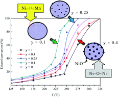
The B-site substitution of LaMnO3 perovskites by Ni was investigated under the oxidation of ethanol. Proper characterization techniques, including BET surface area measurement, XRD, FTIR, XPS, TPR, O2-TPD, and FE-SEM experiments were performed to survey the physicochemical properties of perovskites. The results reveal that up to 25% of Mn can be replaced by Ni; beyond this limit, segregated NiOx can be synthesized. Inserting Ni into the solid solution of perovskite yields unique bridging lattice oxygen sites (Ni–O–Mn) in Ni-doped LaMnO3. Based on catalytic performance in ethanol oxidation, the Ni–O–Mn sites are likely to promote ethanol conversion and the oxidation of acetaldehyde to CO2 at low reaction temperatures. The abatement of intermediates over Ni–O–Mn sites is hypothesized and a plausible reaction pathway is proposed. Moreover, the time on-stream testing revealed that the interaction between Ni and Mn is likely to enhance perovskite's thermal stability in ethanol oxidation.


Introduction

Ethanol can be derived from renewable energy crops and lignocellulosic biomass. Using ethanol as a fuel additive is a potential way to alleviate our fossil-fuel addiction. For example, 10% and 85% ethanol–gasoline blended fuels, i.e., E10 and E85, are commercially available for drivers in the US. As demand for energy continues to increase, the market growth for ethanol fuel is inevitable.1 However, burning ethanol-containing fuels yields different vehicle exhaust compounds from those produced by burning fossil fuels.2 More volatile organic compounds (VOCs) are generated using 10% and 15% ethanol–diesel blends in existing engines when compared to certified diesel fuel.3 Unburned ethanol and its derivatives, such as acetaldehyde, a carcinogen in humans,4 are harmful pollutants. Hence, the development of suitable catalysts for the abatement of ethanol and ethanol derivatives is urgently required.

Perovskite (ABO3) is an effective catalyst for the removal of CO, NOx, VOCs, and hydrocarbons.5,6 In some cases, perovskites perform comparably to, or even better than, conventional (precious metal-based) three-way catalysts, such as strontium-containing perovskites in NOx abatement.7 Daihatsu Motors has successfully implemented trace platinum group metal (pgm)-doped perovskites in their exhaust converter systems.8–11 The self-regeneration behavior of pgm-promoted perovskites under oxidative and reductive conditions makes them more active and durable than platinum-based catalysts. Our group has recently reported on the effectiveness of similar perovskites in methanol partial oxidation12,13 and combustion.14 The unique lattice structure and oxygen nonstoichiometry of perovskite are believed to be important in oxidation reactions.5

Surprisingly, only a few studies of ethanol combustion using perovskites are available. The pioneering study by the Hitachi research group15 tested LnBO3 (Ln = La, Pr, Sm, and Gd; B = Fe, Co and Ni) as ethanol sensors in ethanol oxidation. The short response time at low ethanol concentration makes perovskite a promising candidate in ethanol conversion. This inspires follow-up studies to utilize various compositions and elements at the A- or B-site position.16–22 Wang et al.17 and Najjar and Batis21 reported relatively high reactivities (low T50 and T95, where the former denotes the temperature for 50% ethanol conversion, and the latter is the temperature for 95% ethanol conversion) of their catalysts in ethanol oxidation. Wang et al.17 used Ag-supported La0.6Sr0.4MnO3 in both methanol and ethanol oxidations, and reported that anchored Ag+ on the surface of perovskite not only acts as a catalytic promoter, but also increases the surface adsorbed oxygen-to-lattice oxygen (O2−2/O) ratio; both factors are essential in the conversion of alcohols. Najjar and Batis21 investigated gelling agents (glycine and citric acid) for LaMnO3 synthesis using a combustion method, and discovered that LaMnO3 made by using glycine as the gelling agent had a high thermal stability and activity in ethanol combustion. This is attributed to the decrease of surface La/Mn ratio and the increase of the adsorbed oxygen species on the perovskite's surface.

This study investigates the use of Ni-incorporated LaMnO3 in ethanol oxidation. LaMnO3 is known to have a wide range of oxidative (excess oxygen) and reductive (oxygen vacancy) non-stoichiometries.23 The non-stoichiometric oxygen content markedly affects various oxidation reactions, such as methane24 and propane23 oxidations. This can be ascribed to the oxygen adsorption–activation step on catalyst surface and the enhancement of oxygen mobility.17 Partial substitution of Mn with cations with a valence less than three (such as Cu) is another way to improve the oxidation chemistry either in the bulk or on the surface of LaMnO3-based perovskites.23 In this work, Ni is chosen as the B-site dopant because of its bivalence and reactivity in oxidative environments.25–27 Incorporating Ni in LaMnO3 may have a synergistic effect on the conversion of ethanol and its derivatives in oxidative environments. This study attempts to explore the effect of Ni-substituted LaMnO3 perovskite in ethanol combustion. Physicochemical properties of catalysts were examined, and the relationship between the Ni content and the catalytic behaviors of LaMnO3-based perovskites was elucidated.

Experimental

Catalyst synthesis

Substituted LaMn1−yNiyO3 (y = 0.1, 0.25, and 0.4) and plain LaMnO3 and LaNiO3 were synthesized by the Pechini method.28,29 Metal nitrates precursors (La(NO3)3·6H2O (Merck, 99%), Mn(NO3)2·4H2O (Merck, 99%), and Ni(NO3)2·6H2O (Merck, 99%)) were used during the synthesis of the respective perovskites. These precursors were dissolved in water in the desired stoichiometric ratios. The obtained solution was then added dropwise to a 1 M citric acid solution, which contained approximately four times as many citric acid ions in relation to the metal ions of the precursors. The mixture was stirred and heated to 80 °C in a water bath. After stirring for an hour, an equivalent molar ratio of ethylene glycol (Alfa Aesar, 99%) using citric acid as the reference was poured into the solution. The resulting solution was then stirred at 90 °C for more than four hours until a gel was formed. The gel was transferred to a furnace, and dried at 80 °C for 24 h. The remaining paste was then calcined in air from 80 to 400 °C at a heating rate of 1 °C min−1, and kept isotherm for two hours. Finally, the precursors were calcined from 400 to 900 °C (3 °C min−1) for four hours.

Characterization

Powder X-ray diffraction patterns were obtained using a Shimadzu Labx XRD-6000 with Cu Kα radiation (0.15418 nm). The scan rate was set to 4° min−1 and the 2θ range was 20–80°. The Scherrer relation was employed to estimate the crystallite size of each sample, based on the strongest peak from the (1 1 0) plane of perovskite.

Fourier transform infrared spectroscopy (FTIR) spectra were recorded in the 400–4000 cm−1 region with a Perkin-Elmer Spectrum 100 Optica using the KBr pellet method. The catalyst powder was diluted to about 1 wt% within KBr. The mixture was then pulverized and pelletized. Prior to the test, the pellet was dehydrated at 130 °C for 30 min in a N2 stream (25 mL min−1). All the spectra were obtained with an average of 16 scans and 4 cm−1 resolution at room temperature.

X-ray photoelectron spectroscopy (XPS) was performed using a Thermo Scientific K-Alpha system equipped with a 180° hemispherical sector analyzer and an Al monochromator (Al Kα X-ray sources, 1486.6 eV). The diameter of the X-ray spot was 400 μm. The C 1s signal (284.5 eV) of adventitious carbon was used to correct the energy shift.

Single-point BET surface area measurement, temperature-programmed reduction (TPR), and O2-temperature-programmed desorption (O2-TPD) were carried out using a Micromeritics apparatus model Autochem II 2920 with a thermal conductivity detector (TCD). Approximately 100 mg of sample was consumed per trial. Prior to each test, the sample was dehydrated at 150 °C under an N2 stream (30 mL min−1) for 30 min. In the BET test, a 30% N2/He stream was used for N2 physisorption at −196 °C. The specific surface area of each sample was estimated from the desorbed N2 area using the single-point BET equation. In TPR analysis, a 10% H2/Ar (30 mL min−1) stream was passed through the sample and the temperature was raised from 50 to 900 °C with a ramp of 5 °C min−1. For O2-TPD, the sample was treated in a He stream (30 mL min−1) from 50 to 1000 °C at 5 °C min−1 followed by an isotherm at 1000 °C for half an hour.

The surface morphologies of as-synthesized and used samples were determined using a field-emission scanning electron microscope (FE-SEM, JEOL JSM-6701F). The acquisition time per location was 10 min. The SEM accelerating voltage was 10 kV. The scanned area was ∼4 μm × 3 μm. Both as-synthesized and used catalysts were examined.

Activity evaluation

Catalytic performances were examined in a continuous fixed-bed system.30 Ethanol was injected into the system by a HPLC pump (Jasco PU-2080) at a feeding rate of 3 × 10⁻³ mL min−1. Oxygen and nitrogen were regulated using mass flow controllers (Brooks 5850E). The molar composition of the feed was C2H5OH/O2/N2 = 1.7/18.1/80.2 and the gas hourly space velocity (GHSV) was 10[thin space (1/6-em)]446 h−1. About 10 mg of catalyst with a 40–80 mesh size was used in each trial. To maintain an isothermal condition, the catalyst was diluted with 150 mg SiO2 (40–80 mesh). SiO2 was inert in each trial. Internal and external mass transfer limitations were examined prior to the tests. The former was probed by measuring the catalytic performances using two different catalyst particle sizes (20–40 mesh and 40–80 mesh); the latter was tested by comparing two catalyst bed loadings, 10 mg catalyst mixed with 150 mg SiO2 and 15 mg catalyst mixed with 220 mg SiO2, with different flow rates but the same contact time (GHSV = 10[thin space (1/6-em)]446 h−1). Both internal and external mass transfer resistances could be ignored because the differences of ethanol conversions in these trials were trivial (less than 3%).

The system outlet was connected to a gas chromatograph (GC, SRI 8610) equipped with a TCD, a methanizer, and a flame ionization detector (FID). Two packed columns, 5 Å molecular sieve and Porapak Q, were used to separate reactants and products. The detected compounds were ethanol, acetaldehyde, CO2, CO, CH4, O2, and N2.

Nitrogen also served as the internal standard. All products were measured relative to GC calibration standards. Catalytic results were reported with carbon atom mass balances less than 10% errors. Three to four runs were made for each data point, providing 95% confidence intervals (error bars) for conversion and selectivity. Ethanol conversion was calculated as moles of ethanol reacted divided by moles of ethanol injected. Selectivity of carbon-containing product was defined as 100 × (moles of ethanol converted to product Xi)/(moles of ethanol converted). The data of conversion or selectivity was interpolated with a B-spline function to allow a better comparison.

Results and discussion

Table 1 lists the surface areas of the LaMn1−yNiyO3 catalysts. All catalysts displayed similar surface areas, ranging from 7.6 to 10.5 m2 g−1. Fig. 1 shows the XRD patterns of LaMn1−yNiyO3. Those of LaMnO3 ([JCPDS: 35-1353]) and LaNiO3 ([JCPDS: 33-0711]) were included for reference. All samples yielded the characteristic peaks of ABO3-type perovskite phase. The main diffraction peak of NiO ([JCPDS: 89-5881]) at 2θ = 43.3° was observed on LaNiO3. It reveals the segregation of NiO particles from perovskite solid solution. However, NiO was not identified on Ni-substituted catalysts, possibly because no segregated NiO was present or the NiO clusters were too small to be detected. The crystallite size decreased from 15.1 to 13.0 nm with increasing y values, suggesting the formation of disorder or deformation in LaMnO3 perovskite by adding Ni.
Table 1 Surface area, mean crystallite size, and XPS-derived surface composition of LaMn1−yNiyO3
LaMn1−yNiyO3 SAA (m2 g−1) dXRD (nm) Mn/La Ni/La Ni/Mn La/(Mn + Ni) Ni/(Mn + Ni) (Mn4+/Mn3+) Ox/Olatt
y = 0 8.7 15.1 0.83 0 1.21 0 1.03 0.63
y = 0.1 9.3 14.8 1.21 0.16 0.13 0.73 0.12 1.10 0.66
y = 0.25 10.5 14.5 1.08 0.28 0.26 0.74 0.21 1.21 0.70
y = 0.4 8.2 13.2 0.79 0.37 0.47 0.86 0.32 1.06 0.64
y = 1 7.6 13.0 0.67 1.49 1 2.00


image file: c3ra46323k-f1.tif
Fig. 1 XRD patterns of LaMn1−yNiyO3 perovskites. The diffraction of (1 1 0) plane is zoomed in to show the 2θ angle shift with increasing Ni loading.

A close inspection of the XRD patterns showed that the 2θ angle of each peak increased slightly with increasing Ni amount. This correlation is attributed to the decrease of interplanar distance by Ni substitution: the more Ni incorporated, the more Mn3+ (low-spin 0.58 Å, high-spin 0.65 Å) could be replaced by Ni3+ (low-spin 0.56 Å, high-spin 0.60 Å) ions, resulting in a decrease of the unit cell volume.31 Blasco et al.32 have also observed a similar tendency regarding unit cell volume with increasing Ni content in LaMn1−yNiyO3 perovskites.

Fig. 2 shows (1 1 0), (1 1 1), (2 0 0), and (2 1 1) diffraction shifts as functions of Ni content. The shift gradually increased with Ni content from y = 0 to y = 0.25 then leveled off. Provendier et al.26 and Pecchi et al.31 have observed the same trend of XRD peak shift in the LaFe1−yNiyO3 system. The shift has been ascribed to the degree of Ni substitution, and a certain proportion of segregation has been noted in the high substitution region with the maximum shift value. LaMnO3 has an orthorhombic unit cell. Substituting Mn with smaller Ni ions destabilizes the MnO6 octahedra and enhances the rhombohedral distortion (due to the Jahn–Teller effect).33 This structural phase transition of LaMn1−yNiyO3 may lead to the coexistence of multiple phases (e.g., La2NiMnO6 and LaNiO3) at a high Ni-loading.32,33 Therefore, the complete insertion of Ni in perovskite structure should occur at y < 0.25; NiOx phases should segregate at y = 0.25 and above.31


image file: c3ra46323k-f2.tif
Fig. 2 High angle shifts of the diffraction peaks as functions of Ni substitution content.

Fig. 3 shows the FTIR spectra of the LaMn1−yNiyO3 catalysts. Besides LaNiO3, all perovskites displayed an adsorption band at approximately 600 cm−1. This band is related to Mn–O stretching in octahedral site.34 The band of Mn–O stretching was shifted from 610 to 601 cm−1 (red shift) with an increase of Ni loading. The red shift may be explained by the photon response theory35 since Ni is heavier than Mn in the solid solution. Therefore, the downward-shifted wavenumber suggested the presence of Ni–O–Mn bonding in Ni-containing LaMnO3. Moreover, at y = 0.25 and 0.4, a band at 411 cm−1 and a shoulder at 638 cm−1 were observed. The former is the stretching vibration of Ni–O bond;36,37 the latter, Ni–OH bending.38 This is consistent with the XRD diffraction shifts: NiOx could be formed on the surface at y = 0.25 and above. The IR spectrum of LaNiO3 was not well resolved; a close inspection within 400–600 cm−1 was inserted in Fig. 3. The unclear vibrational modes of LaNiO3 may be related to its low electrical resistivity (10−1 Ω cm).39 At such a low resistivity, the residence time (10−12 s) of charge carriers at a site can be comparable to the lattice vibrational period (10−13 s), at which a localized vibrational mode may not be specified.40 Two tiny bands at 428 cm−1 and 502 cm−1 were identified, in accord with an earlier work.41 However, the assignment of these two responses is unclear.


image file: c3ra46323k-f3.tif
Fig. 3 FTIR spectra of LaMn1−yNiyO3 perovskites.

Fig. 4 displays the XPS spectra of LaMn1−yNiyO3 perovskites. The La 3d spectra included two doublet peaks at 830 eV to 840 eV and 850 eV to 860 eV, corresponding to La 3d5/2 and La 3d3/2 signals, respectively. Notably, the responses of La 3d3/2 and Ni 2p3/2 overlap each other, and are difficult to be differentiated. A small hump at approximately 870 eV, which is associated with Ni 2p1/2, was identified in the spectrum of LaNiO3 (y = 1). Multiple states of Ni (Ni3+/Ni2+) have been noted in LaNiO3-based perovskites.42 The spin-orbit splitting of the La 3d level for each sample was close to 16.8 eV;42 moreover, the La 3d5/2 and La 3d3/2 responses were close to the index peaks of pure La2O3,43 indicating that La ions are in a trivalent state. Mn 2p3/2 and Mn 2p1/2 signals were located at approximately 641.7 eV and 653.3 eV, respectively. Mn2+, Mn3+, and Mn4+ could coexist in the Mn 2p3/2 region;44 however, the lack of a satellite peak of Mn2+ (648.8 eV)45 eliminates the possibility of the presence of Mn2+ ions. The responses of Mn 2p3/2 and Mn 2p1/2 can be deconvoluted into four peaks. The responses at 641.5 eV and 653.0 eV were assigned to Mn3+ cations, while the peaks at 642.9 eV and 654.5 eV were attributed to Mn4+ ions.42 To provide quantitative analysis, the surface atomic ratios (see Table 1) were estimated from the intensities of La 3d5/2, Ni 2p1/2, and Mn 2p3/2 peaks using the atomic sensitivity factors provided by Wagner et al.46 The Mn4+/Mn3+ molar ratio was reported in Table 1 as well.


image file: c3ra46323k-f4.tif
Fig. 4 La 3d, Ni 2p, Mn 2p, and O 1s core level spectra of LaMn1−yNiyO3 perovskites.

The surface enrichment of La can be speculated for plain LaMnO3 and LaNiO3 because the Mn/La and Ni/La ratios at y = 0 and 1 were all less than unity. However, this is not the case for the y = 0.1 to 0.4 samples: the La/(Mn + Ni) ratios were all less than unity. This suggests the surface of Ni-containing LaMnO3 was enriched by Ni and Mn cations. To provide a clear view of the distribution of La, Mn, and Ni on the surface of perovskite, Fig. 5 displays measured surface atomic ratios as functions of nominal surface atomic ratios of Mn/La, Ni/La, Ni/Mn, and Ni/(Mn + Ni). Apparently, the surface became Mn-enriched for Ni-containing LaMnO3 because their experimental Mn/La values were all greater than nominal ones. The measured Ni/La, Ni/Mn, and Ni/(Mn + Ni) all displayed a descending trend with growing y value. Segregation of NiOx on perovskite's surface at y = 0.25 and above is a possible explanation. This is because segregated NiOx particles contained Ni cations in the bulk that cannot be detected by XPS, thereby lowering the surface atomic ratio for Ni with respect to La or Mn. The y = 0.1 sample had all its experimental Ni/La, Ni/Mn, and Ni/(Mn + Ni) ratios greater than nominal ones, indicating the presence of highly dispersed Ni on its surface. The Mn4+/Mn3+ molar ratios were close to 1, decreased followed the order as: y = 0.25 (1.21) > y = 0.1 (1.10) > y = 0.4 (1.06) > y = 0 (1.03). This is in accordance with Zhang et al.,42 who have reported similar values of LaMnO3 at 0.975 and LaMn0.8Ni0.2O3 at 1.216. Apparently, Ni substitution in the B-site position can increase the composition of Mn4+ in LaMnO3 perovskites.


image file: c3ra46323k-f5.tif
Fig. 5 Experimental Mn/La, Ni/La, Ni/Mn, and Ni/(Mn + Ni) atomic ratios versus nominal ones.

Combined signals were obtained from the O 1s state, suggesting the presence of different oxygen species on the surface layer. Lattice oxygen (e.g., La–O–Mn at 529.1 eV),13 (O2) species or low coordinated oxygen ions located at special site or domains of the surface (531.0 eV),47 and hydroxyl/carbonate groups (532.3 eV)31 are believed to coexist on the surface of perovskite. Therefore, the O 1s spectra were deconvoluted based on these three responses. As the Ni content increased, the BE of lattice oxygen shifted from 529.2 eV to 528.1 eV. Tabata et al.45 observed a similar O 1s chemical shift in LaMn1−yCuyO3 (y = 0, 0.1, 0.2, 0.3, and 0.4). They proposed that more defects and oxygen vacancies could be generated as the substitution of Cu ions at the B-site increased. This effect alters the bond strength of the surface lattice oxygen. Similarly, replacing Ni in LaMnO3 generates defects and/or segregated NiO particles, which weakening the lattice oxygen bonds in perovskite. Therefore, the BEs of lattice oxygen declined as the Ni content increased. The molar ratio of (O2) species or low coordinated oxygen ions to lattice oxygen (Ox/Olatt) was presented in Table 1. The Ox/Olatt ratio showed a same trend as that of (Mn4+/Mn3+), declined as: y = 0.25 (0.70) > y = 0.1 (0.66) > y = 0.4 (0.64) > y = 0 (0.63). Accordingly, the more Mn4+ on the surface, the more (O2) species or low coordinated oxygen ions could exist.42 Plain LaNiO3 displayed higher Ox/Olatt value (2.00) than the other samples because La–Ni oxides are prone to form oxygen-deficient perovskites, which can be described as a superstructure with a formula LanNinO3n−1.48 Hence, less lattice oxygens were available on LaNiO3 than other perovskites.

The C 1s spectra were presented in Fig. S1 (ESI). The C 1s signals of all perovskites showed responses of adventitious carbon (284.5 eV),49 lanthanum carbonate (∼286.2 eV),50 and carbonyl group (∼288.6 eV).49,51 This is in agreement with the literature that lanthanum-based perovskites are often contaminated by carbonate species.

Fig. 6 presents the TPR profiles of tested perovskites and Table S1 (ESI) lists the amounts of H2 consumed by them. The TPR patterns of pure LaMnO3 and LaNiO3 included multiple peaks. The reduction of LaMnO3 is complicated: Mn4+ and Mn3+ can coexist at the outset of TPR, and a full reduction of Mn ions to metallic Mn is not possible.52,53 Therefore, the low-temperature responses (<400 °C) are attributed to the reduction of Mn4+ to Mn3+; the high-temperature signals (>400 °C) are referred to the reduction of Mn3+ to Mn2+. Some reduction of Mn3+ to Mn2+ at low temperatures cannot be ruled out.54 The reduction of LaNiO3 was similar to that in an earlier study:55 the reduction of Ni3+ to Ni2+ (i.e., La2Ni2O5) at 347 °C was followed by that of Ni2+ to metallic Ni at 469 °C. The XRD patterns of post-TPR LaMnO3 and LaNiO3 (see Fig. S2 of ESI) further support the aforementioned assertions: the index peaks of La2O3 and MnO were with the reduction of LaMnO3; those of La2O3 and Ni were associated with the reduction of LaNiO3.


image file: c3ra46323k-f6.tif
Fig. 6 TPR profiles of LaMn1−yNiyO3 perovskites.

The first reduction of LaMn1−yNiyO3 occurred in the range of 250 °C to 500 °C, in which the LaMnO3 and LaNiO3 phases were reduced. Hence, Mn and Ni ions were reduced concurrently in this range of temperatures. The second reduction of LaMn1−yNiyO3 may be related to Mn and Ni ions since the amount of H2 consumed in the high temperature range does not increase in proportional to the extent of Ni replacement (see Table S1).

To provide a comparison, Fig. S3 (ESI) presents the TPR results for physical mixtures of LaNiO3 and LaMnO3 with LaNiO3-to-LaMnO3 ratios of 0.1, 0.25, and 0.4. In Fig. 6, Ni-incorporated LaMnO3 exhibited two TPR responses at 200 °C to 500 °C and 650 °C to 850 °C. The high-temperature responses of LaMn1−yNiyO3 shifted to higher temperatures than those of plain LaMnO3 and LaNiO3, especially for the y = 0.1 sample. Tailing of reduction response at 800 °C and above could be observed for the y = 0.1 and 0.25 samples. This suggests an existence of Ni–O–Mn interaction, which retards the reduction process. The TPR profiles of physically mixed LaNiO3 and LaMnO3 further support the above-mentioned hypothesis: three distinct peaks at 331, 466, and 731 °C appear to be the merged responses of LaNiO3 and LaMnO3. Notably, LaMn0.9N0.1O3 exhibited the highest reduction signal at 783 °C, indicating that the interaction within the solid solution of LaMn0.9N0.1O3 is the strongest among all samples. Conversely, the weakest interaction between Ni and Mn was found in LaMn0.6N0.4O3 since it had the lowest high-temperature reduction response at 736 °C.

Fig. 7 shows the O2-TPD profiles. For clarity, the signal of LaNiO3 was multiplied by a factor of 0.1. O2-TPD can be used to estimate the lability of oxygen in tested perovskites. The outset of desorption signal increased in the order of y = 0 (82 °C) < y = 0.1 (114 °C) < y = 0.25 (132 °C) < y = 0.4 (211 °C) < y = 1 (677 °C), suggesting that more substituted Ni at B-site resulted in lower oxygen lability in LaMn1−yNiyO3. Desorbed oxygen is frequently categorized as α- and β-oxygen, using 500 °C as the demarcation. α-oxygen is adsorbed and near-surface oxygen, while β-oxygen is lattice oxygen in the perovskite framework.56 Negligible α-oxygen desorption was detected in LaNiO3. Table 2 summarizes the amounts of desorbed oxygen and the temperatures of peak desorption of tested perovskites. The desorption temperatures of α- and β-oxygen increased with Ni content for LaMn1−yNiyO3. Plain LaNiO3 exhibited a distinct peak at 948 °C. Ni-containing LaMnO3 perovskites had higher α-oxygen-to-β-oxygen ratios than pure LaMnO3 and LaNiO3. The α-oxygen-to-β-oxygen ratio followed the sequence of y = 0.25 (0.47) > y = 0.1 (0.39) > y = 0.4 (0.32) > y = 0 (0.30), in consistent with the trend of the Ox/Olatt ratio observed by XPS. Among all catalysts, LaMn0.9Ni0.1O3 released the most α-oxygen (72.2 μmol/g cat.); LaNiO3 contained the highest β-oxygen (695.9 μmol/g cat.).


image file: c3ra46323k-f7.tif
Fig. 7 O2-TPD profiles of LaMn1−yNiyO3 perovskites.
Table 2 Oxygen desorption characterization of LaMn1−yNiyO3 catalysts
Catalyst O2-TPD
α-O2a β-O2b α-O2/β-O2
a Maximum temperature of oxygen desorption peak below 500 °C.b Maximum temperature of oxygen desorption peak above 500 °C.
y = 0 T (°C) μmol/g cat. 131 686  
69.8 (23.0%) 233.2 (77.0%) 0.30
y = 0.1 T (°C) μmol/g cat. 136 697  
72.2 (28.1%) 184.5 (71.9%) 0.39
y = 0.25 T (°C) μmol/g cat. 205 928  
68.1 (32.1%) 144.0 (67.9%) 0.47
y = 0.4 T (°C) μmol/g cat. 255 1000  
24.8 (24.1%) 78.0 (75.9%) 0.32
y = 1 T (°C) μmol/g cat. 948  
695.9 (100%) 0


Ni substitution negatively affects oxygen lability/oxygen availability in LaMnO3-based perovskites. This can be explained by estimating the oxygen content from the charge balance. According to the XPS results, trivalent and tetravalent Mn ions could coexist in LaMnO3; therefore, its chemical formula is LaMn3+aMn4+1−aO3+δ (δ = (1 − a)/2). Divalent and trivalent Ni ions were present in LaNiO3, yielding the formula LaNi2+bNi3+1−bO3+λ (λ = −(1 − b)/2). Since δ is positive and λ is negative, LaMnO3 should host excess oxygen while oxygen vacancies are formed in LaNiO3. Substituting Ni in LaMnO3 should satisfy the charge balance condition, yielding an oxygen content between δ and λ. The only exception is LaNiO3, which released the most oxygen in O2-TPD. This finding is consistent with the XPS results concerning the inhomogeneity of the surface and unstable Ni3+ in perovskite, which yields weak lattice oxygen bonding in LaNiO3. Still, doping Ni has a positive effect on α-oxygen content, which is optimal at y = 0.1.

Fig. 8 plots ethanol conversions of LaMn1−yNiyO3 as functions of temperature. Each catalyst yields an S-shaped curve besides LaNiO3, which displays an abrupt increase of ethanol conversion at 250 °C. At temperatures below 200 °C, all catalysts converted less than 20% ethanol, except for LaMn0.9Ni0.1O3, which achieved 21% conversion at 200 °C. The T50 increased in the order of LaMn0.9Ni0.1O3 (226 °C) < LaMn0.75Ni0.25O3 (243 °C) < LaMnO3 (249 °C) < LaMn0.6Ni0.4O3 (255 °C) < LaNiO3 (266 °C); the T95, LaMn0.9Ni0.1O3 (252 °C) < LaMn0.75Ni0.25O3 (269 °C) < LaMnO3 (288 °C) < LaMn0.6Ni0.4O3 (309 °C) < LaNiO3 (314 °C). This indicates that LaMn0.9Ni0.1O3 is the most active catalyst in ethanol combustion while LaNiO3 is the least active. Ling et al.57 have observed a sudden increase of conversion at 250 °C for LaNiO3 in ethanol oxidation. By a conductivity test, surface reaction of chemisorbed ethanol and oxygen species was discovered to be promoted at 250 °C. This leads to an increase of LaNiO3 conductivity due to oxygen consumption, and ethanol conversion can be greatly improved on partially reduced surface. They also pointed out that the maximum conductivity change of LaNiO3 occurs in the range of 250 to 350 °C, in accordance with our conversion trend.


image file: c3ra46323k-f8.tif
Fig. 8 Conversion of ethanol as a function of temperature over LaMn1−yNiyO3 perovskites.

Fig. 9 shows acetaldehyde and carbon dioxide selectivities as functions of temperature. Negligible amounts of CO and CH4, both less than 1%, were detected in each catalyst, and are ignored for clarity. At temperatures below 225 °C, the selectivity of acetaldehyde exceeded 80% while that of CO2 was less than 20%. With increasing temperature, the selectivity of acetaldehyde declined and that of CO2 increased. At 235 °C, LaMn0.9Ni0.1O3 generated about 38% acetaldehyde and 62% CO2. The other catalysts produced more than 80% acetaldehyde and less than 20% CO2 at this temperature. At 250 °C, LaMn0.75Ni0.25O3 yielded equal amounts of acetaldehyde (30%) and CO2 (70%) to those of LaMn0.9Ni0.1O3. The remaining catalysts at 260 °C and above yielded less than 40% acetaldehyde and over 60% CO2. LaNiO3 seems to be the least effective in acetaldehyde abatement because more than 40% acetaldehyde selectivity was detected at 275 °C.


image file: c3ra46323k-f9.tif
Fig. 9 Acetaldehyde and carbon dioxide selectivities as functions of temperature over LaMn1−yNiyO3 perovskites.

The oxidation activity and chemistry of alkane and alcohol are known to be strongly associated with the transport and storage of oxygen.23,58 The above catalytic outcomes indicate that adsorbed and near-surface oxygen (α-oxygen) are more effective than lattice oxygen (β-oxygen) in ethanol oxidation. Hence, the α-oxygen-lean LaNiO3 was the least active catalyst whereas the activities of α-oxygen-rich LaMn0.9Ni0.1O3 and LaMn0.75Ni0.25O3 were enhanced. The high onset temperature of O2-TPD and low concentration of α-oxygen of LaMn0.6Ni0.4O3 reflected low reactivity, implying the critical role of α-oxygen in ethanol oxidation. α-oxygen is known to act as an activated oxygen species, which initiates the oxidation of chemisorbed intermediates into carbon oxides and water.59 The role of α-oxygen in ethanol oxidation is as proposed elsewhere.17,21 To provide a better comparison, Fig. S4 (ESI) displays the variations of XPS-derived Ni loading, amounts of α- and β-oxygen, and T50 and T95 of tested LaMn1−yNiyO3.

LaMn0.9Ni0.1O3 possesses the highest activity among all catalysts, even greater than that of LaMn0.75Ni0.25O3, which has the highest Ox/Olatt ratio. This implies that besides α-oxygen effect, other factors should be considered for catalyst's efficacy in ethanol oxidation. Presumably, at a lower Ni loading, Ni can be distributed more evenly in perovskite, generating more Ni–O–Mn linkages on the surface. Increasing the Ni loading in LaMnO3 produces segregated NiOx clusters on the surface of perovskite, reducing the amounts of Ni–O–Mn bonds. The bridging oxygen of Ni–O–Mn should possess an unique interaction, which is effective in promoting ethanol oxidation and intermediate conversion to carbon dioxide. Similar bridging sites have been noted on different perovskites (e.g., Ni2+–O–Ni3+ species of La2−xSrxNiO4,60 Mn3+–O–Mn4+ bonds of LaMn1−yCuyO3,52 and Fe3+–O–Fe5+ pairs of SrFeO361). These sites have been proposed to be effective in promoting charge transfer and oxygen mobility of the catalysts in N2O decomposition60 and CH4 combustion.52,61

Fig. 10 displays the proposed reaction pathway of ethanol conversion over LaMn1−yNiyO3. The strongly basic O–La–O sites are known to be active in ethanol activation;21 however, the pathway for the possibility of ethanol chemisorption on La is excluded when considering only the effect of B-site substitution on LaMnO3. At the outset of the reaction, ethanol chemisorbs on the surface, and forms an ethoxide at the metal site (Mn or Ni) and a hydroxyl at the neighboring lattice oxygen site. Subsequent dehydration yields an acetaldehyde-derived intermediate and an oxygen vacancy. At low temperatures, the reduced surface is re-oxidized, and acetaldehyde is desorbed. At high temperatures, the acetaldehyde intermediate may associate with the surface bridging oxygen (Mn–O–Mn, Ni–O–Mn, or Ni–O–Ni) to form an acetyl, which is then converted to carbon oxides, water, and methane.62 Ethanol dehydrogenation to acetaldehyde was excluded in Fig. 10 because negligible conversion (less than 5%) of ethanol was identified without feeding O2 in our system.


image file: c3ra46323k-f10.tif
Fig. 10 Proposed reaction pathway of ethanol oxidation.

The catalytic results show that acetaldehyde selectivities are nearly identical (above 80%) at temperatures below 225 °C. Acetaldehyde is known to be derived through a redox cycle in ethanol oxidation.21,63 Therefore, the redox properties for LaMnO3, LaNiO3, and LaMn1−yNiyO3 perovskites should be similar. Carbon dioxide selectivities of these catalysts increase differently with increasing temperature, implying changing oxidation power in converting the chemisorbed intermediate (herein acetyl species). In the proposed pathway, acetyl oxidation proceeds on the bridging oxygen in Mn–O–Mn, Ni–O–Mn, or Ni–O–Ni. The amount of CO2 increased abruptly over LaMn0.9Ni0.1O3 at the lowest temperature (235 °C) of any of the tested perovskites, suggesting that its bridging oxygen was the most strongly oxidizing. This is attributed to the unique Ni–O–Mn interaction of LaMn0.9Ni0.1O3, as revealed by the TPR test. To ascertain whether the Ni–O–Mn interaction in perovskite is uniquely responsible for its reactivity and acetaldehyde oxidation, catalytic tests of physically mixed LaNiO3 and LaMnO3 in LaNiO3-to-LaMnO3 ratios of 0.1, 0.25, and 0.4 were conducted (see Fig. S5 and S6 of ESI). The T50 values of the physically mixed samples were in the range of 245 °C to 254 °C – about 20 °C higher than that of LaMn0.9Ni0.1O3 but close to those of LaMn0.75Ni0.25O3 and LaMn0.6Ni0.4O3. However, the T95 values of the physically mixed samples exceeded their respective counterparts, ranging from 300 to 321 °C. For all samples, below 250 °C, acetaldehyde selectivity exceeded 80% and CO2 selectivity was less than 20%. These results demonstrate that the Ni–O–Mn interaction in Ni-doped LaMnO3 promotes the conversion of ethanol and the oxidation of acetaldehyde to CO2.

Fig. 11 illustrates the evolution of time on-stream ethanol conversion over a 36 h period at 300 °C. Initially, ethanol was fully converted over LaMnO3, LaMn0.9Ni0.1O3, and LaMn0.75Ni0.25O3, while 93.7% and 91.9% conversions were achieved by LaMn0.6Ni0.4O3 and LaNiO3, respectively. All catalysts suffered of ageing but at different extents. Ethanol conversion decreased from 100% to 96.2% for LaMnO3 (a decrease of ∼3.8%), 100% to 98.0% for LaMn0.9Ni0.1O3 (a decrease of ∼2.0%), 100% to 97.5% for LaMn0.75Ni0.25O3 (a decrease of ∼2.5%), 93.7% to 89.7% for LaMn0.6Ni0.4O3 (a decrease of ∼4.4%), and 91.9% to 78.6% for LaNiO3 (a decrease of ∼14.5%). Moreover, LaMn0.9Ni0.1O3 remained its activity for more than 25 h, whereas deactivation could be observed within 20 h for other catalysts. Apparently, LaMn0.9Ni0.1O3 has the highest thermal stability while LaNiO3 has the least among tested catalysts. Since coking is less likely to occur under oxygen-rich environments, agglomeration of catalyst particle and/or deconstruction of perovskite structure should be responsible for catalyst deactivation.64 The SEM micrographs of freshly prepared and used LaMn1−yNiyO3 after on-stream testing (see Fig. S7 of ESI) further supported this claim: some welding and particle agglomeration could be identified on used samples; however, no carbon deposition (e.g., carbon nanotubes) was specified. The high thermal stability of LaMn0.9Ni0.1O3 may, again, ascribe to the unique interaction between Ni and Mn, thereby suppressing the destruction of its perovskite framework.


image file: c3ra46323k-f11.tif
Fig. 11 Conversion of ethanol as a function of time on-stream over LaMn1−yNiyO3 perovskites at 300 °C.

Conclusions

Doping Ni into LaMnO3 perovskite can substantially alter its physicochemical properties, and thereby changing its catalytic performances. A limited amount of Ni (less than 25%) can be dispersed in LaMnO3 framework; adding more Ni forms segregated NiOx clusters. Incorporating Ni generates Ni–O–Mn sites, which are associated with a unique interaction, is assumed to play a key role in enhancing catalytic activity, acetaldehyde abatement, and stability in ethanol oxidation. A plausible mechanism based on the surface bridging oxygen site was thereby proposed. Future study should seek to clarify the nature of the bridging oxygen site, possibly through computational chemistry such as density functional theory,65 to provide a molecular-level understanding of perovskite catalyst design.

Acknowledgements

The authors acknowledge the National Science Council (Taiwan) for the financial support under Contract Number 102-2221-E-155-060-MY2. The authors also thank Oscar Kerkenaar for the English language editing.

Notes and references

  1. A. Hira, Energy Policy, 2011, 39, 6925–6935 CrossRef CAS PubMed.
  2. A. C. Hansen, Q. Zhang and P. W. L. Lyne, Bioresour. Technol., 2005, 96, 277–285 CrossRef CAS PubMed.
  3. M. D. Kass, J. F. Thomas, J. M. Storey, N. Domingo, J. Wade and G. Kenreck, SAE Technical Paper 2001-01-2018 (SP-1632), 2001 Search PubMed.
  4. US EPA, http://www.epa.gov/chemfact/s_acetal.txt.
  5. M. A. Peña and J. L. G. Fierro, Chem. Rev., 2001, 101, 1981–2018 CrossRef PubMed.
  6. J. Zhu and A. Thomas, Appl. Catal., B, 2009, 92, 225–233 CrossRef CAS PubMed.
  7. C. H. Kim, G. Qi, K. Dahlberg and W. Li, Science, 2010, 327, 1624–1627 CrossRef CAS PubMed.
  8. Y. Nishihata, J. Mizuki, T. Akao, H. Tanaka, M. Uenishi, M. Kimura, T. Okamoto and N. Hamada, Nature, 2002, 418, 164–167 CrossRef CAS PubMed.
  9. H. Tanaka, Catal. Surv. Asia, 2005, 9, 63–74 CrossRef CAS PubMed.
  10. H. Tanaka, M. Uenishi, M. Taniguchi, I. Tan, K. Narita, M. Kimura, K. Kaneko, Y. Nishihata and J. Mizuki, Catal. Today, 2006, 117, 321–328 CrossRef CAS PubMed.
  11. H. Tanaka, M. Taniguchi, M. Uenishi, N. Kajita, I. Tan, Y. Nishihata, J. Mizuki, K. Narita, M. Kimura and K. Kaneko, Angew. Chem., Int. Ed., 2006, 45, 5998–6002 CrossRef CAS PubMed.
  12. C.-L. Li, C.-L. Wang and Y.-C. Lin, Catal. Today, 2011, 174, 135–140 CrossRef CAS PubMed.
  13. C.-L. Li and Y.-C. Lin, Appl. Catal., B, 2011, 107, 284–293 CrossRef CAS PubMed.
  14. C.-L. Li, B.-S. Jiang, W.-L. Fanchiang and Y.-C. Lin, Catal. Commun., 2011, 16, 165–169 CrossRef CAS PubMed.
  15. H. Obayashi, Y. Sakurai and T. Gejo, J. Solid State Chem., 1976, 17, 299–303 CrossRef CAS.
  16. T. Shimizu, Appl. Catal., 1986, 28, 81–88 CrossRef CAS.
  17. W. Wang, H. Zhang, G. Lin and Z. Xiong, Appl. Catal., B, 2000, 24, 219–232 CrossRef CAS.
  18. N. A. Merino, B. P. Barbero, P. Ruiz and L. E. Cadus, J. Catal., 2006, 240, 245–257 CrossRef CAS PubMed.
  19. B. P. Barbero, J. A. Gamboa and L. E. Cadus, Appl. Catal., B, 2006, 65, 21–30 CrossRef CAS PubMed.
  20. B. Bialobok, J. Trawczynski, W. Mista and M. Zawadzki, Appl. Catal., B, 2007, 72, 395–403 CrossRef CAS PubMed.
  21. H. Najjar and H. Batis, Appl. Catal., A, 2010, 383, 192–201 CrossRef CAS PubMed.
  22. M. Zawadzki and J. Trawczynski, Catal. Today, 2011, 176, 449–452 CrossRef CAS PubMed.
  23. T. Nitadori, S. Kurihara and M. Misono, J. Catal., 1986, 98, 221–228 CrossRef CAS.
  24. I. Rossetti, C. Biffi and L. Forni, Chem. Eng. J., 2010, 162, 768–775 CrossRef CAS PubMed.
  25. T. Hayakawa, A. Andersen, M. Shimizu, K. Suzuki and K. Takehira, Catal. Lett., 1993, 22, 307–317 CrossRef CAS.
  26. H. Provendier, C. Petit, C. Estournes, S. Libs and A. Kiennemann, Appl. Catal., A, 1999, 180, 163–173 CrossRef CAS.
  27. F. B. Noronha, M. C. Durao, M. S. Batista and L. G. Appel, Catal. Today, 2003, 85, 13–21 CrossRef CAS.
  28. M. P. Pechini, US Pat., 3330697, 1967.
  29. P. Courty, H. Ajot, C. Marcilly and B. Delmon, Powder Technol., 1973, 7, 21–38 CrossRef CAS.
  30. C.-L. Li and Y.-C. Lin, Catal. Lett., 2010, 140, 69–76 CrossRef CAS.
  31. G. Pecchi, P. Reyes, R. Zamora, L. E. Cadus and J. L. G. Fierro, J. Solid State Chem., 2008, 181, 905–912 CrossRef CAS PubMed.
  32. J. Blasco, M. C. Sanchez, J. Perez-Cacho, J. Garcia, G. Subias and J. Campo, J. Phys. Chem. Solids, 2002, 63, 781–792 CrossRef CAS.
  33. N. Van Minh, S.-J. Kim and I.-S. Yang, Phys. B, 2003, 327, 208–210 CrossRef CAS.
  34. F. Gao, R. A. Lewis, X. L. Wang and S. X. Dou, J. Alloys Compd., 2002, 347, 314–318 CrossRef CAS.
  35. D. W. Taylor, Photon response theory and the infrared and Raman experiments, Elsvier Science Publishers, Amsterdam, 1988 Search PubMed.
  36. G. Zhou, D.-W. Wang, L.-C. Yin, N. Li, F. Li and H.-M. Cheng, ACS Nano, 2012, 6, 3214–3223 CrossRef CAS PubMed.
  37. S. Mustansar Abbas, S. Tajammul Hussain, S. Ali, N. Ahmad, N. Ali, S. Abbas and Z. Ali, J. Solid State Chem., 2013, 202, 43–50 CrossRef CAS PubMed.
  38. N. Bayal and P. Jeevanandam, J. Alloys Compd., 2012, 537, 232–241 CrossRef CAS PubMed.
  39. T. Vaz and A. V. Salker, Mater. Sci. Eng., B, 2007, 143, 81–84 CrossRef CAS PubMed.
  40. P. Ganguly and N. Y. Vasanthacharya, J. Solid State Chem., 1986, 61, 164–170 CrossRef CAS.
  41. M. Panneerselvam and K. J. Rao, J. Mater. Chem., 2003, 13, 596–601 RSC.
  42. C. Zhang, C. Wang, W. Zhan, Y. Guo, Y. Guo, G. Lu, A. Baylet and A. Giroir-Fendler, Appl. Catal., B, 2013, 129, 509–516 CrossRef CAS PubMed.
  43. M. O'Connell, A. K. Norman, C. F. Huttermann and M. A. Morris, Catal. Today, 1999, 47, 123–132 CrossRef CAS.
  44. M. Kantcheva, M. U. Kucukkal and S. Suzer, J. Mol. Struct., 1999, 482–483, 19–22 CrossRef CAS.
  45. K. Tabata, Y. Hirano and E. Suzuki, Appl. Catal., A, 1998, 170, 245–254 CrossRef CAS.
  46. C. D. Wagner, L. E. Davis, M. V. Zeller, J. A. Taylor, R. H. Raymond and L. H. Gale, Surf. Interface Anal., 1981, 3, 211–225 CrossRef CAS.
  47. S. Rousseau, S. Loridant, P. Delichere, A. Boreave, J. P. Deloume and P. Vernoux, Appl. Catal., B, 2009, 88, 438–447 CrossRef CAS PubMed.
  48. M. T. Anderson, J. T. Vaughey and K. R. Poeppelmeier, Chem. Mater., 1993, 5, 151–165 CrossRef CAS.
  49. M. Cinar, L. M. Castanier and A. R. Kovscek, Energy Fuels, 2011, 25, 4438–4451 CrossRef CAS.
  50. C. K. Jorgensen and H. Berthon, Chem. Phys. Lett., 1972, 13, 186–189 CrossRef.
  51. H. Taguchi, A. Sugita, M. Nagao and K. Tabata, J. Solid State Chem., 1995, 119, 164–168 CrossRef CAS.
  52. L. Lisi, G. Bagnasco, P. Ciambelli, S. De Rossi, P. Porta, G. Russo and M. Turco, J. Solid State Chem., 1999, 146, 176–183 CrossRef CAS.
  53. S. Royer, H. Alamdari, D. Duprez and S. Kaliaguine, Appl. Catal., B, 2005, 58, 273–288 CrossRef CAS PubMed.
  54. H. Ziaei-Azad, A. Khodadadi, P. Esmaeilnejad-Ahranjani and Y. Mortazavi, Appl. Catal., B, 2011, 102, 62–70 CrossRef CAS PubMed.
  55. H. Chen, H. Yu, F. Peng, G. Yang, H. Wang, J. Yang and Y. Tang, Chem. Eng. J., 2010, 160, 333–339 CrossRef CAS PubMed.
  56. J. N. Kuhn and U. S. Ozkan, J. Catal., 2008, 253, 200–211 CrossRef CAS PubMed.
  57. T.-R. Ling, Z.-B. Chen and M.-D. Lee, Appl. Catal., A, 1996, 136, 191 CrossRef CAS.
  58. J. Choisnet, N. Abadzhieva, P. Stefanov, D. Klissurski, J. M. Bassat, V. Rives and L. Minchev, J. Chem. Soc., Faraday Trans., 1994, 90, 1987–1991 RSC.
  59. B. Levasseur and S. Kaliaguine, Appl. Catal., A, 2008, 343, 29–38 CrossRef CAS PubMed.
  60. A. K. Ladavos and P. J. Pomonis, J. Chem. Soc., Faraday Trans., 1991, 87, 3291–3297 RSC.
  61. V. C. Belessi, A. K. Ladavos and P. J. Pomonis, Appl. Catal., B, 2001, 31, 183–194 CrossRef CAS.
  62. K. Rintramee, K. Fottinger, G. Rupprechter and J. Wittayakun, Appl. Catal., B, 2012, 115–116, 225–235 CrossRef CAS PubMed.
  63. B.-S. Jiang, R. Chang and Y.-C. Lin, Ind. Eng. Chem. Res., 2013, 52, 37–42 CAS.
  64. H. Najjar, J.-F. Lamonier, O. Mentre, J.-M. Giraudon and H. Batis, Catal. Sci. Technol., 2013, 3, 1002–1016 CAS.
  65. M. Pishahang, C. E. Mohn, S. Stolen and E. Bakken, RSC Adv., 2012, 2, 10667–10672 RSC.

Footnote

Electronic supplementary information (ESI) available: The reduction properties, C 1s core level spectra, and post-TPR XRD patterns of LaMn1−yNiyO3, TPR profiles of physically mixed LaMnO3 and LaNiO3 particles, ethanol conversion and product selectivity as functions of temperature of physically mixed LaMnO3 and LaNiO3 particles, the summary of XPS-derived Ni loading, amounts of α- and β-oxygen, and T50 and T95 of tested LaMn1−yNiyO3, the SEM images of as-synthesized and used samples. See DOI: 10.1039/c3ra46323k

This journal is © The Royal Society of Chemistry 2014
Click here to see how this site uses Cookies. View our privacy policy here.