DOI:
10.1039/C3RA46267F
(Paper)
RSC Adv., 2014,
4, 10990-10996
Simple and surfactant free synthesis and characterization of CdS/ZnS core–shell nanoparticles and their application in the removal of heavy metals from aqueous solution
Received
30th October 2013
, Accepted 3rd January 2014
First published on 6th January 2014
Abstract
In the current study, CdS/ZnS core–shell nanoparticles were successfully synthesized from CdSO4·8H2O and Zn(NO3)2·6H2O starting reagents in the presence of ultrasonic irradiation. The effects of preparation parameters such as ultrasonic power, irradiation time, and precursor concentration on the morphology of the CdS/ZnS core–shell nanoparticles and the removal of heavy metals (Hg+2, Pb+2) were studied by SEM and batch adsorption studies. The as-synthesized products were characterized by powder X-ray diffraction (XRD), transmission electron microscopy (TEM), high-resolution field-emission transmission electron microscopy (HRTEM), photoluminescence spectroscopy (PL), scanning electronic microscopy (SEM), energy dispersive X-ray spectroscopy (EDS), and ultraviolet-visible (UV-Vis) spectroscopy.
Introduction
Since the concept of “size quantization effects” was introduced in 1980s by Brus,1 semiconductor nanocrystals have attracted great attention in scientific circles because of their unique optical and electronic properties.2 In 1993, Bawendi's group introduced the organometallic method to synthesize uniform sized CdE (E = S, Se, Te) nanocrystals.3 In the 2000s, Peng introduced CdO as the new Cd source to replace the dangerous dimethyl cadmium (Cd(CH3)2)4 and a noncoordinating solvent was used to replace the traditional coordinating solvents.5 The syntheses of semiconductor nanocrystals are widely investigated,3–6 especially for II–VI semiconductor nanocrystals such as CdSe, CdS, ZnSe, ZnS, etc.3–7 The optical properties of these nanomaterials depend strongly on their size and surface quality, which can be improved by passivating the bare surface with a suitable coating or shell material.8–11 Several synthetic routes such as, chemical precipitation, sol gel, microemulsion, and inverse micelles have been used to grow core–shell chalcogenide nanoparticles with an emphasis on better control over size, shape, and size distribution.12–14 Industrialization and urbanization have seriously and rapidly led to problems with the disposal of wastewater streams containing heavy metals. Cadmium, zinc, copper, nickel, lead, mercury, and chromium are often detected in industrial wastewaters originating from metal plating, mining, smelting, battery manufacture, tanneries, petroleum refining, paint manufacture, pesticides, pigment manufacture, printing and photographic industries, etc.15 The concentration of these metals in wastewater may therefore rise to a level that can be hazardous to human health, livestock, and the aquatic environment. Lead is of particular interest because of its toxicity and its widespread presence in the environment.16 All Pb compounds are considered to be cumulative poisons. Pb poisoning can affect the gastrointestinal track and nervous system.17 To eliminate such environmental hazards associated with heavy metals, wastewater streams should be treated using robust techniques. Various treatment methods such as chemical precipitation, reverse osmosis, ion exchange, solvent extraction, coagulation, and adsorption are utilized to remove metal ions from aqueous solutions. Different types of core@shell nanoparticles have been applied in water treatment.18–20 The efficiency of adsorption depends on many factors, including the surface area, pore size distribution, polarity, and functional groups of the adsorbent.21 In comparison with other similar work, the sulfide groups on the surface of the synthesized particles in the current experiment are appropriate trapping sites, so the particles need no further modification for metal absorbing. The adsorption technique, suffers from massive mass transport resistance due to the size of the adsorbents. To overcome this limitation, advanced techniques in material design are required. This can be achieved through nanotechnology. Characteristics such as large surface area, potential for self assembly, high specificity, high reactivity, and catalytic potential make nanoparticles excellent candidates for water treatment applications. Nano adsorbents are quite efficient for the fast adsorption of heavy-metal ions and organic molecules from aqueous solutions because of their high specific surface areas and the absence of internal diffusion resistance.22 Materials with high adsorption capacities are very attractive from an economical point of view. Different solids such as zeolites, clay minerals, metal oxides, organic polymers, etc., have been tested as insoluble adsorbents.23 In this paper, we report the synthesis of CdS/ZnS core–shell nanoparticles in the presence of ultrasonic irradiation. The present study aimed to investigate Pb(II) and Hg(II) sorption characteristics of CdS/ZnS nanoparticles using a batch sorption mode. Furthermore, the effects of ultrasonic power, reaction time, and precursor concentration on the sorption rate of the CdS/ZnS core–shell nanoparticles and the morphology and particle size of CdS/ZnS core–shell nanoparticles were investigated.
Experimental
Characterization
X-ray diffraction (XRD) patterns were recorded by a Philips-X'PertPro, X-ray diffractometer using Ni-filtered Cu Kα radiation at a scan range of 10 < 2θ < 80. Scanning electron microscopy (SEM) images were obtained on LEO-1455VP equipped with an energy dispersive X-ray spectroscopy. Transmission electron microscope (TEM) images were obtained on a Philips EM208S transmission electron microscope with an accelerating voltage of 100 kV. Room temperature photoluminescence (PL) properties were studied on a Perkin-Elmer (LS 55) fluorescence spectrophotometer. The energy dispersive spectrometry (EDS) analysis was studied with an XL30, Philips microscope. UV-Vis diffuse reflectance spectroscopy analysis (UV-Vis) was carried out using a Shimadzu UV-Vis scanning spectrometer.
Synthesis of CdS core nanoparticles
All the chemicals used in this method were of analytical grade and used as received without further purification. In a typical procedure, 0.5 mmol of CdSO4·8H2O was dissolved in 50 mL of hot distilled water under magnetic stirring. Then, 1 mmol of thioacetamide (TAA) was dissolved in 50 mL of distilled water. Afterwards, the TAA solution was added to the CdSO4·8H2O solution under magnetic stirring. The solution was stirred while the pH was kept constant at about 12 with NaOH 1.00 M. Finally, the solution was sonicated. The purification process of the yellowish product was performed as follows: 40 mL of ethanol was added to the mixture, and the yellowish product was precipitated and separated via centrifugation. Later, the ethanol impurities separated and a yellowish precipitation was dispersed in distilled water. Finally, the synthesized CdS cores were used to produce CdS/ZnS core–shell nanoparticles.
Synthesis of the ZnS shell
The synthesis of ZnS nanoparticles as a shell was performed by the following procedure: 0.5 mmol of Zn(NO3)2·6H2O was dissolved in 50 mL of hot distilled water containing CdS nanoparticles under magnetic stirring then, a solution containing TAA was added with sonication. The mixture was sonicated under different conditions. The prepared yellowish powder was then centrifuged and washed several times with distilled water and dried at 50 °C for 5 h under vacuum. Table 1 shows the conditions of the reactions in details.
Table 1 Reaction conditions for CdS/ZnS core–shell nanoparticles
| Sample |
2CdSO4·8H2O (mg) |
Zn(NO3)2·6H2O (mg) |
TAA (mg) |
Time (min) |
Power (w) |
| 1 |
156 |
450 |
63 |
40 |
60 |
| 2 |
156 |
675 |
63 |
40 |
60 |
| 3 |
156 |
450 |
63 |
50 |
60 |
| 4 |
156 |
450 |
63 |
70 |
60 |
| 5 |
156 |
450 |
63 |
40 |
0 |
| 6 |
156 |
450 |
40 |
40 |
60 |
| 7 |
156 |
450 |
80 |
40 |
60 |
| 8 |
156 |
450 |
63 |
40 |
50 |
| 9 |
156 |
450 |
63 |
40 |
70 |
Results and discussion
Fig. 1 shows the UV-Vis absorption spectra of CdS/ZnS core–shell and CdS nanoparticles. The fundamental absorption edge in most semiconductors follows the exponential law. Using the absorption data, the band gap was estimated by Tauc's relationship:
where α is the absorption coefficient, hν is the photon energy, α0 and h are constants, Eg is the optical band gap of the material, and n depends on the type of electronic transition and can be any value between ½ and 3.24 The absorption edge of CdS/ZnS core shells shows a blue shift compared to the CdS (core), which can be explained as the overall result of the size effect and the potential-well effect. When a shell is formed on the surface of the core, the total size is larger than that of the core, which allows a further delocalization of the electronic wave function and creates a red-shift of the band edge transition (size effect). On the other hand, because both the conduction and the valence band of CdS are located within the energy gap of the ZnS, type I, electron–hole pairs tend to localize in a semiconductor with a lower band gap, i.e., CdS, which provides the lowest energy states for both electrons and holes. However, the potential wells of CdS/ZnS become deeper than those of the pure CdS nanoparticles. As a result, for CdS/ZnS core shell nanoparticles, the increased potential well can lead to an increase in the transition energies with respect to the CdS core and blue-shifts of the absorption spectra (potential well effect). Moreover, the stress induced by the lattice mismatch of ZnS and CdS results in an asymmetric internal electric field across the interface, which affects the electronic states and absorption properties of core shell nanoparticles. The optical properties of the CdS/ZnS core–shell were characterized by photoluminescence (PL) spectroscopy. The room temperature PL spectrum of the as-obtained samples excited at 250 nm is presented in Fig. 2. The PL spectrum of the CdS particles shows a red maximum emission around 650 nm and a blue maximum emission for ZnS around 540 nm as shown in Fig. 2a and b, respectively. The PL spectrum of CdS/ZnS core–shell is around 598 nm as shown in Fig. 2c. The blue-shift observed in the CdS/ZnS core–shell particles compared to the CdS particles can be attributed to the possible presence of the ZnS disordered shell coated on the surface of the CdS core, as observed by the XRD and HRTEM images.24 Another interesting effect in the PL spectra is the enhancement in the emission for the CdS/ZnS core–shell that can be associated with an inter band connection between the interface of the CdS core and the ZnS disordered shell, where the ZnS confines the photo generated electron–hole pairs to the interface with the CdS core where they are modified by the quantum confinement effect, leading to the passivation of non-radiative transitions, thus enhancing the luminescence intensity.25,26 Thus, the core–shell interface can be monitored directly by PL. XRD patterns of the as-synthesized CdS nanoparticles and CdS/ZnS core–shell particles at 60 W for 40 min (sample 1) are presented in Fig. 3a and b, respectively. The XRD pattern of the CdS nanoparticles (Fig. 3a) indicates the formation of a hexagonal phase CdS (space group P63mc, JCPDS No. 75–0581). The XRD pattern of pure CdS/ZnS core–shell (sample 1) is shown in Fig. 3b. Extremely broad reflection peaks were observed in Fig. 3b, which indicated a fine particle nature of the obtained CdS/ZnS core–shell nanoparticles. These peaks can be indexed as (220), (311), (400), (331), (422), (511), (440), and (620) diffraction lines with space group of Fd3m (JCPDS card no. 77–2100). The EDS elemental analysis of CdS/ZnS core–shell was performed for its detailed characterization. A typical EDS spectrum of CdS/ZnS core–shell nanoparticles, as shown in Fig. 4, indicates the presence of Cd, S, and Zn in the product. In addition, neither N nor C signals were detected in the EDS spectrum. Therefore, both XRD and EDS analyses show that pure CdS/ZnS core–shell nanoparticles are successfully produced via a sonochemical synthetic route. The effects of the ultrasonic power, irradiation time and precursor concentration on the morphology of CdS/ZnS core–shell nanoparticles prepared using the sonochemical method have been investigated by SEM. SEM images of samples 1–2 prepared at 60 W for 40 min are shown in Fig. 5a and b, respectively. According to the Fig. 5a and b, when the amount of zinc precursor was increased from 450 to 675 mg, the morphology of CdS/ZnS core–shell nanoparticles (sample 2) consisted of sphere-like nanostructures composed of agglomerated nanoparticles. In addition, the increase in ratio did not lead to separation of particles. It can be concluded that when the concentration of zinc nitrate hexahydrate increases, the concentration of Zn2+ increases, too. These free Zn2+ ions resulted from an excess of zinc nitrate hexahydrate molecules binding to the surface of the ZnS, and Zn–Zn bonds were formed between zinc nitrate hexahydrate molecules. The ZnS nanoparticles cross-link together via Zn–Zn bond interactions. An SEM image of sample 3 prepared at 60 W for 50 min is shown in Fig. 5c. The morphology of all samples is particle-like, but with increasing the reaction time from 40 to 50 min, the size of the as-produced CdS/ZnS core–shell nanoparticles increased slightly. Therefore, with prolonged reaction conditions under ultrasonication, smaller particles cannot be produced. The increase in particles size might be due to two processes discussed by Morsali et al.27 for AgI nanoparticles. One of these processes is the diffusion process of the reactants at the surface of the growing crystallite and the other one is the reaction at these surfaces to incorporate the reactant as a part of the growth process. Both these processes cause an increase of the size of the particles. Fig. 5d shows the SEM image of sample 4 that is related to the effect of the ultrasonic power on the morphology of CdS/ZnS core–shell nanoparticles prepared using the sonochemical method. Apparently, by increasing the ultrasonic power from 60 to 70 W, the particle size of the as-produced CdS/ZnS core–shell nanoparticles increases. The blank reaction was performed without ultrasound irradiation to investigate its effect on the product. Without sonication, it was not possible to obtain nanoparticles (Fig. 5e). Further morphological characterization of the CdS/ZnS core–shell nanoparticles was carried out by TEM. The TEM and HRTEM images of CdS core and CdS/ZnS core–shell nanoparticles are shown in Fig. 6. According to Fig. 6a, the morphology of CdS is sphere-like and well-isolated. Fig. 6b shows the TEM image of the as-synthesized CdS/ZnS core–shell nanoparticles with well-isolated spherical shapes. Based on the synthetic process, a distinct contrast of the TEM image between the core and shell region is due to the different electron penetration efficiency of CdS and ZnS. Fig. 6c and d show the HRTEM images of CdS nanoparticles and CdS/ZnS core–shell nanoparticles, respectively. The HRTEM image of the CdS nanoparticles is shown in Fig. 6c, where the lattice fringe is measured to be 0.24 nm corresponding to the (101) lattice plane of tetragonal CdS. After the growth of ZnS shells, the d-spacing changed to 0.31 nm, corresponding to the (111) planes of the zinc blende structured ZnS. Also it can be seen in these images that the size of the CdS core and the ZnS shell are 3 and 1 nm, respectively. Fig. 7 shows the effect of time and concentration of TAA on the morphology of the products. According to Fig. 7, time and concentration of TAA had slight effect on the morphology of the products.
 |
| | Fig. 1 UV-Vis diffuse reflectance spectra of CdS nanoparticles and CdS/ZnS core–shell nanoparticles. | |
 |
| | Fig. 2 Room temperature PL spectra of (a) CdS (b) ZnS (c)/ZnS core–shell nanoparticles. | |
 |
| | Fig. 3 XRD patterns of (a) CdS nanoparticles (b) CdS/ZnS core–shell nanoparticles. | |
 |
| | Fig. 4 EDS pattern of CdS/ZnS core–shell nanoparticles (sample 1). | |
 |
| | Fig. 5 SEM images of CdS/ZnS core–shell nanoparticles (a) sample 1, (b) sample 2, (c) sample 3 (d) sample 4 (e) sample 5. | |
 |
| | Fig. 6 TEM images of CdS (a) and subsequent CdS/ZnS core–shell nanoparticles (b). (c) and (d) Are the corresponding HRTEM images of (a) and (b), respectively. | |
 |
| | Fig. 7 SEM images of CdS/ZnS core–shell nanoparticles (a) sample 6, (b) sample 7, (c) sample 8 (d) sample 9. | |
Application
The experiment was carried out in a 1 L batch reactor with the initial Pb(II) and Hg(II) concentrations of 20 mg L−1. The adsorbent mass was fixed at 0.1 g under stirring at room temperature. At preordained time intervals, 15 mL of the sample was taken from the reactor and filtered. Afterwards, residual Pb(II) and Hg(II) concentration were measured using an atomic absorption spectrophotometer (AAS). By performing appropriate material balance, the quantities of Pb(II) and Hg(II) adsorbed at the selected time intervals were determined. The effects of the ultrasonic power, irradiation time and precursor concentration on the sorption of Pb(II) and Hg(II) by CdS/ZnS core–shell nanoparticles prepared by using sonochemical method have been investigated. Based on the experimental results, a plausible mechanism for metal ion removal of Pb and Hg can be interpreted as Scheme 1.
 |
| | Scheme 1 Schematic diagram illustrating experimental condition. | |
It is believed that due to the presence of the sulfide sites on the surface of the core@shell nanoparticles they can act as a trapping site for metal ions and there is no necessity for further modification of the final particle. The presence of the electron pair on the sulfide groups of the products can interact with the metal ions and trap them on the surface of the particles.
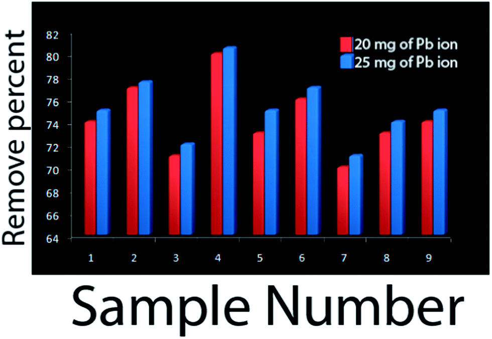
Fig. 8a and b show the adsorption kinetics of Pb(II) and Hg(II) ions onto the CdS/ZnS core–shell nanoparticles. It seems that the increase in reaction time and ultrasonic power result in an increase in adsorption of Pb(II) and Hg(II) ions, which is due to the increase in size of CdS/ZnS core–shell nanoparticles. Furthermore, an increase in concentration of Zn(NO3)2·6H2O results in a decrease in adsorption of Pb(II) and Hg(II) ions due to agglomeration of the nanoparticles. It seems Hg(II) ions, due to their atomic radius, are absorbed more than Pb(II). To investigate the concentration of Pb ion on the remove percent, we used a 25 mg L−1 solution of Pb ions. By increasing the ion concentration, the percentage of Pb removed was increased (Fig. 9).
 |
| | Fig. 8 Adsorption kinetics of (a) Pb(II) (b) Hg(II) ions onto CdS/ZnS core–shell nanoparticles. | |
 |
| | Fig. 9 Adsorption kinetics of Pb(II) ions onto CdS/ZnS core–shell nanoparticles with different concentrations of Pb(II) ions. | |
Conclusions
In summary, Cd/ZnS core–shell nanoparticles were successfully synthesized by sonochemical reactions at 60 W for 40 min. CdSO4.8H2O and Zn(NO3)2·6H2O were used as starting reagents in distilled water. Their PL emissions were at approximately 650 nm for pure CdS nanoparticles, 540 nm for ZnS, and 598 nm for CdS/ZnS core–shell nanocomposites. In this paper, we investigated the effect of preparation parameters such as ultrasonic power, irradiation time, and precursor concentration on the morphology of CdS/ZnS core–shell nanoparticles. Furthermore, the removal efficiency was controlled by ultrasonic power, irradiation time, and precursor concentration. Cd/ZnS core–shell nanoparticles were characterized by XRD, PL, TEM, HRTEM, EDX, and SEM.
Acknowledgements
Authors are grateful to council of Young Research Club, Kashan Branch for supporting this work by grant no. (159271/19) and TEM section, SAIF, NEHU, Shillong, Meghalaya, India, for providing financial support to undertake this work.
References
- L. Brus, J. Phys. Chem., 1986, 90, 2555 CrossRef CAS.
- S. M. Hosseinpoor-Mashkani, F. Mohandes, M. Salavati-Niasari and K. Venkateswara-Rao, Mater. Res. Bull., 2012, 47, 3148 CrossRef PubMed.
- C. B. Murray, D. J. Norris and M. G. Bawendi, J. Am. Chem. Soc., 1993, 115, 8706 CrossRef CAS.
- Z. A. Peng and X. Peng, J. Am. Chem. Soc., 2001, 123, 183 CrossRef CAS.
- W. W. Yu and X. Peng, Angew. Chem., Int. Ed., 2002, 41, 2368 CrossRef CAS.
- J. J. Li, Y. A. Wang, W. Guo, J. C. Keay, T. D. Mishima, M. B. Johanson and X. Peng, J. Am. Chem. Soc., 2003, 125, 12567 CrossRef CAS PubMed.
- Y. Wang, H. Yang, Z. Xia, Z. Tong and L. Zhou, Bull. Korean Chem. Soc., 2011, 32, 2316 CrossRef CAS.
- Y. J. Lee, T. G. Kim and Y. M. Sung, Nanotechnology, 2006, 17, 3539 CrossRef CAS.
- P. Reiss, J. Bleuse and A. Pron, Nano Lett., 2002, 2, 781 CrossRef CAS.
- J. J. Li, Y. A. Wang, W. Guo, J. C. Keay, T. D. Mishima, M. B. Johnson and X. Peng, J. Am. Chem. Soc., 2003, 125, 12567 CrossRef CAS PubMed.
- D. V. Talapin, A. L. Rogach, A. Kornowski, M. Haase and H. Weller, Nano Lett., 2001, 1, 207 CrossRef CAS.
- B. Yu Xia, S. Ding, H. Bin Wu, X. Wang and X. Wen, RSC Adv., 2012, 2, 792–796 RSC.
- X. Wang, W. Li, B. Zhao, D. Zhang, K. Sun, X. An, Z. Zhang and Z. Shen, RSC Adv., 2013, 3, 3553–3556 RSC.
- B. T. Huy, M. H. Seo, P. T. Phong, J. M. Lim and Y. I. Lee, Chem. Eng. J., 2014, 236, 75–81 CrossRef CAS PubMed.
- W. S. W. Ngah and M. A. K. M. Hanafiah, Bioresour. Technol., 2008, 99, 3935 CrossRef PubMed.
- S. H. Abdel-Halim, A. M. A. Shehata and M. F. El-Shahat, Water Res., 2003, 37, 1678 CrossRef CAS.
- K. Zhang, W. H. Cheung and M. Valix, Chemosphere, 2005, 60, 1129 CrossRef CAS PubMed.
- X. Han, L. Gai, H. Jiang, L. Zhao, H. Liu and W. Zhang, Synth. Met., 2013, 171, 1–6 CrossRef CAS PubMed.
- C. M. Babu, B. Palanisamy, B. Sundaravel, M. Palanichamy and V. Murugesan, J. Nanosci. Nanotechnol., 2013, 13, 2517–2527 CrossRef CAS PubMed.
- Y. Tang, S. Liang, J. Wang, S. Yu and Y. Wang, J. Environ. Sci., 2013, 25, 830–837 CrossRef CAS.
- A. Ewecharoena, et al., Journal of Hazardous Materials Chemosphere, 2009, 171, 335 CrossRef PubMed.
- S. Zhang, J. Alloys Compd., 2006, 426, 281 CrossRef CAS PubMed.
- D. Carriazo, Appl. Clay Sci., 2007, 37, 231 CrossRef CAS PubMed.
- N. Mir and M. Salavati-Niasari, Mater. Res. Bull., 2013, 48, 1660 CrossRef CAS PubMed.
- L. Wang, H. W. Wei, Y. J. Fan, X. Z. Liu and J. H. Zhan, Nanoscale Res. Lett., 2009, 4, 558 CrossRef CAS PubMed.
- R. G. Xie, U. Kolb, J. X. Li, T. Basche and A. Mews, J. Am. Chem. Soc., 2005, 127, 7480 CrossRef CAS PubMed.
- A. R. Abbasi and A. Morsali, Ultrason. Sonochem., 2010, 17, 572 CrossRef CAS PubMed.
|
| This journal is © The Royal Society of Chemistry 2014 |
Click here to see how this site uses Cookies. View our privacy policy here.