A simple one-step solution deposition process for constructing high-performance amorphous zirconium oxide thin film

Yongjuan Miab, Jinqing Wang*a, Zhigang Yanga, Zhaofeng Wanga, Honggang Wang*a and Shengrong Yanga
aState Key Laboratory of Solid Lubrication, Lanzhou Institute of Chemical Physics, Chinese Academy of Sciences, Lanzhou, 730000, P. R. China. E-mail: jqwang@licp.cas.cn; hgwang@licp.cas.cn; Fax: +86-931-8277088; Tel: +86-931-4968076
bUniversity of Chinese Academy of Sciences, Beijing, 100080, P. R. China

Received 28th October 2013 , Accepted 4th December 2013

First published on 5th December 2013


Abstract

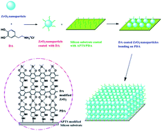
Based on the magic substance dopamine (DA), a simple and effective one-step solution deposition strategy has been developed for constructing high-performance amorphous zirconium oxide (ZrO2) nanocomposite thin film. DA can be auto-polymerized to form a polydopamine (PDA) coating, which adheres strongly to the silicon substrate and serves as an active platform to induce the subsequent deposition of ZrO2. The obtained ZrO2 nanocomposite thin films with the thickness of about one hundred nanometers presents outstanding mechanical and tribological performances even without a subsequent high-temperature annealing process. More importantly, the preparation process is definitely low-consumption and environmentally friendly. This work overcomes the claim that ZrO2 films obtained by aqueous solution deposition process generally possess low mechanical properties and thus avoid the defects of focusing mainly on nature and performances of heat-treated and crystalline ZrO2. Based on this work, it is believed that the as-deposited ZrO2 nanocomposite film can be widely used as a protecting coating and lubricating material for micro- and nano-electromechanical systems (MEMS/NEMS) and many other systems working in similar conditions.


1. Introduction

Due to the unique combination of excellent thermal, chemical and mechanical stability as well as excellent tribological property, ZrO2 can be used as protective coatings for micro- and nano-electromechanical systems (MEMS/NEMS) working in harsh, corrosive, friction and wear environments. However, most of these studies are mainly focused on high-temperature treated and crystalline ZrO2, due to the poor performances of the unannealed amorphous ZrO2,1–3 such as low mechanical and poor tribological performances. As to be discussed in the next paragraph, the preparation of such widely-researched ZrO2 usually needs vacuum environment and special apparatus or mild environment accompanied by a severe high-temperature post-treatment process; apparently, both of which are not the ideal routes to fabricate ZrO2 due to the above mentioned disadvantages. So, it is quite necessary to develop a simple and mild preparation method which does not need high-temperature post-treatment for ZrO2 film with good mechanical and tribological performances.

However, to the best of our knowledge, till now, such a process has not been proposed. The conventional methods used to fabricate ZrO2 film can mainly be divided into two categories, viz., vacuum-based and solution-based techniques. The formers, including physical vapor deposition (PVD),4 chemical vapor deposition (CVD),5 sputtering,6,7 ion-beam-assisted evaporation,8 and electron beam evaporation,9 are effective to fabricate ZrO2 films with good mechanical and tribological performances; however, special apparatus and vacuum environment are required. The latter ones, including the sol–gel deposition,10–14 the so-called “biomimetic” approach,15–21 as well as the self-assembled and layer-by-layer (LbL) self-assembled techniques,3,19,22–24 are impressive for simple solution deposition process and mild conditions (such as low process temperature and atmospheric pressure) but limited by their tedious operation process and poor low-temperature performances (in other words, a high-temperature post-treatment is generally needed to improve the performances).

Considering the advantages and disadvantages of the preparation methods mentioned above, the aim of this research is to develop a novel solution deposition process which promotes the advantages and overcomes the disadvantages of the aforementioned preparation techniques, namely, to develop ZrO2 film with good mechanical and tribological performances by a mild deposition process without the aid of special apparatus and avoid the high-temperature post-treatment step. In this work, such a preparation strategy for high-performance amorphous ZrO2 film is proposed. The key factor for this novel process is attributed to the introduction of dopamine (DA), which plays multiple roles in our present work. Specifically, on one hand, DA is auto-polymerized to form polydopamine (PDA), which adheres strongly to various substrates and serves as an active platform to induce the subsequent deposition of ZrO2. On the other hand, DA molecules are supposed to chelate with the as-formed ZrO2 particles (the formation mechanism is to be discussed later) in the bulk solution and results in a stable particle dispersion.25–27 These two functions of DA induce the formation of ZrO2 films with outstanding mechanical and tribological performances, even without a subsequent high-temperature annealing process which is indispensable to improve the mechanical and tribological performances of ZrO2 obtained by a similar solution-based process.

From above discussions, it is obvious that the biggest bright spot of this work is the creative use of DA in the one-step solution deposition process, by virtue of which ZrO2 thin film with high performances is obtained without the need of high-temperature post-treatment. In the light of these significant advantages of the as-proposed one-step solution deposition process, it is expected to accelerate the production and application of ZrO2 film in various fields.

2. Materials and methods

2.1. Materials

N-type polished single-crystal silicon (100) wafers were purchased from GRINM Semi-conductor Materials. 3-hydroxytyramine hydrochloride (dopamine hydrochloride, 99%), 3-aminopropyl triethoxysilane (APTS, 99%), and tris(hydroxymethyl) aminomethane (Tris, 99.8%) were purchased from Acros Organics and used as received. All other reagents are analytical grade and used as received. Ultrapure water (>18 MΩ cm) was used for rinsing and as the solvent as well.

2.2. Deposition of ZrO2 thin film

The Si-APTS-PDA coated wafers (the details of the preparation procedure are described in ESI) were immersed into a mixed solution of freshly prepared 4 mM Zr(SO4)2/0.2 M HCl and 10 mM DA and then kept at 50 °C for a desired time. Subsequently, the samples were taken out, ultrasonically cleaned and rinsed with ultrapure water, followed by completely drying with N2 flow. For comparison, homogenous ZrO2 thin film was also fabricated by immersion of the Si-APTS-PDA coated substrates into 4 mM Zr(SO4)2/0.2 M HCl solution kept at 50 °C for the same time.

2.3. Characterization of ZrO2 films

The water contact angle (WCA) was recorded on a DSA100 contact angle meter (Krüss, Germany). The listed results for each sample are the average values of at least five repeat measurements. X-ray photoelectron spectroscopy (XPS) was performed on a PHI-5702 (Physical Electronics, USA) spectrometer with monochromatic Al Kα irradiation and the chamber pressure of ∼3 × 10⁻⁸ Torr to detect the chemical composition and binding energy state of the films. Peak deconvolution with Gaussian curves of elements was accomplished by Origin 7.0 and the binding energies in all the XPS spectra were all calibrated using that of C 1s (284.8 eV). The field emission transmission electron microscopy (FE-TEM) image was recorded on a TF20 FE-TEM (FEI, USA). The section morphology and the thickness of films were obtained using a field emission scanning electron microscopy (FE-SEM, JSM-6701F, JEOL, Japan). The surface morphology of different samples was observed with a Nanoscope IIIa multimode atomic force microscope (AFM, Veeco, USA) in tapping mode and the FE-SEM. The information of the crystallographic structure was determined on an X-ray diffractometer D/MAX-2400 (Rigaku, Japan) operating with Cu Kα (λ = 1.54056 Å) radiation and graphite monochrometer.

The mechanical properties of samples were investigated by TI 950 Triboindenter in situ nanomechanical test system (Hysitron, USA) with an indentation depth of 30 nm. The macrotribological properties of different films were measured on the UMT-2MT tribometer (CETR) in a ball-on-plate contact configuration. Commercially available steel ball (φ = 3 mm, announced mean roughness = 0.02 μm) was used as the stationary upper counterpart, whereas the lower tested samples were mounted onto the flat base and driven to slide reciprocally at a distance of 0.5 cm. The friction coefficient-versus-time curve was recorded automatically. At least three repetitive measurements were performed for each frictional pair. The friction coefficient and antiwear life were measured at a relative error of ±10 and ±5%, respectively.

3. Results and discussion

3.1. The formation of ZrO2 film

According to literatures,17,19,23 ZrO2 nanoparticles can be formed by controlling hydrolysis of zirconium sulfate and the proposed major equilibrium reactions are listed as follows:
2[Zr(SO4)2]n + 3nH2O ↔ n[Zr2(OH)3(SO4)4]3− + 3nH+

[Zr2(OH)3(SO4)4]3− + 3OH ↔ Zr2(OH)6SO4 + 3SO42−

In zirconium sulfate precursor solution, ionic species and oligomers present may experience polymerization and condensation to form ZrO2 nanoparticles, which may be further modified by DA molecules due to its high affinity. Although the detailed binding mechanism between DA and ZrO2 has not been fully understood, binding does occur and can be evidenced by XPS analysis (to be discussed later) and the previous works.28,29 Such modification of DA can restore a more stable configuration for ZrO2 nanoparticles,30–36 thus allowing to form a stable particle dispersion. To confirm this supposition, FE-TEM observation of the precursor solutions for depositing both films (homogenous ZrO2 film and the DA modified ZrO2 nanocomposite film) were performed to detect the state of particles and the result confirms this supposition, which will be discussed in part 3.2.

Then, ZrO2 nanoparticles modified by DA can be deposited onto PDA surface. The detailed mechanism for such deposition may be understood by following analyses. Specifically, deposition on PDA may occur in two ways, viz., heterogeneous nucleation process or homogeneous nucleation process (accompanied by a subsequent surface bonding). Heterogeneous nucleation is facilitated by the hydroxyl groups on PDA surface, which are supposed to chelate with Zr4+ in solution and then facilitate the nucleation and growth of ZrO2. Homogeneous nucleation process involves the formation of ZrO2 particles in the bulk solution and the subsequent surface bonding process, the driving force for which may be Van der Waals force, π–π interaction or hydrogen bond interaction between DA modified ZrO2 particles and PDA-SAM.37–39 Considering the low surface density of free catechols on PDA and our experimental conditions, which favor the formation of particles in the bulk solution (as discussed earlier), the homogeneous nucleation may be the main deposition mechanism.19,22,40 The process and possible deposition mechanism for construction of ZrO2-based nanocomposite thin film are shown in Scheme 1.


image file: c3ra46169f-s1.tif
Scheme 1 The process for construction of ZrO2-based nanocomposite thin film.

3.2. Characterization of the film

WCA measurement was used to characterize the variation of surface wettability. As listed in Table 1, the hydroxylated silicon surface is highly hydrophilic with the WCA of about 0°. Once the APTS and PDA films prepared on the surface, the WCA greatly increased to ∼50°. Due to the as-deposited ZrO2 nanocomposite film shows a similar WCA value with the PDA sublayer, ellipsometric thickness measurement and cross-sectional FE-SEM image analysis (as inserted in Fig. 3a) were combined together to ensure the formation of ZrO2 nanocomposite films on the substrate. The significant increase of film thickness to 105.2 nm indicates the formation of ZrO2 nanocomposite film on the surface of PDA.
Table 1 The surface WCA and thickness for various films
Samples WCA/° Thickness/nm
Si/SiO2 ∼0 ∼4.0
APTS-SAM 49.6 ± 1.7 ∼0.75
APTS-PDA 50.0 ± 1.1 ∼14.3
ZrO2 nanocomposite film 48.3 ± 6.2 ∼105.2


The chemical composition and corresponding chemical state of the as-deposited film were investigated by XPS. As shown in Fig. 1a, peaks corresponding to zirconium (Zr), oxygen (O), carbon (C), and nitrogen (N) elements are clearly recognized on the survey spectrum, indicating that the as-deposited film is consisted of zirconium-related substances. The presence of N further validates the affinity of DA with ZrO2 nanoparticles (XPS is used to detect surface composition and its detection depth is about ∼10 nm, which is far less than the thickness of the as-deposited ZrO2 film, so the influence of N in PDA underlayer can be eliminated). As depicted in Fig. 1b, the binding energies for Zr 3d 5/2 and Zr 3d 3/2 are 182.5 and 184.9 eV, respectively, which can be identified as Zr(IV) in the oxide.41,42 Deconvolution of O1s core-level spectrum results in four components: Zr–O–Zr (530.2 eV), Zr–O–H (531.8 eV), H–O–H (532.6 eV),43,44 and oxygen from DA (533.2 eV),45 as clearly displayed in Fig. 1d.


image file: c3ra46169f-f1.tif
Fig. 1 XPS spectrum of the as-deposited ZrO2 nanocomposite film: (a) XPS survey spectrum; (b) XPS spectrum of Zr 3d; (c) XPS spectrum of N 1s and (d) deconvolution result of XPS O 1s core-level spectra.

In order to evaluate the effect of annealing process on surface composition and the corresponding chemical state of the as-deposited ZrO2 nanocomposite film, XPS analysis was also carried out for the 500 °C annealed sample, as shown in Fig. S1. In comparison with the as-deposited film, significantly enhanced Zr 3d is recorded and the binding energies for Zr 3d 5/2 and Zr 3d 3/2 show slight shift to 182.3 and 184.6 eV, respectively. Such variation of Zr 3d is attributed to the elimination of partial surface species, such as DA (the disappearance of N 1s can be regarded as a direct evidence, as illustrated in Fig. S1a and S1c), which results in an increase of Zr exposed on the surface. This viewpoint can be further verified from the O1s XPS spectrum of the annealed film in which the oxygen state is mainly composed of three kinds of oxygen bonds: Zr–O–Zr (530.2 eV), Zr–O–H (531.8 eV) and H–O–H (532.7 eV), as illustrated in Fig. S1d. Comparing with the unannealed film, oxygen bond attributed to DA (533.2 eV) disappears and that there is an increase in proportion of the oxide (Zr–O–Zr), which also tallys well with the proposition.

The structure information for the surface film was determined by grazing incident angle X-ray diffraction (GIAXRD) with an incident angle of 2°. Fig. 2d shows GIAXRD patterns of the as-prepared ZrO2 nanocomposite film and the 500 °C annealed sample. Obviously, no diagnostic peaks belonging to crystalline ZrO2 are recorded, indicating the as-deposited ZrO2 is amorphous. And the pattern for 500 °C annealed sample is nearly identical with the as-deposited film, revealing that the as-constructed ZrO2 nanocomposite film sustains its amorphous framework up to 500 °C. For homogenous ZrO2 film, similar results are recorded and no diagnostic peaks belonging to crystalline ZrO2 for both films (homogenous ZrO2 film before and after annealing) are observed, as shown in Fig. S2. These results are deviated from previously reported results where the annealing of these films at 500 °C led to crystallization of amorphous ZrO2.1,17,19,41,46,47 Through fully analysis of experimental conditions, we think that it maybe the surface species, such as DA, or the SAM coating that serve as “heat absorber” and prevent crystallization of ZrO2 during annealing,44 and further research is needed to find out the specific reasons.


image file: c3ra46169f-f2.tif
Fig. 2 (a) FE-TEM image of the precursor solution for the homogenous ZrO2 after being kept at 50 °C for 10 h and the corresponding SAED pattern as inset in (a); (b) and (c) FE-TEM images of the precursor solution for ZrO2 nanocomposite film after being kept at 50 °C for 10 h and the corresponding SAED pattern as inset in (c); (d) GIAXRD patterns of the as-prepared ZrO2 nanocomposite film and the film annealed at 500 °C for 1 h.

Composition, structure and morphology are important factors affecting film performance. FE-TEM images present the state of particles in the precursor solutions for the homogenous ZrO2 film and the DA modified ZrO2 nanocomposite film after being kept at 50 °C for 10 h. It can be seen from Fig. 2a that in the precursor solution for homogenous ZrO2 film, aggregates from large particles are formed; however, for the precursor solution of DA modified ZrO2 film, small grains are produced and well dispersed with no big aggregate observed, as shown in Fig. 2b and c. This result confirms that the introduction of DA is indeed in favor of stable particle dispersion. The corresponding SAED patterns for homogenous ZrO2 and DA modified ZrO2 both present characteristic results of an amorphous phase, thus the obtained homogenous ZrO2 and DA modified ZrO2 are all amorphous, which consists with the GIAXRD result (as inserted in Fig. 2a and c).

The morphology of the as-constructed film was observed by FE-SEM and AFM. Fig. 3a and b provide FE-SEM images of the as-prepared ZrO2 nanocomposite film and the homogeneous ZrO2 film. It is obvious that both films are composed of densely packed ZrO2 nanoparticles and all surfaces are integrated and defect-free.


image file: c3ra46169f-f3.tif
Fig. 3 (a) FE-SEM image of the as-prepared ZrO2 nanocomposite film. The corresponding cross-sectional FE-SEM image is shown as inset; (b) FE-SEM image of the as-prepared homogeneous ZrO2 film and (c) AFM morphology of the as-prepared ZrO2 nanocomposite film with a scanning area of 1 μm × 1 μm and the corresponding section analysis.

Remarkably, for the 500 °C annealed homogenous ZrO2 film, obvious cracks and hole defects are visible; however, no surface defects are observed for the annealed ZrO2 nanocomposite film, as depicted in Fig. S3a and S3b. This result supports that the modification of ZrO2 with DA enhances the packing density of ZrO2 nanoparticles, which can be attributed to the strong interaction between adjacent ZrO2 nanoparticles (such as Van der Waals force, π–π interaction and hydrogen bond interaction), thus avoiding the generation of surface defects caused by the extending of voids during annealing process.

To get more accurate and intuitionistic surface information of the as-deposited ZrO2 nanocomposite film, AFM image of the obtained film was also taken, as displayed in Fig. 3c and S4a. A surface film composed of huge numbers of nanoparticles is observed with a particle diameter of 15–20 nm. As the FE-SEM image displays, the obtained film is integrated and crack-free with the root-mean-square (RMS) surface roughness of 2.652 nm. For the 500 °C annealed ZrO2 nanocomposite film, it takes similar surface morphology accompanied with slightly increased particle size of 20–30 nm and significantly reduced RMS surface roughness of 1.265 nm, as depicted in Fig. S3c and S4b.

3.3. Mechanical properties

As is well known, ceramic materials gaining access to wide application is largely dependent on its excellent mechanical and tribological properties. In this work, the mechanical performances (microhardness and reduced modulus) of various films were evaluated by the Triboindenter in situ nanomechanical test system at an indentation depth of 30 nm and the results are listed in Table 2.
Table 2 Hardness and reduced modulus of ZrO2 nanocomposite films at an indentation depth of 30 nm
Samples H (GPa) Er (GPa)
As-deposited 8.105 218.465
300 °C 12.825 220.210
400 °C 13.460 191.630
500 °C 15.935 214.250


Compared with the as-deposited homogenous ZrO2 film (Table S1) and previous reported results,1–3,23 the mechanical performance of the as-deposited ZrO2 nanocomposite film exhibits remarkable improvement with an enhanced hardness (H) and an reduced modulus (Er) of ∼8.1 GPa and ∼218 GPa, respectively. For the applied test system, the values of Er and elastic modulus (Es) abide by the following equation:

image file: c3ra46169f-t1.gif

Here, Es, Vs and Ei, Vi are the elastic modulus and Poisson's ratio for the tested sample and the diamond indenter, respectively. By computation, the value of image file: c3ra46169f-t2.gif is close to zero, thus Er and Es have an approximately linear relationship. Therefore, to some extent, the variation of Er can represent the change trend of Es. The outstanding mechanical properties of the as-deposited ZrO2 nanocomposite film can be ascribed to the formation of organic–inorganic hybrid microstructure. Namely, the modification of ZrO2 with DA molecules, which functionalize as the stabilizer and adjuster for ZrO2, gives additional stability (as has been discussed on the FE-TEM images) and high particle packing density due to the enhanced cohesion between particles, which can be confirmed from the FE-SEM images of the 500 °C annealed homogenous ZrO2 film and the DA modified ZrO2 nanocomposite film (as have been discussed in FE-SEM analysis). Thus the applied surface pressure can be well spread and scattered in the whole coating. For the annealed sample, the hardness of the obtained film gains obvious improvement. The 500 °C annealed sample presents an H of 15.935 GPa that is similar to 500 °C annealed homogenous sample (See Table S1). This may be a result of the burnout of surface species and the decrease of the microvoids in the film. On the other hand, this result confirms that the outstanding mechanical properties of the as-obtained nanocomposite film may be largely ascribed to the introduction of DA.

3.4. Tribological properties

In this work, the tribological properties of the as-deposited ZrO2 film under various conditions were performed and recorded in Fig. 4. In addition, the tribological performances of the as-deposited homogenous ZrO2 film were also carried out under the same condition for comparison.
image file: c3ra46169f-f4.tif
Fig. 4 Variation in friction coefficient with time for various film samples under different applied loads and a fixed sliding frequency of 1 Hz: the as-deposited homogenous ZrO2 at the load of 0.1 N (a); the as-deposited ZrO2 nanocomposite film under the loads of 0.3 N (b), 0.5 N (c), and 1.0 N and (d). The stable friction coefficient (FC) was given above the corresponding curve.

As shown in Fig. 4a, the as-deposited homogenous ZrO2 film shows poor tribological properties with a rapidly increased friction coefficient (FC) even under mild testing conditions of 0.1 N and 1 Hz. In sharp contrast, the as-deposited ZrO2 nanocomposite film exhibits significantly improved tribological behaviors. Within the significantly extended operation period (6000 s) and under the progressively lifted loads, the friction coefficients steadily maintain at a constant low level. Specifically, under the applied load of 0.3 N, the friction coefficient of the as-deposited nanocomposite film keeps stably at the value of ∼0.08, as shown in Fig. 4b. Further increasing the applied load to 0.5 N and 1.0 N, the friction coefficients still maintain at low levels of 0.097 and ∼0.1187, respectively. These results manifest that the as-deposited film has possessed excellent friction-reduction property with long anti-wear life and high load carrying capacity compared with the as-deposited homogenous ZrO2 film and some annealed samples.3,48 The outstanding tribological performances can be attributed to the modification of DA, which not only improves the dispersibility and stability of ZrO2 particles (which has been illustrated in the discussion part for the FE-TEM results) but also endows high particle packing density and cohesion (which can be reflected from the FE-SEM images of 500 °C annealed homogenous ZrO2 film and the DA modified ZrO2 film).

To explore the effect of heat treatment on the tribological performances of the as-deposited films, tribological test of the 500 °C annealed samples was also conducted under the same condition. As exhibited in Fig. S5, the 500 °C annealed homogenous ZrO2 film still presents poor performance; nevertheless, the annealed ZrO2 nanocomposite film still has significant advantage in tribological performance. To be specific, under lower loads, the annealed nanocomposite film displays good experiment results as reports;3,48 notably, further lifting the applied load, such annealed ZrO2 nanocomposite film begins to show advantages in stability on both friction coefficient and antiwear life. This result confirms that the obtained ZrO2 nanocomposite film has excellent anti-friction stability.

Fig. 5 presents the micrographs of the worn surfaces for the as-deposited and 500 °C annealed ZrO2 nanocomposite films after the pre-configured experimental period of 6000 s. As shown in Fig. S6, the worn surfaces of the as-deposited and 500 °C annealed homogeneous ZrO2 thin films were also observed for comparison, and the signs of severe peeling off are visible accompanied with the generation of a mass of wear debris on the surfaces, which lead to the invalidation of films even at a quite small load of 0.1 N. Obviously, contrary to the homogeneous ZrO2 thin films, the as-deposited and 500 °C annealed ZrO2 nanocomposite films show mild scuffing signs even after sliding against the steel ball for 6000 s at high load of 1.0 N and little wear debris is visible on the wear tracks, which agrees well with the good wear-resistance of the ZrO2 nanocomposite thin film.


image file: c3ra46169f-f5.tif
Fig. 5 SEM micrographs of the worn surfaces of ZrO2 nanocomposite thin films sliding against steel ball under the applied load of 1.0 N and a fixed sliding frequency of 1 Hz: the as-deposited ZrO2 nanocomposite thin film (a) and (c), the 500 °C annealed ZrO2 nanocomposite thin film (c) and (d).

Based on the above analyses, it is clear that the as-deposited ZrO2 nanocomposite film possesses competitive tribological performance characterized by low friction coefficient, high load-carrying capacity, long antiwear life and high anti-friction stability. More significantly, the friction coefficient, antiwear performance and the load-carrying capacity get improvement synchronously. These results ensure the as-deposited ZrO2 nanocomposite film an excellent lubricating film that can work as protective coatings for MEMS/NEMS applied in harsh friction and wear environments.

4. Conclusions

In this work, a simple one-step solution deposition process was proposed for the construction of ZrO2 nanocomposite film with significantly improved performances on SAM-modified silicon substrate. Compared with other solution-based methods, the pre-modification of PDA coating makes this method a versatile substitute with wider applicability and greater simplicity. Meanwhile, different from the homogeneous ZrO2 thin film, the introduction of DA into precursor solution endows the obtained film significant improvement in mechanical and tribological performances. More importantly, the preparation process is definitely low-consumption and environmentally friendly, solving the troubles of multifarious experimental operation and harsh experiment conditions (such as special apparatus, high temperature and high vacuum, etc.). Thusly, the as-prepared ZrO2 nanocomposite film with splendid mechanical and tribological performances is expected to be a good candidate for coating protection and lubricating material in MEMS/NEMS and other systems working in harsh environments such as high-temperature, friction and wear. In a word, this work not only offers us a simple route for constructing high-performance ZrO2 nanocomposite film on various substrates but also enriches the research points of ceramic materials.

Acknowledgements

We thank the financial support from the National Natural Science Foundation of China (Grant nos. 51075384, 51375474 and 51205385) and the “Funds for Distinguished Young Scientists of Gansu Province” scheme.

Notes and references

  1. I. Zlotnikov, I. Gotman and E. Y. Gutmanas, Appl. Surf. Sci., 2008, 255, 3447–3453 CrossRef CAS PubMed.
  2. A. Rosidian, Y. Liu and R. O. Claus, Adv. Mater., 1998, 10, 1087–1091 CrossRef CAS.
  3. J. Wang, X. Liu, S. Ren, F. Guan and S. Yang, Tribol. Lett., 2005, 18, 429–436 CrossRef CAS PubMed.
  4. A. S. James and A. Matthews, Surf. Coat. Technol., 1990, 41, 305–313 CrossRef CAS.
  5. J. S. Kim, H. A. Marzouk and R. J. Rencroff, Thin Solid Films, 1995, 254, 33–38 CrossRef CAS.
  6. Y. M. Sun, J. Lozano, H. Ho, H. J. Park, S. Veldaman and J. M. White, Appl. Surf. Sci., 2000, 161, 115–122 CrossRef CAS.
  7. D. H. Kuo and C. H. Chien, Thin Solid Films, 2003, 429, 40–45 CrossRef CAS.
  8. M. Matsuoka, S. Isotani, S. Miyake, S. Setsuhara, K. Ogata and N. Kuratani, J. Appl. Phys., 1996, 80, 1177–1181 CrossRef CAS PubMed.
  9. K. H. Muller, R. P. Netterfield and P. J. Martin, Phys. Rev. B: Condens. Matter Mater. Phys., 1987, 35, 2934–2941 CrossRef CAS.
  10. J. S. Lee, T. Matsubara, T. Sei and T. Tsuchiya, J. Mater. Sci., 1997, 32, 5249–5256 CrossRef CAS.
  11. M. J. Paterson, P. J. K. Paterson and N. B. Ben, J. Mater. Res., 1998, 13, 388–395 CrossRef CAS.
  12. R. Brenier, J. Mugnier and E. Mirica, Appl. Surf. Sci., 1999, 143, 85–91 CrossRef CAS.
  13. M. C. Caracoche, P. C. Rivas and M. M. Cervera, J. Am. Ceram. Soc., 2000, 83, 377–384 CrossRef CAS.
  14. K. Izumi, M. Murakami, T. Deguchi and A. Morita, J. Am. Ceram. Soc., 1989, 72, 1465–1468 CrossRef CAS.
  15. K. Chitre, Q. Yang, T. O. Salami, S. R. Oliver and J. Cho, J. Electron. Mater., 2005, 34, 528–533 CrossRef CAS PubMed.
  16. B. C. Bunker, P. C. Rieke, B. J. Tarasevich, A. A. Campbel, G. E. Fruxell, G. L. Graff, L. Song, J. W. Virden and G. L. McVay, Science, 1994, 264, 48–55 CAS.
  17. M. Agarwal, M. R. De Guire and A. H. Heuer, J. Am. Ceram. Soc., 1997, 80, 2967–2981 CrossRef CAS.
  18. A. D. Polli, T. Wagner, A. Fischer, G. Weinberg, F. C. Jentoft, R. Schloegl and M. Ruehle, Thin Solid Films, 2000, 379, 122–127 CrossRef CAS.
  19. J. Wang, S. Yang, X. Liu, S. Ren, F. Guan and M. Chen, Appl. Surf. Sci., 2004, 221, 272–280 CrossRef CAS.
  20. V. V. Roddatis, D. S. Su, E. Beckmann, F. C. Jentoft, U. Braun, J. Krohnert and R. Schlogl, Surf. Coat. Technol., 2002, 151–152, 63–66 CrossRef CAS.
  21. T. O. Salami, Q. Yang, K. Chitre, S. Zarembo, J. Cho and S. R. J. Oliver, J. Electron. Mater., 2005, 34, 534–540 CrossRef CAS PubMed.
  22. J. Ou, J. Wang, Y. Qiu, L. Liu and S. Yang, Surf. Interface Anal., 2011, 43, 803–808 CrossRef CAS.
  23. G. Zhang, J. Y. Howe, D. W. Coffey, D. S. Blom, L. F. Allard and J. Cho, Mater. Sci. Eng., C, 2006, 26, 1344–1350 CrossRef CAS PubMed.
  24. G. Freiman, P. Barboux, J. Perrière and K. Giannakopoulos, Thin Solid Films, 2009, 517, 2670–2674 CrossRef CAS PubMed.
  25. H. Lee, S. M. Dellatore, W. M. Miller and P. B. Messersmith, Science, 2007, 318, 426–430 CrossRef CAS PubMed.
  26. M. Guvendiren, P. B. Messersmith and K. R. Shull, Biomacromolecule, 2008, 9, 122–128 CrossRef CAS PubMed.
  27. M. J. Sever, J. T. Weisser, J. Monahan, S. Srinivasan and J. Wilker, Angew. Chem., Int. Ed., 2004, 43, 448–450 CrossRef CAS PubMed.
  28. R. Y. Floriolli, J. V. Langen and J. H. Waite, Mar. Biotechnol., 2000, 2, 352–363 CAS.
  29. J. H. Waite and X. Qin, Biochemistry, 2001, 40, 2887–2893 CrossRef CAS PubMed.
  30. B. Malisova, S. Tosatti, M. Textor, K. Gademann and S. Zürcher, Langmuir, 2010, 26, 4018–4026 CrossRef CAS PubMed.
  31. P. Tarakeshwar, F. D. Shapiro, S. J. Hurst, T. Rajh and V. Mujica, J. Phys. Chem. C, 2011, 115, 8994–9004 CAS.
  32. N. Lee, D. R. Hummer, D. A. Sverjensky, T. Rajh, R. M. Hazen, A. Steele and G. D. Cody, Langmuir, 2012, 28, 17322–17330 CrossRef CAS PubMed.
  33. S. J. Hurst, H. C. Fry, D. J. Gosztola and T. Rajh, J. Phys. Chem. C, 2011, 115, 620–630 CAS.
  34. T. Rajh, L. X. Chen, K. Lukas, T. Liu, M. C. Thurnauer and D. M. Tiede, J. Phys. Chem. B, 2002, 106, 10543–10552 CrossRef CAS.
  35. S. Zürcher, D. Wäckerlin, Y. Bethuel, B. Malisova, M. Textor, S. Tosatti and K. Gademann, J. Am. Chem. Soc., 2006, 128, 1064–1065 CrossRef PubMed.
  36. X. L. Wang, Z. Y. Jiang, J. F. Shi, Y. P. Liang, C. H. Zhang and H. Wu, ACS Appl. Mater. Interfaces, 2012, 4, 3476–3483 CAS.
  37. Q. Ye, F. Zhou and W. M. Liu, Chem. Soc. Rev., 2011, 40, 4244–4258 RSC.
  38. H. C. Gao, Y. M. Sun, J. J. Zhou, R. Xu and H. W. Duan, ACS Appl. Mater. Interfaces, 2013, 5, 425–432 CAS.
  39. S. L. Phua, L. P. Yang, C. L. Toh, S. Huang, Z. Tsakadze, S. K. Lau, Y. W. Mai and X. H. Lu, ACS Appl. Mater. Interfaces, 2012, 4, 4571–4578 CAS.
  40. J. Ryu, S. H. Ku, H. Lee and C. B. Park, Adv. Funct. Mater., 2010, 20, 2132–2139 CrossRef CAS.
  41. K. Kuratani, M. Uemura, M. Mizuhata, A. Kajinami and S. Deki, J. Am. Ceram. Soc., 2005, 88, 2923–2927 CrossRef CAS.
  42. S. Ardizzone and C. L. Bianchi, Appl. Surf. Sci., 1999, 152, 63–69 CrossRef CAS.
  43. P. Ragupathy, D. H. Park, G. Campet, H. N. Vasan, S. J. Hwang, J. H. Choy and N. Munichandraiah, J. Phys. Chem. C, 2009, 113, 6303–6309 CAS.
  44. N. D. Kim, H. J. Yun, I. Nam and J. Yi, J. Mater. Chem., 2011, 21, 15885–15888 RSC.
  45. Z. Y. Xi, Y. Y. Xu, L. P. Zhu, Y. Wang and B. K. Zhu, J. Membr. Sci., 2009, 327, 244–253 CrossRef CAS PubMed.
  46. F. Namavar, G. H. Wang, C. L. Cheung, R. F. Sabirianov, X. C. Zeng, W. N. Mei, J. Bai, J. R. Brewer, H. Haider and K. L. Garvin, Nanotechnology, 2007, 18, 1–6 CrossRef.
  47. J. H. Park, Y. B. Yoo, K. H. Lee, W. S. Jang, J. Y. Oh, S. S. Chae and H. K. Baik, ACS Appl. Mater. Interfaces, 2013, 5, 410–417 CAS.
  48. Y. X. Chen and W. M. Liu, J. Am. Ceram. Soc., 2002, 85, 2367–2369 CrossRef CAS.

Footnote

Electronic supplementary information (ESI) available. See DOI: 10.1039/c3ra46169f

This journal is © The Royal Society of Chemistry 2014
Click here to see how this site uses Cookies. View our privacy policy here.