Pure gold nanocages by galvanic replacement reaction of magnesium nanoparticles

Xiang Li a, Hui Liu a, Jing Yang a, Shi-Zhang Qiao *ab and Xi-Wen Du *a
aTianjin Key Laboratory of Composite and Functional Materials, School of Materials Science and Engineering, Tianjin University, Tianjin 300072, P.R. China. Fax: +86 22 87894128; Tel: +86 22 27406794
bSchool of Chemical Engineering, The University of Adelaide, Adelaide, SA 5005, Australia. E-mail: s.qiao@adelaide.edu.au; xwdu@tju.edu.cn; Fax: +61-8-83134373; Tel: +61-8-83136443

Received 21st October 2013 , Accepted 14th November 2013

First published on 14th November 2013


Abstract

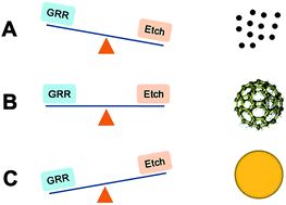
We develop an integrative-gas-liquid strategy to produce Au nanocages with high purity, where Mg nanoparticles are first generated by laser ablation and then blown into aqueous solution for growing Au nanostructures. The Au nanocages exhibit a high DOX-loading capacity, which favors biomedical applications.


Gold nanostructures with tuneable morphologies have received much attention in recent years due to their biocompatibility, non-cytotoxicity, ease of surface modification, and unique localized surface plasmon resonance (LSPR) property.1–6 These materials have been widely applied in the biomedical fields, such as imaging,1,2,5 diagnostics,2 sensing,4,5 photothermal therapy,2,7,8 drug delivery and release.2,5,9–14 Especially, gold nanocages (NCs) with hollow interiors and porous surface usually exhibit LSPR peaks in the near-infrared (NIR) region, particularly from 700 to 900 nm, where the absorption by hemoglobin and water in the blood is negligible.15 Therefore, such nanostructures were highly anticipated as ideal candidates for chemotherapy2,12 and photothermal therapy12 that require deeper penetration in soft tissues.

The most popular synthetic strategy towards Au NCs is based on galvanic replacement reaction (GRR) of metal nanoparticles (NPs). For example, Xia's group synthesized Au NCs by the GRR of silver nanocubes via two distinctive steps, which include alloying and dealloying.16–19 Yu's group utilized cobalt NPs as sacrificial templates to synthesize Au NCs.20 So far, all approaches for Au NCs involve two-step reactions, the sacrificial templates are limited to noble (e.g. Ag) or less active metal (e.g. Co), and the products usually contain the impurity from template materials due to incomplete dealloying in the second step.16–19

To simplify GRR synthesis and obtain pure products, we imagine employing superactive metal NPs, for example, Mg NPs, as sacrificial seeds for GRR. In comparison with Ag template, Mg is not mutually soluble with Au product, which forbids the formation of an alloy intermediate and thus permits a one-step synthesis of pure Au NCs. On the other hand, due to the sensitivity of Mg nanoseeds to the environment, the reaction process and then the final nanostructures can be controlled by simply changing the reaction conditions. Nevertheless, Mg NPs are superactive and hard to be fabricated via conventional wet-chemical routes. In our previous works, pure active metal (such as Mg and Zn) NPs were synthesized by laser ablation of an active-metal target in argon and then transferred into reactive solutions for the synthesis of Au and Ag nanostructures (such a technique was named as separated-gas-liquid (SGL) process).21–25 Owing to their superactivity, active metal NPs are oxidized immediately in the transfer procedure, leading to a surface oxide layer. The oxide layer makes a remarkable influence on the final morphology of the product. Resultantly, spongy nanospheres or solid NPs were obtained.25 So far, Au NCs have never been synthesized by using superactive metal template.

In the present work, we developed a novel technology to generate oxygen-free Mg NPs, where laser ablation was performed in protective gas, and the generated Mg NPs product were blew into a reactive liquid medium by insert gas. The clean Mg NPs then reacted with aqueous chloroauric acid solution, giving rise to Au nanocage. The above process was named as integrated-gas-liquid (IGL) route. The experimental set-up was shown in Fig. S1. We found that, unlike Ag, Mg element was not detected in Au NCs, and raw Mg NPs could be etched out by water, thus the products are always pure Au NCs, which is beneficial for the further photothermal or drug delivery applications.2 Furthermore, the morphology and porosity of the Au NCs were easily manipulated by adjusting the relative rates of GRR and etching reaction of Mg NPs.

As shown in Fig. 1a and b, the IGL technology produces many NCs which possess a hollow interior and a porous shell. In contrast, the SGL process can cause Au NPs merely (see ESI Fig. S3). The selected-area electron diffraction (SAED) pattern in Fig. 1a indicates that the product owns a face-centered cubic structure. High-resolution TEM (HRTEM) image shows that the shell is composed of many grains with interplanar spacing of 0.235 nm, which corresponds to (111) gold planes (Fig. 1c). Energy dispersive X-ray spectroscopy (EDS) analysis (Fig. 1d) performed on an Au nanocube illustrates that the structure only consists of gold element, Cu and C signals arise from the Cu grid and C supporting film, respectively.


image file: c3ra45995k-f1.tif
Fig. 1 Characterizations on Au NCs. (a) TEM image, the inset is a SAED pattern. (b) STEM image, (c) HRTEM image and (d) EDS spectrum.

The AuCl4 concentration and pH value of the solution make significant impact on the morphology of gold nanostructures. Fig. 2a–d show TEM images of the samples obtained at different the concentration of AuCl4 but at a fixed pH value of 7.50. At a low AuCl4 concentration of 0.005 mM, Au NPs were obtained with an average diameter of 3 nm (Fig. 2a). As the concentration was elevated to 0.01 mM, the product consists of loose aggregates composed of 5 nm sized Au NPs (Fig. 2b). Further increase in AuCl4 concentration to 0.025 mM induced gold NCs with a porous shell (Fig. 2c). Finally, Au NCs with a continuous shell are synthesized at a high concentration (0.05 mM), as shown in Fig. 3d. Moreover, Au NCs with a denser shell were produced by fixing the AuCl4 concentration at 0.05 mM but raising the pH value to 8.56 or 9.45 (Fig. 2e and f).


image file: c3ra45995k-f2.tif
Fig. 2 TEM images of Au nanostructures prepared at different experimental conditions. (a) AuCl4 concentration = 0.005 mM, pH = 7.40, (b) AuCl4 concentration = 0.01 mM, pH = 7.40, (c) AuCl4 concentration = 0.025 mM pH = 7.40, (d) AuCl4 concentration = 0.05 mM, pH = 7.40, (e) AuCl4 concentration = 0.05 mM, pH = 8.56, (f) AuCl4 concentration = 0.05 mM, pH = 9.45.

image file: c3ra45995k-f3.tif
Fig. 3 Schematic illustration of competition between GRR and etching and corresponding products.

Next, we discuss the formation mechanisms of Au NCs. When Mg NPs entered the aqueous solution of chloroauric acid, two chemical reactions took place simultaneously. The first one is the GRR between Mg and AuCl4 ions. Since the standard reduction potential of the Mg2+/Mg couple (−2.37 V) is much lower than that of the AuCl4/Au couple (0.99 V), AuCl4 could be reduced into atomic Au by Mg NPs.

 
3Mg + 2AuCl4 → 3Mg2+ + 2Au + 8Cl(1)

The second change is the chemical etching of Mg NPs by the aqueous solution of chloroauric acid, Mg NPs can react fiercely with H+ ions as well as water.

 
Mg + 2H+ → Mg2+ + H2(2)
 
Mg + 2H2O → Mg (OH)2 + H2(3)

The product morphology depends on the competition of galvanic GRR and etching. As schematically illustrated in Fig. 3, when the etching rate is higher than that of GRR (at low AuCl4 concentrations), only a small part of Mg NPs is involved in the GRR for generating tiny Au NPs before the Mg NPs are etched out, and the Au NPs are then dispersed in the solution due to the absence of the structure-directing scaffold, as shown in pathway A, Fig. 3. By increasing the concentration of chloroauric acid or the pH value of the solution, GRR and etching can reach a balance, where many Au nanocrystals are immediately deposited outside the Mg NPs and connect into a network. Meanwhile, the sacrificial template, Mg NPs, was etched out, resulting in Au NCs, as shown in pathway B, Fig. 3. Further increase in the concentration of chloroauric acid or the pH leads to higher GRR rate than that of ER, and compact gold nanospheres are produced by rapid GRR before the elimination of Mg templates by ER, as shown in pathway C, Fig. 3.

We then tested the drug loading properties of the as-prepared Au NCs. DOX is a popular water-soluble drug used for cancer chemotherapy, and its amino group can link with gold nanstructures.26 We attempted two ways for the DOX loading, namely adding DOX into the solution of chloroauric acid before the addition of Mg NPs (in situ way) and after the Au NCs formation (ex situ way), respectively. As shown in the insert of Fig. S4, the plasma resonance absorption for Au NCs emerges in a broad range 600–900 nm. Such a NIR absorption is very crucial for the photothermal therapeutic application,27 because the NIR light can penetrate through skin and heat the lesion tissues. Besides, an intensive absorption peak from DOX appears around 500 nm (Fig. S4).

Common methods for determining the content of the hybrid inorganic–organic nanomaterials are based on thermogravimetric analysis or optical absorption.26,28,29 Here we adopt the method reported in ref. 26 to measure the DOX loading capacity (CL) of Au NCs, and the formula can be expressed as follows,

 
image file: c3ra45995k-t1.tif(4)
where mAu is the weight of the Au NCs which was calculated based on the amount of starting material, chloroauric acid, madsorbed DOX is the weight of DOX adsorbed on Au NCs which is the difference between the weight of total DOX used (mtotal DOX, 0.12 mg) and that of the free DOX in solution (mfree DOX). mfree DOX was determined by conducting numerous dialysis until the DOX absorption peak completely disappears in dialyzate and calculating the sum of the DOX weight in each dialysate (mi), as shown in eqn (5),
 
image file: c3ra45995k-t2.tif(5)
where Ci is the DOX concentration in each dialysate, and V is the volume of dialyzate which was fixed to 200 ml each time. Ci can be determined by the peak intensity in absorption spectrum (Ii) of the dialysate, because the absorption intensity is linearly related to the DOX concentration, as shown in the concentration–absorption curve of DOX in Fig. S6:
 
Ci = 44.31 × (Ii − 0.00291)(6)

Therefore, the weight of free DOX (mfree DOX) can be estimated on the basis of the absorption intensity shown in Fig. S5, and they are 98.28 μg and 36.24 μg for ex situ and in situ samples, respectively. According the eqn (4), the in situ loading capacity of Au NCs is 58.63%, which is more than twice that by the ex situ method (26.87%). Therefore, the in situ process is promising for improving DOX loading capacity of Au NCs.

In summary, an IGL strategy was developed and demonstrated to be very effective for producing Au NCs with high purity at room temperature. In such a process, clean Mg nanoparticles were generated by laser ablation of a Mg target in insert gas, and then they were blown into aqueous solution of chloroauric acid for growing Au nanostructures by GRR. This experimental set-up could avoid forming the oxide layer on the surface of Mg NPs, which is very critical for the formation of Au NCs. The morphology and porosity of Au nanostructures can be facilely controlled by adjusting the relative rates of GRR and etching, and Au NCs can be produced when the two reactions are well balanced. Moreover, Au NCs can load DOX efficiently, especially the in situ loading process can induce a high loading capacity, which is six times that by ex situ loading technique.

This work was supported by The National Basic Research Program of China (2014CB931703), the Natural Science Foundation of China (no. 51171127, 51102176, 51271129 and 11272229), Natural Science Foundation of Tianjin City (no. 11ZCKFGX01300, 11JCYBJC02000 and 09JCZDJC22600) and Seed Foundation of Tianjin University.

Notes and references

  1. V. A. Khanadeev, B. N. Khlebtsov, S. A. Staroverov, I. V. Vidyasheva, A. A. Skaptsov, E. S. Ileneva, V. A. Bogatyrev, L. A. Dykman and N. G. Khlebtsov, J. Biophotonics, 2011, 4, 74–83 CrossRef CAS PubMed.
  2. Y. N. Xia, W. Y. Li, C. M. Cobley, J. Y. Chen, X. H. Xia, Q. Zhang, M. X. Yang, E. C. Cho and P. K. Brown, Acc. Chem. Res., 2011, 44, 914–924 CrossRef CAS PubMed.
  3. Y. D. Jin, Adv. Mater., 2012, 24, 5153–5165 CrossRef CAS PubMed.
  4. L. Vigderman, B. P. Khanal and E. R. Zubarev, Adv. Mater., 2012, 24, 4811–4841 CrossRef CAS PubMed.
  5. Y. C. Yeh, B. Creran and V. M. Rotello, Nanoscale, 2012, 4, 1871–1880 RSC.
  6. C. M. Cobley, J. Y. Chen, E. C. Cho, L. V. Wang and Y. N. Xia, Chem. Soc. Rev., 2011, 40, 44–56 RSC.
  7. J. Y. Chen, D. L. Wang, J. F. Xi, L. Au, A. Siekkinen, A. Warsen, Z. Y. Li, H. Zhang, Y. N. Xia and X. D. Li, Nano Lett., 2007, 7, 1318–1322 CrossRef CAS PubMed.
  8. L. Au, D. S. Zheng, F. Zhou, Z. Y. Li, X. D. Li and Y. N. Xia, ACS Nano, 2008, 2, 1645–1652 CrossRef CAS PubMed.
  9. M. S. Yavuz, Y. Y. Cheng, J. Y. Chen, C. M. Cobley, Q. Zhang, M. Rycenga, J. W. Xie, C. Kim, K. H. Song, A. G. Schwartz, L. H. V. Wang and Y. N. Xia, Nat. Mater., 2009, 8, 935–939 CrossRef CAS PubMed.
  10. W. Y. Li, X. Cai, C. H. Kim, G. R. Sun, Y. Zhang, R. Deng, M. X. Yang, J. Y. Chen, S. Achilefu, L. V. Wang and Y. N. Xia, Nanoscale, 2011, 3, 1724–1730 RSC.
  11. G. D. Moon, S. W. Choi, X. Cai, W. Y. Li, E. C. Cho, U. Jeong, L. V. Wang and Y. N. Xia, J. Am. Chem. Soc., 2011, 133, 4762–4765 CrossRef CAS PubMed.
  12. P. Shi, K. G. Qu, J. S. Wang, M. Li, J. S. Ren and X. G. Qu, Chem. Commun., 2012, 48, 7640–7642 RSC.
  13. L. Vigderman and E. R. Zubarev, Adv. Drug Delivery Rev., 2013, 65, 663–676 CrossRef CAS PubMed.
  14. A. E. Nel, L. Madler, D. Velegol, T. Xia, E. M. V. Hoek, P. Somasundaran, F. Klaessig, V. Castranova and M. Thompson, Nat. Mater., 2009, 8, 543–557 CrossRef CAS PubMed.
  15. R. Weissleder, Nat. Biotechnol., 2001, 19, 316–317 CrossRef CAS PubMed.
  16. Y. G. Sun and Y. N. Xia, Nano Lett., 2003, 3, 1569–1572 CrossRef CAS.
  17. X. M. Lu, L. Au, J. McLellan, Z. Y. Li, M. Marquez and Y. N. Xia, Nano Lett., 2007, 7, 1764–1769 CrossRef CAS PubMed.
  18. Q. Zhang, C. M. Cobley, J. Zeng, L. P. Wen, J. Y. Chen and Y. N. Xia, J. Phys. Chem. C, 2010, 114, 6396–6400 CAS.
  19. H. Wu, P. Wang, H. He and Y. Jin, Nano Res., 2012, 5, 135–144 CrossRef CAS PubMed.
  20. Y. Lu, Y. Zhao, L. Yu, L. Dong, C. Shi, M. J. Hu, Y. J. Xu, L. P. Wen and S. H. Yu, Adv. Mater., 2010, 22, 1407–1411 CrossRef CAS PubMed.
  21. K. Y. Niu, J. Yang, S. A. Kulinich, J. Sun and X. W. Du, Langmuir, 2010, 26, 16652–16657 CrossRef CAS PubMed.
  22. K. Y. Niu, J. Yang, S. A. Kulinich, J. Sun, H. Li and X. W. Du, J. Am. Chem. Soc., 2010, 132, 9814–9819 CrossRef CAS PubMed.
  23. K. Y. Niu, J. Yang, J. Sun and X. W. Du, Nanotechnology, 2010, 21, 295604 CrossRef PubMed.
  24. K. Y. Niu, S. A. Kulinich, J. Yang, A. L. Zhu and X. W. Du, Chem.–Eur. J., 2012, 18, 4234–4241 CrossRef CAS PubMed.
  25. H. Liu, J. J. Li, S. A. Kulinich, X. Li, S. Z. Qiao and X. W. Du, Nanotechnology, 2012, 23, 365601 CrossRef PubMed.
  26. J. You, G. D. Zhang and C. Li, ACS Nano, 2010, 4, 1033–1041 CrossRef CAS PubMed.
  27. D. Pissuwan, S. M. Valenzuela and M. B. Cortie, Trends Biotechnol., 2006, 24, 62–67 CrossRef CAS PubMed.
  28. L. Vigderman, P. Manna and E. R. Zubarev, Angew. Chem., Int. Ed., 2012, 51, 636–641 CrossRef CAS PubMed.
  29. S. Khatua, P. Manna, W. S. Chang, A. Tcherniak, E. Friedlander, E. R. Zubarev and S. Link, J. Phys. Chem. C, 2010, 114, 7251–7257 CAS.

Footnote

Electronic supplementary information (ESI) available. See DOI: 10.1039/c3ra45995k

This journal is © The Royal Society of Chemistry 2014
Click here to see how this site uses Cookies. View our privacy policy here.