Moving towards individualized medicine with microfluidics technology

Peiyi Song, Rui Hu, Danny Jian Hang Tng and Ken-Tye Yong*
School of Electrical and Electronic Engineering, Nanyang Technological University, Singapore 639798, Singapore. E-mail: ktyong@ntu.edu.sg; Tel: +65-6790-5444

Received 7th October 2013 , Accepted 12th February 2014

First published on 12th February 2014


Abstract

The development of microfluidics technology has enabled the biomedical research community to create novel strategies for applications ranging from diagnostics to therapy of various human diseases. Recent advances in microfluidic technology will aid in providing new sets of solutions to overcome the shortcomings of conventional detection and treatment methods available in clinics and hospitals. Microfluidic technology is equipped with the ability to precisely control and manipulate fluids and allow medical researchers to engineer a translational medicine platform for rapid biological sample analysis and controlled drug delivery therapy. In addition, the dimensions of microfluidic device can be miniaturized to a desirable size thereby offering the convenience of embedded implant treatment. These unique features of microfluidic technology are valuable assets for advancing individualized medicine plans such as new treatment protocols and diagnosis approaches. Individualized medicine research has been recently explored for applications such as point-of-care testing and individualized drug therapy. By carefully fusing microfluidic technology into these applications, we would be able to improve the effectiveness in detecting biomolecules and monitoring drug delivery profiles in vivo. In this review, we report and discuss the recent development, advancement, and future trends of using microfluidic technology for individualized diagnosis and therapy studies in vitro and in vivo.
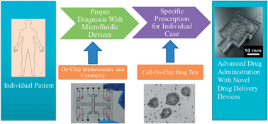

1. Introduction

In recent years, individualized diagnosis and therapy of human diseases have gained great attention due to their potential for biomedical research and clinical applications.1,2 Unlike conventional methods where patients having the same diseases will be treated with a standardized medicine plan without carefully considering individual patient response to the drug treatment, individualized diagnosis and therapy are intended to comprehensively optimize the treatment strategy for specifically treating the patient at the most effective level. For example, chronic diseases such as chronic pain syndrome, melancholia, ocular disease, and cancer will require the help of individualized long-term therapeutic plans to treat and cure. This can be achieved through a continuous or pulsatile drug administration approach.3–5 Infectious disease is another important healthcare area that needs the support of individualized detection and treatment plans. For instance, AIDS, malaria, measles and tuberculosis are the major cause of death in many developing countries, if one could rapidly detect these diseases at their early stage using individualized diagnosis methods, this will allow the medical doctors to have sufficient time to carefully prescribe optimized medicine plans to control and treat the illnesses.6 However, current diagnostic assessments require tedious steps to process the samples and long hours of analysis are needed before any conclusive information can be drawn. In addition, majority of diagnostic techniques rely heavily on non-portable and bulky equipment that is based in professional laboratories and such tools are not suitable for on-site patient screening, such as in clinics or at patient homes. Thus, new reliable, low-cost, rapid, and easy to use diagnostic methods are urgently needed.7,8 In addition to developing new diagnostics methods, we need to improve the current available therapeutics methods at the same time as well since both areas must work hand in hand together to achieve the ultimate goal: to cure the disease. Current conventional therapeutic strategies commonly use oral, topical, or injectable administration routes for delivering drugs in vivo. These methods generally require the needs for high repetition of drug administration and large dosage of drugs for treating many human diseases such as cancers and AIDS.5,9 The consequences of using such aggressive approaches in treating the diseases can result harmful side effects to the patients and in turn impacting their life quality.5,10

Microfluidics is a technology that manipulates and controls micro volume of fluids ranging from 10−9 to 10−18 liter. The research of microfluidics was first started around 1980s and mainly it involved the fabrication of microscale channels (tens to hundreds of microns in size), microvalves, sensors, and actuators for studying the fluid dynamics within the space of microscale volume.11 Recent advancements in microfluidic technology have opened new avenues for improving the methods of individualized disease diagnosis and therapy.12,13 For the example of diagnosis, microfluidic devices require much lower volume of biological samples for screening disease biomarkers within a shorter period of time upon comparing with conventional methods. Also, parallel assays analysis based on one microfluidic device can be easily be achieved by using multichannel detection technique and this will provide statistical meaningful data for analysis.14,15 In addition, the use of microfluidic devices for designing the individualized treatment plan will allows one to precisely control and program drug delivery profiles that are tailored specifically with a set of drug administration approaches for each individual patient.3 More importantly, these devices can be miniaturized for implanted drug delivery applications.16

The fabrication of microfluidic devices is based on the MicroElectroMechanical System (MEMS) technology. MEMS technology provides set of tools to fabricate mechanical components in micrometer scale, such as micro-channels, micro-pumps, micro-valves, and micro-mixers. The integration of different parts of MEMS components into the device is generally depending on the specific type of biological usage. Microfluidic processes usually involve a series of physical reactions such as fluids actuation, reagents mixing, flow direction, and detection of molecules. Through careful design and fabrication processes, MEMS technology will allow one to integrate different microscale components into one functional device for applications ranging from analyzing biological samples to drug delivery.17 For example, MEMS infusion insulin pumps were designed for treating diabetes disease.18 Also, these MEMS devices can be coupled with smartphones, laptops, or microscopes that will allow clinical doctors or biomedical researchers to access and understand patient symptoms in a much shorter time frame and thereby increasing the chances of successful treatment of the patients. Metals, silicon, glass, and polymers are generally used as the raw materials in preparing the microfluidic devices. Many of these newly created devices are currently being tested for biomedical applications.19,20 It is worth highlighting that poly(dimethylsiloxane) (PDMS) polymer are commonly employed as the main material to prepare functional implant devices.21 PDMS has many attractive properties for biomedical studies. For example, PDMS is optically transparent in both visible and UV wavelength and allowing one to perform sterilization of medical devices as well as to observe samples in PDMS device through optical equipment.22 Also, PDMS is a FDA approved biocompatible material and thus it is suitable to be used in making and packaging implant devices for in vivo use.5,21 To date, many PDMS-based microfluidics devices are made by using soft-lithography method. Basically, this method relies on both molding and casting processes. This approach involves the casting of PDMS polymer solution in a mold with a negative pattern of desired patterns at high temperature and thereby creating the desired microstructures for specific fluidic applications.14

Biomedical microfluidic device possesses several unique features such as they are easy to use, they can be made with low cost, they have long hours of usage time and their small device size allows the ease of portability.13,23 These features will come to good use in setting the platform of engineering microfluidic devices for individualized medicine. It is proposed that the future vision of individualized medicine will be focusing on both early disease diagnosis and therapy areas (Fig. 1). In the near future, individualized diagnosis, also known as point-of-care testing, will allow the clinical staffs to perform an on-site and real-time analysis of patient's biological fluid sample with miniaturized microfluidic devices and the analyzed data will be conveniently transmitted to the clinicians, doctors, and even patient themselves to discuss and determine the best therapeutic strategy for each treating the disease.24 In the case of the individualized treatment, we envision that MEMS advanced drug administration devices can be applied for long term sustainable drug delivery therapy applications ranging from glaucoma to diabetes treatment. The addition of microfluidic detection techniques and MEMS-based drug administration methods into the healthcare sector will certainly provide new opportunities for improving individualized medicine in the years to come.7,25 In this review, we will discuss the recent advances of tailored-made microfluidic devices for diagnosis and therapy studies such as immunoassay, flow cytometry, drug testing, and implantable drug delivery. Furthermore, we will highlight the advantages of each microfluidic techniques and the improvements needed to be made in translating them for individualized medicine.


image file: c3ra45629c-f1.tif
Fig. 1 Future vision on the basic steps of individualized medicine with microfluidic devices.

2. Lab on a chip (LOC) immunoassays

Immunoassay is an important technique for various diseases diagnosis and they are usually used in many laboratories and clinics.25,26 To date, traditional immunoassay has been regarded as the gold standard for disease diagnoses because of its high sensitivity, reliability and accuracy.13 However, there are many limitations for currently used immunoassays. Basically, it requires a sequence of sample/reagent solution processes which are usually handled by experienced professionals in a well-equipped laboratories.27 Also, it needs long hours of analysis time, reactive reagents, and experienced personnel to obtain useful result. These are major roadblocks in implementing them for individualized diagnosis applications at homes and remote areas.8,13,26 Recently, there are numbers of lab-on-a-chip (LOC) immunoassay devices been developed and tested. For example, Chin et al. presented a microfluidics based chip for the diagnosis of infectious diseases.25 The reported microchip has a microchannel network which was structured by a molding process on polystyrene material. The polystyrene material is transparent and it is suitable for easy readout tests using simple microscope setup or naked eyes. In their study, a sequence of solutions was transported into the microchannels and digitized with small air gaps. The driving force for the fluid flow was originated from the microfluidic syringe pump besides the device. The entire testing process, including sample flow, gold-labeled antibody flow, silver reagents flow and a series of buffer washes, was accomplished in less than 30 minutes. In another example, Weigl et al. demonstrated the use of a simple microfluidic chip for sensing enteric pathogens.28 Similarly, they have used a microchip with microfluidic networks for immunoassays analysis. Using the chip, polymerase chain reaction (PCR) was completed in eight minutes with accuracy comparable to the bench top based PCR machine. In addition, the fabricated LOC immunoassays possesses rapid detection speed and they required only small volume of samples for analysis.8,13,29 It is worth noting that such LOC devices can possibly be prepared with low cost and apply them for rapid on-site diagnostic tests for patients who are living in resource-poor environment. The development of future LOC devices will be focusing on optimization of the system's sensitivity and integrating automatic on-chip testing processes into the device with equivalent functions as compared to the laboratory equipment. For a typical LOC immunoassay, the actuation and manipulation of the sample/reagent solutions are the two major challenges that needs to be overcome for obtaining comparable sensitivity by using traditional immunoassay measurements.30

The actuation provides the main driving force for the fluid flow. Currently, several different actuation mechanisms have been developed to drive the flow of the fluid. Table 1 listed different actuation methods for LOC biomedical studies. Among them, the pressure-driven method is the most widely employed in developing LOC devices for testing biological fluids.25,26,31–33 Pressure-driven fluid flow is typically achieved by creating a pressure difference between the two ends of a microchannel using a vacuum pump or applying a positive pneumatic force from a syringe pump.30 The pressure-driven method can be easily integrated into a wide range of substrates for driving organic and aqueous fluids. Chin et al. have developed a micro immunoassay chip for analyzing infectious disease based on a simple syringe actuation.25 However, in practical applications, pressure-driven flow displays the characteristic of a non-linear velocity profile that causes sample plug dispersion to occur and this may disrupt the consistency of the fluid flow.8,30 Another major problem arising from this method is that the fluid resistance will dramatically increase when the dimension of the microchannels is reduced and this will require ultra-high driving pressure to push the fluid to move forward.30 However, such high-pressure pumps are expensive and bulky. These are the challenges that need to be overcome in releasing the use of microfluidic immunoassays device for clinical phase usage and research. Electrokinetic-driven method is commonly used for fluidic actuation and manipulation in LOC immunoassays.34–37 In this method, ionic particles in the fluids will cause the fluid to move in the direction parallel to the applied electrical field.30,32 No pumps or valves are needed in this method and a fluid with uniform velocity profile can be achieved by carefully controlling the applied electric field.38 Electrokinetic-driven process provides high controllability in moving a very small amount of solutions within the device channels. Xiang et al. have reported a microfluidic immunoassay system that utilized electrokinetic force to control the reagents flow profile.37 It is worth noting that this approach can also be used to drive discrete droplets to move along a microchannel and this phenomenon is served as the main principle for the development of digital microfluidic (DMF) immunoassays.39–41 For example, Miller et al. shown that DMF device was able to conduct heterogeneous immunoassay with high throughput and low volume of samples were needed.41 The disadvantage of electrokinetic-driven method is that only conductive fluids can be used within the system. Also, the Joule heating from this method may cause the fluid to evaporate and impacting the reactivity of the reagents used in the immunoassays system.8,32 In addition to pressure and electrokinetic driven methods, passive driven method is another popular technique being used to control the fluid flow in LOC immunoassay devices due to the simplicity in the implementation and fabrication of the system.8,29 Commonly, both capillary29,42 and gravitational43 forces are employed as the means to provide fluid flow manipulation. For example, Li et al. have demonstrated a device equipped with capillary pump for blood analysis study.29 Upon comparing to other methods, passive driven technique does not require external pumps and this approach can be applied to different microfluidic devices. But, this method does come along with some limitations. For example, this technique has low controllability of flow rate and long period of time is needed for detecting desired biomolecules. However, these limitations will not affect the system performance for specific applications such as long term drug delivery therapy of cancer, skin disease or glaucoma.

Table 1 Comparison of microfluidic actuation methods for LOC biomedical studies
Item ref. (i) System miniaturization for on-chip actuation (ii) Biomedical test compatibility (iii) Controllability (iv) Power consumption (v) Consistency & reliability References
Integration on-chip MEMS fabrication compatibility Approx. flow rate Rate controllability Response speed
Pressure driven Syringe pump No, off-chip pump required Low High 10−1 to 103 μl min−1 Fair (∼10−1 μl min−1) High (few seconds) Fair (101 to 103 mW) Long-term 25, 26, 29 and 33
Electrochemical pump Yes High Fair, may cause sample oxidation 10−1 to 102 μl min−1 Fair (∼10−1 μl min−1) High (few seconds) Low (10−1 to 101 mW) Long-term, but the efficiency may drop by electrodes damage 61
Electrical driven Electrokinetic driven Yes High Fair, Joule heating induced 10−2 to 102 μl min−1 High (∼10−2 μl s−1) High (few microseconds) Very high (few Watt) Long-term 34–37
Electrowetting (digital microfluidics) Yes High Fair, for conductive solutions usage only 10−3 to 10−1 μl min−1 High (∼10−2 μl s−1) High (few microseconds) High (∼10−1 Watt) Long-term 39–41
Passive driven Capillary driven Yes High High 10−3 to 10−2 μl min−1 Low (fixed rate) Low (few minutes–few hours) No power consumption One-time 29 and 42
Gravitational driven Yes High High 10−3 to 10−1 μl min−1 Low (fixed rate) Low (few minutes) No power consumption One-time 43
Centrifugal force driven Rotatory motor No, off-chip motor required Low High 10−3 to 10−1 μl min−1 High (∼10−2 μl s−1) Fair (depends, from few seconds to minutes) High (∼10−1 Watt) Long-term 27 and 62


Besides the fluids actuation, manipulation of sample fluid is another major concern for achieving high accuracy of immunoassays system. The homogenous mixing of the biological fluid samples and reagents in immunoassays system is important since successful analytical analysis of samples strongly relies on the effective interaction between the samples and reagents. Due to the laminar flow nature of fluid in microchannels, fluids are not able to be mixed homogenously even when they are flowing within the same microchannel.44 Unsuccessful mixing of the samples and reagents may lead to false assay result. Both active and passive methods have been used to mix solutions within the microfluidic devices.44,45 Active microfluidic mixer generates displacement of fluid flow by using either stirring or agitating approach. In the case of agitating approach, acoustic wave,46 electronic,47 pressure-perturbation,48 magnetic field45 and thermal radiation49 are commonly used to create a homogeneous mixture. For many individualized medical devices, passive mixing methods will more suitable to be utilized because they do not depend on “off-chip” equipment or external power supplies. It is worth noting that MEMS microfabrication technology does offer an unique approach to define desired geometries of the microchannels and thus leading the significant advancement in the development of passive mixers for achieving the expected homogenous state of the mixture.50 Intersecting channels such as zigzag channels, 3-D serpentine channels, and twisted channels are often used for fluids mixing purpose.51–54 Another key area in microfluidic immunoassay system that has been greatly improved over the years is the simplicity of the samples and reagents preparation using the developed device. For example, successful plasma separation and extraction from whole blood is a critical step for obtaining high sensitivity signals from the immunoassays system. The current protocol for plasma extraction requires sophisticated and bulky equipment in the laboratories. This is a relatively slow process and the samples may be contaminated if the process is operated in the absence of clean laboratory environment.29 Recently, various “on-chip” solutions have been proposed and applied for samples preparation in LOC immunoassays system. These solutions utilize dielectrophoretic separation,55 capillary filtration,56 microbead filtration,29,57 and acoustic force separation58 for performing biological extraction process. Based on these techniques and integrating them into microfluidic immunoassay devices, the whole blood analysis is made possible now.

In recent years, there is a growing interest in the diagnostic community to develop novel approaches for multiple antigens detection using low volume of samples and reagents with high speed.35,44 For conventional detection methods, each method is designed to diagnose a specific type of disease. Thus, multiple diseases detection scheme generally requires a tedious series of tests and costly examination fees. Studies have shown that some LOC immunoassays allow parallel analysis of multiple samples and the device is able to simultaneously process the assays to analyze the availability of specific proteins in the samples. These studies are useful examples and able to provide a platform to further improve the device performance in screening and detecting multiple diseases biomarkers through a single flow of multiple samples into the device.8 For example, Chin et al. developed a method to separate samples, reagents, antibodies, and buffer using a single channel where air spacers were created within the flowing section.25 Shi et al. demonstrated the selective immobilization of antibodies on substrate was achieved through an electrochemical process using strategically located microelectrodes.59 Heyries et al. presented a process to transfer microarrays of macromolecules to PDMS substrate for selective antibody immobilization and this technique was used for allergen-specific antibodies detection.60 Herrmann et al. reported a strategy for multiple samples detection by employing parallel microchannels on a single chip. This strategy basically offers the uniqueness of separating each immunoassay processes into different microchannels thereby allowing multiple detections to take place within the same device.44 Using all these techniques and blending them together, future microfluidic assays can be integrated with the capability to determine different diseases biomarkers simultaneously by simply flowing patient biological fluid to the device.

3. Lab on a chip (LOC) flow cytometry

Being able to rapid analyze the physical and chemical properties of cells is an important key area for screening infectious diseases.13 Flow cytometry is a powerful tool for cell counting, cell sorting, biomarker detection and protein analysis by suspending them in a stream of fluid and passing them through a series of electronic-optical detection apparatus and this tool has been widely used in hospitals and clinics today.63 For example, for accessing HIV-infected patients, flow cytometry was used to determine the counts of CD4+ lymphocytes in the samples and these counts will serve as an important indicator for the clinicians to assign appropriate treatment methods to the patients.64 To date, many commercially available flow cytometers are bulky, difficult to use, non-portable, and costly.63–65 Moreover, they can only be used in common laboratories since these machines require additional set of supporting electronic apparatus to work effectively. These disadvantages have limited their applications for on-site and in-time analysis that is important for individualized diagnosis and monitoring of infected patient. Recently, several microfluidic flow cytometers have been developed63 and demonstrating their potentials to be employed for individualized medical use. The advantages of using LOC flow cytometry for clinical sampling include low consumption of biological fluids, easy to use, high sensitivity, and shorter time for analysis, and these are the crucial factors for developing effective individualized medical care. In the near future, with the advancements of the capability to integrate multiple on-chip optical components into the device, to design novel functional microchannel geometries on the device substrate, and to engineer new microfluidic technology, many microfluidics and biomedical research groups are hopeful that these improvements in LOC device will offer a new paradigm shift for treating and curing diseases.63,66,67

Recent demonstration of on-chip CD4+ cell counts using LOC flow cytometry are excellent examples in laying the foundation for the individualized diagnosis and therapy. CD4+ cell counts in adults are estimated by counting the absolute number of CD4+ T-lymphocytes per microliter of blood.64,65 Cheng et al. reported a simple chip that was able to immobilize CD4+ cells in microchannels and the CD4+ cell counts was obtained by using a microscope to perform manual counting on the cells.65 This cell counting method is simple and straightforward but no fluorescence labelling processes are involved. Thus, this might result in miscounting of cells since this technique is solely relying on the naked eyes to examine the cells. Alyassin et al. reported an automated cell counting method to avoid human errors in cell counting.68 Basically, a CCD camera attached to a microscope is used to obtain images of suspended cells flowing through a microfluidic chip and image analysis is performed to determine the exact cell counts. This method offers high accuracy in cell counting. However, this method does come with some drawbacks such as tedious preparation steps and the need for cell labelling before performing the experiments. Also, it is known that only a fix section of view can be acquired and transform to images under the microscope lenses. Therefore, samples that have very low concentration of cells will affect the device detection performance.69 Moon et al. and Ozcan et al.67,70 have demonstrated the use of lensless shadow imaging technique to obtain large view of images with portable CCD/CMOS detectors (Fig. 2). Indeed, this technique was successfully demonstrated by using built-in cameras on smartphones or tablets and showing its potential for portable individualized medical devices. As shown in Fig. 3, Zhu et al. designed a microfluidic chip for cell capturing and the device was integrated with a smartphone for imaging.66 The image processing and analysis for CD4+ counts were accomplished by using programmed software available in the smartphone. The similar approach was also employed for monitoring the morphology and viability of cultured cells on the chip.71


image file: c3ra45629c-f2.tif
Fig. 2 Lensless approach with integrating CCD chip with microfluidic device was applied for cells imaging and analysis: (a) working principle for the lensless CD4+ cell detect system. (b) Photograph of the chip. (c) Cell image taken from the lensless microfluidic chip. Images and picture were reprinted from ref. 67 with permission of Elsevier.

image file: c3ra45629c-f3.tif
Fig. 3 CD4+ cells were imaged via lens and CMOS on a smartphone: (A–C) diagrams of optofluidic designs on the cell phone and cell imaging principle. (D) Photograph of the platform. Reprinted with the permission from ref. 66. Copyright (2011) American Chemical Society.

Another key advantage of LOC based flow cytometry technique is that it can be integrated to other microfluidic systems for developing a multifunctional portable device for rapid diagnosis of patient samples. For instance, LOC based flow cytometry can be engineered with functions such as cell separation, cell sorting, cell focusing and centralization of the samples information. Recently, Green et al.38 investigated the use of microfluidic cell separation and enrichment chip for understanding the cell bonding on microchannel surfaces within the device. A uniform and predictable adhesion profile can be obtained by using microchannels with smoothly curved turns. Other methods like electrochemical T switching,72 electric force sorting,73 and optical force switching74 have also been reported for cell separation on chip. Cell focusing is also an important step for acquiring higher detection sensitivity, which can be achieved by using on-chip microfluidics.63,75 Conventional cell focusing utilizes a complex, bulky, and expensive fluidic system to induce sheath flows to surround the sample flow, whereby aligning cells into the center of the stream. In the microfluidic scale, cell focusing can be achieved by designing different microchannel geometries for aligning the sample flow. For example, Bhagat et al. have reported the effects of Dean drag and inertia lift force generated by spiral microchannels can be employed to focus cells in the fluid flow.75 Bang et al. demonstrated another approach to accomplish the cell focusing in the flow stream by integrating an expansion channel inside the focusing channel.76 Using this geometry, the particles within the flow path are focused at the stream center. As a result, the signal from the particles was significantly enhanced and in turn increases the overall detection limit. More importantly, this MEMS based technique can be inserted into the conventional flow cytometry machine for increasing the throughput and detection speed.

4. Microfluidic cell-on-chip drug test platforms

In the process of discovering a new drug, a series of tests are required to determine the benefits and risks of the particular drug treatment.77 As the response to drugs varies from person to person, careful and tedious evaluation of new drugs is needed to optimize the treatment protocol for patients with different medical backgrounds.78 Current drug testing methods are extremely time consuming and costly. Generally, a large volume of experiments such as in vitro, in vivo, ex vivo, pre-clinical, phase 1, 2 and 3 clinical trials studies will be conducted before a new drug is approved by FDA and eventually launched in the market for therapy of patients. The entire time frame for this whole process to complete usually takes 10 to 15 years.78,79 Thus, there is an urgent need to develop new techniques to shorten the period of discovery of new drugs and drug testing in various biological models whereby allowing the pharmaceutical professions to determine ultimate faith of the new drugs for clinical applications. Recently, some reports have shown that microfluidic devices can be designed to have the capability for cell culturing and this system have drawn great attention from pharmaceutics and biological sciences community.78 This microfluidic cell culturing platform is useful to create a well-defined microenvironment for homing different disease cell lines under a single chip. By further exploiting the microfluidic cell culturing system, we foresee that many new drug formulations can be tested effectively in diseases cell models within a shorter period of time and more importantly the optimized and potential drugs can be selected for animals trial experiments.77,78

There are several key factors that need to be considered carefully for developing microfluidic on-chip drug tests. The first factor is to be able to mimic the in vivo microenvironment within the chip. This is of great importance for acquiring meaningful biological responses when evaluating the effects from the drug formulations.78 With this in mind, some research groups have proposed the development of three dimensional (3-D) cell culturing system. Classic two dimensional (2-D) cell culturing system has been suggested as a fundamental biological model for understanding cell biology study but they are insufficient to simulate in vivo environment for drug testing applications.80 Also, mechanical forces and signaling between cells can be hardly achieved using the 2-D culturing system and they are crucial in cell proliferations. Recently, it is demonstrated that the design of new complex microfluidic structure can be used for achieving 3-D cell culturing and this system will be useful for in vivo drug testing applications. Among these methods, culturing cells on biocompatible nanostructure scaffolds gained the most attention due to their high resemblance of an in vivo model.80–82 Secondly, different biological parameters like solutions (e.g. medium, buffer and plasma), gases (CO2,83 O2 (ref. 84) and etc.), and temperature85 need to be carefully setup in the chip for maintaining the desirable biological microenvironment during the drug test. A few on-chip solutions have been proposed for maintaining these parameters in the device. For example, Maharbiz et al. developed an electrochemical oxygen generator for on chip cell culturing applications.84 Specifically, water electrolysis reaction was initiated with patterned Ti/Pt electrodes and generating oxygen gas. This is useful for supplying specific concentration of oxygen gas to the desired microfluidic cell culture media. In addition, Lin et al. reported the fabrication of an on-chip indium tin oxide (ITO) based micro heater to maintain the temperature required for cell culturing study.85 The last, but not the least factor is about the cell immobilization and growth on the substrate surface. For common drug testing experiments, the drug is delivered to the cells that are positioned in the defined areas of the device and the substrate surface characteristics are important to be investigated.78 It has been shown that the surface wettability86,87 and roughness87,88 will greatly impact the cell adhesion on the substrate. For this purpose, surface modification techniques such as plasma treatment, UV treatment, deposition of water-likeable materials, and chemical treatment have been used to create hydrophilic surfaces for better cell culturing environment in the devices.89–91 Fuard et al. and Peterson et al. have demonstrated that the use of plasmas treatment to optimize the PDMS surface wettability for supporting cell adhesion on the device substrate.86,92 Steele et al. reported the improvement of cell adhesion in the device by coating neutralized perfluorosulphonic acid (Nafion®) and polytetrafluoroethylene (Teflon®) on PDMS surface.93 In another way, Mata et al. shown that PDMS surface with patterned microtextures was able to improve connective tissue progenitor cells adhesion and proliferation (Fig. 4).87 Also, specially patterned microstructures on substrate can be used to trap single cells at certain positions. Carlo et al. have successfully demonstrated a single cell culture array which utilized a PDMS microstructure on glass for trapping and culturing HeLa cells.94


image file: c3ra45629c-f4.tif
Fig. 4 (a) Microscope brightfield image shows live connective tissue progenitor cells attaching on PDMS surface. (b) SEM image shows the cells adhesion and proliferation on PDMS microtextures. Images were reprinted from ref. 87 with the permission of John Wiley and Sons.

The combination of microfluidics and cell-on-chip technologies is a useful platform for conducting drug screening and testing for patients. Although there are more rooms for improvements in developing this platform for clinical applications, recent achievements have shown promising results in this research area. Torisawa et al. demonstrated the fabrication of an on chip anticancer drug testing device.81 Specifically, human breast cancer (MCF-7) cells were cultured within the PDMS microchannels and subsequently a multidrug chemosensitivity assay was conducted in the device. The result showed that the drug testing device was applicable for evaluating the performance of the drugs against the cancer cells. Importantly, by separating each small drug test in numbers of isolate microchannels, adequate data from the large quantity of drug testing can be acquired and used for statistical evaluation of the samples. Similarly, Fujii et al. reported that they were able to culture stomach cancer cells (SH-10-TC) within PDMS microchannels and conduct drug assays using the same microchip.95 As most of cancer drugs will cause serious side effects, it is important to carry out pre-treatment drug tests to evaluate the risks and benefits ratio for the specific patient. For this reason, parallel on-chip drug test on human organ cells have been proposed and demonstrated.96,97 Different types of organ cells were cultured and linked to the microchannels for mimicking the in vivo circulation system. In recent studies, microfluidic drug test devices have shown the ability to carry out treatments with carefully designed drug delivery profiles.98–100 Kim et al. have demonstrated the fabrication of a LOC anticancer drug test device.100 On chip microchannel network was defined and cooperated with a syringe pump system to control the drug dosage to be delivered to cell microwells. Cancer drug with four different concentrations were tested and they have shown different inhibition rates on the cell proliferation. In the near future, microfluidic drug test devices will allow clinicians to able to design the best treatment scheme for patient to achieve maximized therapeutic effects.

5. Implanted drug delivery strategies

Individualized drug therapy generally requires the need of carefully controlled drug administration parameters such as timing and dosage of drugs.3,4 This remains a great challenge to overcome for many therapies that are relying on conventional drug delivery routes such as oral, topical, nasal, pulmonary, transmucosal, and injection.101 For instance, a high dosage of drug is commonly required when oral and topical drugs are used to treat various human diseases because only a relatively small fraction of drugs can be absorbed by the body and in turns providing sufficient medication to battle the diseases.5,9 As a result, side effects from overdose of drugs will occur in majority patients body upon using these conventional methods mentioned above.102 In the case of drug injection, some patients such as children will experience the sensation of physiological trauma at the injection site when repeated injections are required to treat the diseases such as serious cases of asthma and diabetes.3 Moreover, the lack of methods to precisely control the drug release to the disease sites is a major roadblock to achieve optimized therapeutic effects.103 Recently, implantable device therapy based on localized drug delivery was proposed as a new treatment approach to overcome challenges indicated earlier and the exploration of using such device in medical applications has led significant advancement in preparing highly versatile implantable drug delivery devices for in vivo use.5 Such devices will be valuable for individualized disease therapy where programmable drug delivery profile is needed to enhance the therapeutic impacts for diseases such as glaucoma, corneal disease and anxiety disorders. To date, multi-reservoirs and single reservoir drug delivery methods have been studied for individualized drug delivery applications.

For multi-reservoirs drug delivery method, a series of arrays of drug filled micro-reservoirs were fabricated on a single microchip.104 These microchips featured implantable designs and programmable reservoir openings whereby enabling the controlled-release of drugs to specific sites. Most recently, the first clinical human test using multiple reservoirs drug delivery device with wireless control was demonstrated by Farra et al. (Fig. 5).16 In this study, it was shown that the opening of the micro-reservoirs plays an important role in regulating the desired drug release profile in the body. So far, a number of mechanisms have been used in designing multi-reservoirs drug delivery microchip and they include membrane dissolution, sensitive hydrogels/polymers, magnetically triggered particles, and electrothermal effects.105–108 Membrane dissolution is the most widely used mechanism for making drug delivery microchips. For example, Santini et al. have fabricated a microchip with controlled release drug delivery system. Gold membrane was used to seal the reservoirs (Fig. 6a). Upon applying a potential difference across the gold membrane, the nearby electrode will initiate the electrochemical dissolution of the gold membrane that eventually opens up the reservoir and releasing the drug to the local environment (Fig. 6b).106 Such devices showed a low power consumption and a rapid response time of 1 to 2 minutes.109 Biodegradable polymer materials are also excellent materials to be used for fabricating the dissoluble membranes.105,110,111 Many of these polymer membranes are biocompatible and very often they are used for packing the device for implants use. In recent years hydrogels have been used in making devices that have the capability of active controlled release of drug formulations.107 The rapid swelling and deswelling of the hydrogels can be engineered to respond to specific changes in the environment, such as pH, glucose concentration, temperature, and light.112 Such swelling and deswelling feature is useful for incorporating drugs into the hydrogels for passive delivery therapy. In general, tumors or inflamed tissues have a different pH value than that of the normal one. Under such scenario, one can use multi-reservoirs drug delivery arrays with hydrogel membranes and attach them to the tissue surface where the device can deliver a self-controlled drug delivery profile to the local environment by picking up the subtle changes in pH from the tissue surface. It is worth noting that the development of self-controlled drug delivery MEMS system is possible in near future since both diagnostic and therapeutic functions can be combined into one device. Such devices, also termed as “smart devices” or “closed-loop devices”, have been extensively researched for diabetes therapy.18,113 Naturally, one can foresee that these devices will be translated for individualized medicine applications as this will reduce the cost of diagnosis and therapy for patients.


image file: c3ra45629c-f5.tif
Fig. 5 The first clinical in-human testing drug delivery device with multiple reservoirs: (A) photograph of the drug delivery device. (B) Design of micro reservoirs on chip. (C) Working principle of the reservoir opening and drug delivery. (D) Timeline of each dosage. Image and photograph were reprinted from ref. 16 with the permission of The American Association for the Advancement of Science.

image file: c3ra45629c-f6.tif
Fig. 6 SEM images of a gold membrane covering a micro reservoir. (a) Before electrochemical dissolution reaction. (b) After electrochemical dissolution reaction, membrane was dissolved. Images were reprinted from ref. 106 with the permission of Nature Publishing Group.

Although various advantages can be obtained by using multi-reservoirs drug delivery system, one of its major limitations is that drug refilling to the device is not achievable using the current design.114 As such, multi-reservoirs drug delivery system is not suitable to be employed for long term therapy use. To overcome this problem, an additional type of implantable drug administration method has been proposed and designed.9 In this configuration, a relatively large reservoir is employed and a micro-pump is integrated to actuate the drug fluid. For such devices, the drug refilling process can be easily performed by using syringes via a refill port on reservoir.9,115 This method can be applied to refill devices that is implanted on the eye, nose, or under the skin. Inspired by the dry reagent storage method from LOC technology26 and dry powder inhalers formulation,116 we proposed that dry drug storage in the MEMS implantable drug delivery device may serve as a possible solution for overcoming the drug refilling problem. Compared to drug dispersion, the same dosage of dried drug powder will consume much lower volume in the reservoir of the device. Using this approach, implanted MEMS device could sustainably delivery drug formulations to patient at a longer time period and thus relieving the needs of frequent refills to the device.

The core component of such devices is the integration of micro-actuator/micropump into the system that controls the release of the drugs. There are two main categories of micro-actuators and both are MEMS-based systems: (i) mechanical actuators such as electrostatic, piezoelectric, thermopneumatic, or bimetallic pumps that generates displacement for actuation, and (ii) dynamic, non-mechanical actuators like electro-osmotic, electrowetting, and capillary pumps.3,4 Due to their high actuation efficiency, controllability, and low power consumption, mechanical actuators are generally preferred to be utilized in designing the individualized drug delivery devices. For example, electrochemical actuators using the gas pressure based on simple water electrolysis has been used for drug delivery study.5,9 However, the electrochemical reaction may cause the drug to oxidize and impacting the effectiveness of the drug formulation. To overcome this challenge, Li et al. developed a two-chamber system, where a bellow was used to separate the electrolyte (DI water) and the drug solution during the experimental study.114,117,118 Instead of directly pressurizing the drug reservoir, the electrolysis generated gas deflects the bellows and transfers the pneumatic forces to push the drug out from the reservoir.5,114,118 The biocompatibility, low power consumption (few mini Watts) and large operation range (from pl min−1 to μl min−1) of such devices were demonstrated in both in vitro and in vivo models and they show great promise for future implantable drug devices.5,102,115,119 Based on such platform, our group has successfully engineered an electrochemically actuated-based implantable drug delivery device for studying individualized drug delivery therapy of pancreatic cancer.120 Anticancer drug doxorubicin (Dox) was delivered to cultured pancreatic cancer cell (MiaPaCa-2 and Panc-1) colonies using the designed device (Fig. 7a and b). Our results have shown that the growth of cancer cell colonies was successfully inhibited (Fig. 7a). We have also demonstrated the controllability over the therapeutic scheme through customizing the drug dose profiles using the developed device. As shown in Fig. 7c, the growth of pancreatic cancer cell colonies was inhibited for both the two treatment programs (program I: 6 μg of doxorubicin delivery in day 0, day 1 and day 2, program II: 9 μg of doxorubicin delivery in day 0 and day 3). The colony size decreases in different patterns for the two different therapeutic programs. More importantly, the MiaPaCa-2 pancreatic cancer cell colonies and Panc-1 pancreatic cancer cell colonies have demonstrated different response under the same treatment schedule. As shown in Fig. 7c (Miapaca-2) and Fig. 7d (Panc-1), under the treatment program I, the drug dosage required to inhibit the growth of cancer cells was different in each group (6 μg and 9 μg). This result opens up the new possibility for designing and optimizing appropriate therapeutic effects to treat and cure diseases at their early stages towards each individualized patient. And this will be the future vision that needs to be achieved for upgrading the individualized medical care in medical centers or hospitals.


image file: c3ra45629c-f7.tif
Fig. 7 (a) Illustration of the individualized cancer treatment study. The darkness of cancer cell colonies after treatment was observed with microscope images showing the death of cancer cells. (b) Photograph of the electrochemical actuated drug delivery device.120 (c) Size change of MiaPaCa-2 cancer cell colonies under different treatment programs (program I, II), and without treatment (control). (d) Size change of Panc-1 cancer cell colonies under different treatment programs (program I, II), and without treatment (control).

6. Conclusion

The recent developments in microfluidic devices have shown great promise in paving the way for realizing the individualized medical diagnosis and therapy. In this review, we have discussed and analyzed various types of advances in engineering microfluidic devices for medicinal related applications such as lab-on-a-chip (LOC) diagnosis devices, LOC-based flow cytometry, the on chip drug testing platform, and the implantable drug delivery devices. The benefits and risks of using each device for individualized medicine purpose have been discussed. We have also highlighted the recent engineered lab-on-a-chip (LOC) diagnosis and flow cytometry device provides the advantages of rapid and high accuracy in screening patient biological fluid samples that do not rely on professional, expensive, and bulky equipment. This will be important for future medical healthcare in terms of monitoring the patients from long distance at home or rural areas. For on chip drug testing platform, they may be used to replace the formal expensive and time consuming drug testing systems since many of these systems relies on tedious and complex processing steps to complete a comprehensive study of a drug formulation. In addition, the on chip drug testing platform will help to speed up in identifying the optimal drug formulation for treating patients with minimal side effects. Implantable drug delivery devices provide many benefits over using the traditional oral dosage forms of medicine. Most implantable microfluidic devices provide site specific administration of drug formulations to the infected area where the drugs can have optimized effects in treating the disease. The use of implantable devices will also lower drug dosage for patient treatment whereby minimizing the side effects. Also, implantable drug delivery devices can be designed for long term release of drug formulations and this function will be crucial for disease treatment such as brain tumor, pancreatic tumor and glaucoma. In the near future, we envision that many of these microfluidic device technologies discussed here will be further improved and can be selectively integrated into a single platform for individualized medicine ranging from rapid diagnosis of patient samples to treating the patient with effective minimal dosage of drug formulation.

Acknowledgements

This study was supported by the Start-up grant (M4080141.040) of Nanyang Technological University and partially from the Singapore Ministry of Education under Tier 2 Research Grant MOE2010-T2-2-010 (4020020.040 ARC2/11) and Tier 1 Academic Research Funds (M4010360.040 RG29/10).

References

  1. X. L. Mao and T. J. Huang, Lab Chip, 2012, 12, 1412–1416 RSC.
  2. Y. Rosen and P. Gurman, Curr. Pharm. Biotechnol., 2010, 11, 366–375 CAS.
  3. N. M. Elman and U. M. Upadhyay, Curr. Pharm. Biotechnol., 2010, 11, 398–403 CAS.
  4. N. C. Tsai and C. Y. Sue, Sens. Actuators, A, 2007, 134, 555–564 CrossRef CAS PubMed.
  5. H. Gensler, R. Sheybani, P. Y. Li, R. Lo Mann and E. Meng, Biomed. Microdevices, 2012, 14, 483–496 CrossRef CAS PubMed.
  6. P. Yager, T. Edwards, E. Fu, K. Helton, K. Nelson, M. R. Tam and B. H. Weigl, Nature, 2006, 442, 412–418 CrossRef CAS PubMed.
  7. R. W. Hart, M. G. Mauk, C. Liu, X. Qiu, J. A. Thompson, D. Chen, D. Malamud, W. R. Abrams and H. H. Bau, Oral Dis., 2011, 17, 745–752 CrossRef CAS PubMed.
  8. T. G. Henares, F. Mizutani and H. Hisamoto, Anal. Chim. Acta, 2008, 611, 17–30 CrossRef CAS PubMed.
  9. P. Y. Li, J. Shih, R. Lo, S. Saati, R. Agrawal, M. S. Humayun, Y. C. Tai and E. Meng, Sens. Actuators, A, 2008, 143, 41–48 CrossRef CAS PubMed.
  10. L. Lafleur, D. Stevens, K. McKenzie, S. Ramachandran, P. Spicar-Mihalic, M. Singhal, A. Arjyal, J. Osborn, P. Kauffman, P. Yager and B. Lutz, Lab Chip, 2012, 12, 1119–1127 RSC.
  11. G. M. Whitesides, Nature, 2006, 442, 368–373 CrossRef CAS PubMed.
  12. B. Ziaie, A. Baldi, M. Lei, Y. D. Gu and R. A. Siegel, Adv. Drug Delivery Rev., 2004, 56, 145–172 CrossRef CAS PubMed.
  13. W. G. Lee, Y. G. Kim, B. G. Chung, U. Demirci and A. Khademhosseini, Adv. Drug Delivery Rev., 2010, 62, 449–457 CrossRef CAS PubMed.
  14. E. Verpoorte and N. F. De Rooij, Proc. IEEE, 2003, 91, 930–953 CrossRef CAS.
  15. C. D. Chin, V. Linder and S. K. Sia, Lab Chip, 2012, 12, 2118–2134 RSC.
  16. R. Farra, N. F. Sheppard, L. McCabe, R. M. Neer, J. M. Anderson, J. T. Santini, M. J. Cima and R. Langer, Sci. Transl. Med., 2012, 4, 122ra21 Search PubMed.
  17. J. Wu and M. Gu, J. Biomed. Opt., 2011, 16, 080901 CrossRef PubMed.
  18. E. Renard, Curr. Opin. Pharmacol., 2002, 2, 708–716 CrossRef CAS.
  19. P. Abgrall and A. M. Gue, J. Micromech. Microeng., 2007, 17, R15–R49 CrossRef.
  20. G. Kotzar, M. Freas, P. Abel, A. Fleischman, S. Roy, C. Zorman, J. M. Moran and J. Melzak, Biomaterials, 2002, 23, 2737–2750 CrossRef CAS.
  21. H. Becker and C. Gartner, Anal. Bioanal. Chem., 2008, 390, 89–111 CrossRef CAS PubMed.
  22. B. H. Jo, L. M. Van Lerberghe, K. M. Motsegood and D. J. Beebe, J. Microelectromech. Syst., 2000, 9, 76–81 CrossRef CAS.
  23. J. Y. Pan, Proc. IEEE, 2004, 92, 174–184 CrossRef CAS.
  24. A. J. Tudos, G. A. J. Besselink and R. B. M. Schasfoort, Lab Chip, 2001, 1, 83–95 RSC.
  25. C. D. Chin, T. Laksanasopin, Y. K. Cheung, D. Steinmiller, V. Linder, H. Parsa, J. Wang, H. Moore, R. Rouse, G. Umviligihozo, E. Karita, L. Mwambarangwe, S. L. Braunstein, J. van de Wijgert, R. Sahabo, J. E. Justman, W. El-Sadr and S. K. Sia, Nat. Med., 2011, 17, 1015–U1138 CrossRef CAS PubMed.
  26. D. Y. Stevens, C. R. Petri, J. L. Osborn, P. Spicar-Mihalic, K. G. McKenzie and P. Yager, Lab Chip, 2008, 8, 2038–2045 RSC.
  27. B. S. Lee, J. N. Lee, J. M. Park, J. G. Lee, S. Kim, Y. K. Cho and C. Ko, Lab Chip, 2009, 9, 1548–1555 RSC.
  28. B. H. Weigl, J. Gerdes, P. Tarr, P. Yager, L. Dillman, R. Peck, S. Ramachandran, M. Lemba, M. Kokoris, M. Nabavi, F. Battrell, D. Hoekstra, E. J. Klein and D. M. Denno, Microfluidics, BioMEMS, and Medical Microsystems IV, San Jose, CA, 2006 Search PubMed.
  29. C. Y. Li, C. Liu, Z. Xu and J. M. Li, Microfluid. Nanofluid., 2012, 12, 829–834 CrossRef CAS.
  30. A. Ng, U. Uddayasankar and A. Wheeler, Anal. Bioanal. Chem., 2010, 397, 991–1007 CrossRef CAS PubMed.
  31. L. Zhou, K. Wang, W. Tan, Y. Chen, X. Zuo, J. Wen, B. Liu, H. Tang, L. He and X. Yang, Anal. Chem., 2006, 78, 6246–6251 CrossRef CAS PubMed.
  32. S. K. Sia and G. M. Whitesides, Electrophoresis, 2003, 24, 3563–3576 CrossRef CAS PubMed.
  33. S. Sun, M. Yang, Y. Kostov and A. Rasooly, Lab Chip, 2010, 10, 2093–2100 RSC.
  34. A. Dodge, K. Fluri, E. Verpoorte and N. F. de Rooij, Anal. Chem., 2001, 73, 3400–3409 CrossRef CAS.
  35. Y. L. Gao, G. Q. Hu, F. Y. H. Lin, P. M. Sherman and D. Q. Li, Biomed. Microdevices, 2005, 7, 301–312 CrossRef CAS PubMed.
  36. C. X. Qiu and D. J. Harrison, Electrophoresis, 2001, 22, 3949–3958 CrossRef CAS.
  37. Q. Xiang, G. Q. Hu, Y. L. Gao and D. Q. Li, Biosens. Bioelectron., 2006, 21, 2006–2009 CrossRef CAS PubMed.
  38. J. V. Green, T. Kniazeva, M. Abedi, D. S. Sokhey, M. E. Taslim and S. K. Murthy, Lab Chip, 2009, 9, 677–685 RSC.
  39. R. Sista, Z. Hua, P. Thwar, A. Sudarsan, V. Srinivasan, A. Eckhardt, M. Pollack and V. Pamula, Lab Chip, 2008, 8, 2091–2104 RSC.
  40. R. S. Sista, A. E. Eckhardt, V. Srinivasan, M. G. Pollack, S. Palanki and V. K. Pamula, Lab Chip, 2008, 8, 2188–2196 RSC.
  41. E. M. Miller, A. H. C. Ng, U. Uddayasankar and A. R. Wheeler, Anal. Bioanal. Chem., 2011, 399, 337–345 CrossRef CAS PubMed.
  42. L. Gervais and E. Delamarche, Lab Chip, 2009, 9, 3330–3337 RSC.
  43. P. Morier, C. Vollet, P. E. Michel, F. Reymond and J. S. Rossier, Electrophoresis, 2004, 25, 3761–3768 CrossRef CAS PubMed.
  44. M. Herrmann, T. Veres and M. Tabrizian, Lab Chip, 2006, 6, 555–560 RSC.
  45. L. J. Lucas, J.-H. Han and J.-Y. Yoon, Colloids Surf., B, 2006, 49, 106–111 CrossRef CAS PubMed.
  46. R. H. Liu, R. Lenigk, R. L. Druyor-Sanchez, J. Yang and P. Grodzinski, Anal. Chem., 2003, 75, 1911–1917 CrossRef CAS.
  47. D. G. Yan, C. Yang, J. M. Miao, Y. C. Lam and X. Y. Huang, Electrophoresis, 2009, 30, 3144–3152 CrossRef CAS PubMed.
  48. N. Xize and L. Yi-Kuen, J. Micromech. Microeng., 2003, 13, 454 CrossRef.
  49. J.-H. Tsai and L. Lin, Sens. Actuators, A, 2002, 97–98, 665–671 CrossRef CAS.
  50. C. Y. Lee, C. L. Chang, Y. N. Wang and L. M. Fu, Int. J. Mol. Sci., 2011, 12, 3263–3287 CrossRef CAS PubMed.
  51. J. Melin, G. Gimenez, N. Roxhed, W. van der Wijngaart and G. Stemme, Lab Chip, 2004, 4, 214–219 RSC.
  52. V. Mengeaud, J. Josserand and H. H. Girault, Anal. Chem., 2002, 74, 4279–4286 CrossRef CAS.
  53. R. A. Vijayendran, K. M. Motsegood, D. J. Beebe and D. E. Leckband, Langmuir, 2003, 19, 1824–1828 CrossRef CAS.
  54. S. Hardt, H. Pennemann and F. Schonfeld, Microfluid. Nanofluid., 2006, 2, 237–248 CrossRef CAS.
  55. J. M. Yang, J. Bell, Y. Huang, M. Tirado, D. Thomas, A. H. Forster, R. W. Haigis, P. D. Swanson, R. B. Wallace, B. Martinsons and M. Krihak, Biosens. Bioelectron., 2002, 17, 605–618 CrossRef CAS.
  56. Y. C. Kim, S. H. Kim, D. Kim, S. J. Park and J. K. Park, Sens. Actuators, B, 2010, 145, 861–868 CrossRef CAS PubMed.
  57. J. S. Shim, A. W. Browne and C. H. Ahn, Biomed. Microdevices, 2010, 12, 949–957 CrossRef PubMed.
  58. A. Lenshof, A. Ahmad-Tajudin, K. Jaras, A. M. Sward-Nilsson, L. Aberg, G. Marko-Varga, J. Malm, H. Lilja and T. Laurell, Anal. Chem., 2009, 81, 6030–6037 CrossRef CAS PubMed.
  59. M. H. Shi, Y. Y. Peng, J. Zhou, B. H. Liu, Y. P. Huang and J. L. Kong, Biosens. Bioelectron., 2007, 22, 2841–2847 CrossRef CAS PubMed.
  60. K. A. Heyries, M. G. Loughran, D. Hoffmann, A. Homsy, L. J. Blum and C. A. Marquette, Biosens. Bioelectron., 2008, 23, 1812–1818 CrossRef CAS PubMed.
  61. H. Suzuki and R. Yoneyama, Sens. Actuators, B, 2003, 96, 38–45 CrossRef CAS.
  62. N. Honda, U. Lindberg, P. Andersson, S. Hoffman and H. Takei, Clin. Chem., 2005, 51, 1955–1961 CAS.
  63. T. D. Chung and H. C. Kim, Electrophoresis, 2007, 28, 4511–4520 CrossRef CAS PubMed.
  64. W. R. Rodriguez, N. Christodoulides, P. N. Floriano, S. Graham, S. Mohanty, M. Dixon, M. Hsiang, T. Peter, S. Zavahir, I. Thior, D. Romanovicz, B. Bernard, A. P. Goodey, B. D. Walker and J. T. McDevitt, PLoS Med., 2005, 2, 663–672 CrossRef CAS PubMed.
  65. X. H. Cheng, D. Irimia, M. Dixon, K. Sekine, U. Demirci, L. Zamir, R. G. Tompkins, W. Rodriguez and M. Toner, Lab Chip, 2007, 7, 170–178 RSC.
  66. H. Y. Zhu, S. Mavandadi, A. F. Coskun, O. Yaglidere and A. Ozcan, Anal. Chem., 2011, 83, 6641–6647 CrossRef CAS PubMed.
  67. S. Moon, H. O. Keles, A. Ozcan, A. Khademhosseini, E. Haeggstrom, D. Kuritzkes and U. Demirci, Biosens. Bioelectron., 2009, 24, 3208–3214 CrossRef CAS PubMed.
  68. M. A. Alyassin, S. Moon, H. O. Keles, F. Manzur, R. L. Lin, E. Haeggstrom, D. R. Kuritzkes and U. Demirci, Lab Chip, 2009, 9, 3364–3369 RSC.
  69. U. A. Gurkan, S. Moon, H. Geckil, F. Xu, S. Q. Wang, T. J. Lu and U. Demirci, Biotechnol. J., 2011, 6, 138–149 CrossRef CAS PubMed.
  70. A. Ozcan and U. Demirci, Lab Chip, 2008, 8, 98–106 RSC.
  71. G. A. Zheng, S. A. Lee, Y. Antebi, M. B. Elowitz and C. H. Yang, Proc. Natl. Acad. Sci. U. S. A., 2011, 108, 16889–16894 CrossRef CAS PubMed.
  72. C.-T. Ho, R.-Z. Lin, H.-Y. Chang and C.-H. Liu, Lab Chip, 2005, 5, 1248–1258 RSC.
  73. B. Yao, G.-a. Luo, X. Feng, W. Wang, L.-x. Chen and Y.-m. Wang, Lab Chip, 2004, 4, 603–607 RSC.
  74. M. M. Wang, E. Tu, D. E. Raymond, J. M. Yang, H. Zhang, N. Hagen, B. Dees, E. M. Mercer, A. H. Forster, I. Kariv, P. J. Marchand and W. F. Butler, Nat. Biotechnol., 2005, 23, 83–87 CrossRef CAS PubMed.
  75. A. A. S. Bhagat, S. S. Kuntaegowdanahalli, N. Kaval, C. J. Seliskar and I. Papautsky, Biomed. Microdevices, 2010, 12, 187–195 CrossRef PubMed.
  76. H. W. Bang, H. Y. Yun, W. G. Lee, J. Park, J. Lee, S. Chung, K. Cho, C. Chung, D. C. Han and J. K. Chang, Lab Chip, 2006, 6, 1381–1383 RSC.
  77. P. S. Dittrich and A. Manz, Nat. Rev. Drug Discovery, 2006, 5, 210–218 CrossRef CAS PubMed.
  78. M. H. Wu, S. B. Huang and G. B. Lee, Lab Chip, 2010, 10, 939–956 RSC.
  79. M. Bertau, E. Mosekilde and H. V. Westerhoff, Biosimulation in Drug Development, Wiley-VCH Verlag GmbH & Co. KGaA, 2008, pp. I–XXVIII Search PubMed.
  80. J. W. Haycock, in T 3D Cell Culture, 2010, vol. 695, pp. 1–15 Search PubMed.
  81. Y. Torisawa, H. Shiku, T. Yasukawa, M. Nishizawa and T. Matsue, Biomaterials, 2005, 26, 2165–2172 CrossRef CAS PubMed.
  82. M. W. Tibbitt and K. S. Anseth, Biotechnol. Bioeng., 2009, 103, 655–663 CrossRef CAS PubMed.
  83. M. M. Maharbiz, W. J. Holtz, R. T. Howe and J. D. Keasling, Biotechnol. Bioeng., 2004, 85, 376–381 CrossRef CAS PubMed.
  84. M. M. Maharbiz, W. J. Holtz, S. Sharifzadeh, J. D. Keasling and R. T. Howe, J. Microelectromech. Syst., 2003, 12, 590–599 CrossRef CAS.
  85. J.-L. Lin, M.-H. Wu, C.-Y. Kuo, K.-D. Lee and Y.-L. Shen, Biomed. Microdevices, 2010, 12, 389–398 CrossRef PubMed.
  86. S. L. Peterson, A. McDonald, P. L. Gourley and D. Y. Sasaki, J. Biomed. Mater. Res., Part A, 2005, 72, 10–18 CrossRef PubMed.
  87. A. Mata, C. Boehm, A. J. Fleischman, G. Muschler and S. Roy, J. Biomed. Mater. Res., 2002, 62, 499–506 CrossRef CAS PubMed.
  88. A. Mata, C. Boehm, A. J. Fleischman, G. Muschler and S. Roy, Biomed. Microdevices, 2002, 4, 267–275 CrossRef CAS.
  89. J. W. Zhou, A. V. Ellis and N. H. Voelcker, Electrophoresis, 2010, 31, 2–16 CrossRef CAS PubMed.
  90. H. Makamba, J. H. Kim, K. Lim, N. Park and J. H. Hahn, Electrophoresis, 2003, 24, 3607–3619 CrossRef CAS PubMed.
  91. A. D. Tserepi, M. E. Vlachopoulou and E. Gogolides, Nanotechnology, 2006, 17, 3977 CrossRef CAS.
  92. D. Fuard, T. Tzvetkova-Chevolleau, S. Decossas, P. Tracqui and P. Schiavone, Microelectron. Eng., 2008, 85, 1289–1293 CrossRef CAS PubMed.
  93. J. G. Steele, G. Johnson, W. D. Norris and P. A. Underwood, Biomaterials, 1991, 12, 531–539 CrossRef CAS.
  94. D. D. Carlo, L. Y. Wu and L. P. Lee, Lab Chip, 2006, 6, 1445–1449 RSC.
  95. S. Fujii, M. Uematsu, S. Yabuki, M. Abo, E. Yoshimura and K. Sato, Anal. Sci., 2006, 22, 87–90 CrossRef CAS.
  96. J. F. Leary, J. Key, P.-A. Vidi, C. L. Cooper, A. Kole, L. M. Reece and S. A. Lelièvre, Microfluidics, BioMEMS, and Medical Microsystems XI, San Francisco, CA, 2013 Search PubMed.
  97. J. B. Lee and J. H. Sung, Biotechnol. J., 2013, 8, 1258–1266 CrossRef CAS PubMed.
  98. S. Fujii, M. Uematsu, S. Yabuki, M. Abo, E. Yoshimura and K. Sato, Anal. Sci., 2006, 22, 87–90 CrossRef CAS.
  99. D. J. Hang Tng, P. Song, R. Hu, C. Yang and K.-T. Yong, Analyst, 2014, 139, 407–415 RSC.
  100. C. Kim, J. H. Bang, Y. E. Kim, S. H. Lee and J. Y. Kang, Lab Chip, 2012, 12, 4135–4142 RSC.
  101. R. Lo, P. Y. Li, S. Saati, R. Agrawal, M. S. Humayun and E. Meng, Lab Chip, 2008, 8, 1027–1030 RSC.
  102. R. Lo, P. Y. Li, S. Saati, R. N. Agrawal, M. S. Humayun and E. Meng, Biomed. Microdevices, 2009, 11, 959–970 CrossRef CAS PubMed.
  103. I. Kim, H. J. Byeon, T. H. Kim, E. S. Lee, K. T. Oh, B. S. Shin, K. C. Lee and Y. S. Youn, Biomaterials, 2012, 33, 5574–5583 CrossRef CAS PubMed.
  104. M. Staples, Wiley Interdiscip. Rev.: Nanomed. Nanobiotechnol., 2010, 2, 400–417 CrossRef CAS PubMed.
  105. A. C. R. Grayson, I. S. Choi, B. M. Tyler, P. P. Wang, H. Brem, M. J. Cima and R. Langer, Nat. Mater., 2003, 2, 767–772 CrossRef CAS PubMed.
  106. J. T. Santini, M. J. Cima and R. Langer, Nature, 1999, 397, 335–338 CrossRef CAS PubMed.
  107. A. Baldi, Y. D. Gu, P. E. Loftness, R. A. Siegel and B. Ziaie, J. Microelectromech. Syst., 2003, 12, 613–621 CrossRef.
  108. X. Q. Yang, B. A. Moosa, L. Deng, L. Zhao and N. M. Khashab, Polym. Chem., 2011, 2, 2543–2547 RSC.
  109. J. T. Santini, A. C. Richards, R. Scheidt, M. J. Cima and R. Langer, Angew. Chem., Int. Ed., 2000, 39, 2397–2407 CrossRef.
  110. D. T. Ge, X. D. Tian, R. Qi, S. Q. Huang, J. Mu, S. M. Hong, S. F. Ye, X. M. Zhang, D. H. Li and W. Shi, Electrochim. Acta, 2009, 55, 271–275 CrossRef CAS PubMed.
  111. S. Geetha, C. R. K. Rao, M. Vijayan and D. C. Trivedi, Anal. Chim. Acta, 2006, 568, 119–125 CrossRef CAS PubMed.
  112. J. Chen, M. Chu, K. Koulajian, X. Y. Wu, A. Giacca and Y. Sun, Biomed. Microdevices, 2009, 11, 1251–1257 CrossRef CAS PubMed.
  113. D. Elleri, D. Dunger and R. Hovorka, BMC Med., 2011, 9, 120 CrossRef CAS PubMed.
  114. P. Y. Li, R. Sheybani, C. A. Gutierrez, J. T. W. Kuo and E. Meng, J. Microelectromech. Syst., 2010, 19, 215–228 CrossRef.
  115. S. Saati, R. Lo, P. Y. Li, E. Meng, R. Varma and M. S. Humayun, Curr. Eye Res., 2010, 35, 192–201 CrossRef CAS PubMed.
  116. S. P. Newman, A. Hollingworth and A. R. Clark, Int. J. Pharm., 1994, 102, 127–132 CrossRef CAS.
  117. P. L. Huang, P. H. Kuo, Y. J. Huang, H. H. Liao, Y. J. J. Yang, T. Wang, Y. H. Wang and S. S. Lu, IEEE Trans. Ind. Electron., 2012, 59, 1578–1587 CrossRef.
  118. P. Y. Li, R. Sheybani, J. T. W. Kuo and E. Meng, Solid-State Sensors, Actuators and Microsystems Conference, 2009, TRANSDUCERS 2009. International, 2009 Search PubMed.
  119. H. Gensler, R. Sheybani, P. Y. Li, R. Lo, S. T. Zhu, K. T. Yong, I. Roy, P. N. Prasad, R. Masood, U. K. Sinha and E. Meng and Ieee, in Mems 2010: 23rd Ieee International Conference on Micro Electro Mechanical Systems, Technical Digest, 2010, pp. 23–26 Search PubMed.
  120. P. Song, D. J. H. Tng, R. Hu, G. Lin, E. Meng and K.-T. Yong, Adv. Healthcare Mater., 2013, 2, 1170–1178 CAS.

This journal is © The Royal Society of Chemistry 2014
Click here to see how this site uses Cookies. View our privacy policy here.