Tailored dual coating of magnetic nanoparticles for enhanced drug loading†
Lichao Songa,
Mingjing Sunb,
Yanjun Zhao*c and
Zheng Wang*c
aDepartment of Pharmacy, National Clinical Research Center of Cancer, Tianjin Key Laboratory of Cancer Prevention and Therapy, Tianjin Medical University Cancer Institute and Hospital, Huanhuxi Road, Hexi District, Tianjin 300060, China
bCollege of Science, Tianjin University of Science & Technology, 29 TEDA 13th Avenue, Tianjin 300457, China
cTianjin Key Laboratory for Modern Drug Delivery & High Efficiency, School of Pharmaceutical Science & Technology, Tianjin University, 92 Weijin Road, Tianjin 300072, China. E-mail: zhaoyj@tju.edu.cn; wangzheng2006@tju.edu.cn; Fax: +86-22-27404018; Tel: +86-22-27404018
First published on 10th December 2013
Abstract
A novel type of superparamagnetic iron oxide nanoparticles (SPIONs) with dual coatings was reported to address the problem of poor loading capability of traditional SPIONs. It was found that the loading of a model drug, paclitaxel, in such SPIONs increased up to ca. 190 times.
As a major life-threatening disease, cancer ranks top in terms of both mortality and morbidity globally.1 In recent years, the employment of nanoparticles for cancer treatment and diagnosis has been prevalent aiming for improved efficacy and reduced toxicity; a few products have been approved for clinical use with many still in clinical trials or under development.2 Ideal anti-cancer nanotherapy should integrate the imaging, tumor targeting, and drug delivery into one platform. Finely engineered magnetic nanoparticles is an excellent example to combine the above three features together.3
Among the diverse range of magnetic nanomaterials, superparamagnetic iron oxide nanoparticles (SPIONs), composed of a magnetic core and a surface coating, provide a flexible solution to effective cancer therapies due to their intrinsic appealing characteristics.4 SPIONs exhibit a biodegradable superparamagnetic iron core rendering them suitable as T2 contrast agents for non-invasive magnetic resonance imaging (MRI), which demonstrates the advantage of high spatial resolution and superior contrast differentiation between soft tissues.5 The surface coating not only affords a protective layer to maintain the physical stability of SPIONs, but also enables the loading of therapeutic agents and the attachment of targeting ligands, fluorescent probes, and other functional moieties.6 Therefore, surface engineering is crucial for developing multifunctional anticancer SPIONs; various types of coating materials have been previously utilized including proteins, polysaccharides, fatty acids, and various types of polymers.7
The superparamagnetism of SPIONs only occurs for particles below a threshold core size (ca. 25 nm for Fe3O4), when the thermal energy outcompetes the energy barrier of magnetic flipping.8 However, such size restriction of SPIONs led to the very limited particle surface area, slim surface coating, and thus poor drug loading. For example, the loading of a model anti-cancer agent, 17-DMAG, in SPIONs with monolayer surface coating was reported less than 1% (w/w).9 Increasing the thickness of SPIONs surface coating is a common approach to solve the loading problem. It was reported that the method of sequential deposition of multi-layer different coating polymers could increase the loading of the model drug, doxorubicin, to ca. 6% (w/w). Nevertheless, the layer-by-layer process is tedious, laborious and less productive. An alternative approach was the generation of hybrid systems where Fe3O4 was loaded in polymeric nanoparticles, but such systems would suffer from premature Fe3O4 release and non-uniform magnetic responsiveness.10 In the current study, the aim was to design novel “proof-of-concept” SPIONs with tailored dual coating for enhanced drug loading (Fig. 1). The Fe3O4 core was covered with an inner SiO2 layer to enlarge the core surface area. Then the modified core was further coated with an outer polymer layer, i.e. methoxy poly(ethylene glycol)–poly(lactic acid) (mPEG–PLA), where the model drug, paclitaxel, was located.
 | ||
| Fig. 1 The schematic illustration of SPIONs with dual coating for enhanced drug loading. | ||
The production details of a Fe3O4 core, SPIONs with a single silica coating (i.e. Fe3O4–SiO2), SPIONs with both silica and polymer dual coating (i.e. Fe3O4–SiO2–Polym) were provided in ESI (Fig. S1–S3†). Fe3O4 was generated by the simple coprecipitation method with citric acid as the stabilizer to prevent oxidation and aggregation. The deposition of silica was based on the in situ hydrolysis and condensation of tetraethyl orthosilicate, a sol–gel precursor. Then an amine group was introduced to the Fe3O4–SiO2 surface followed by the conjugation with activated mPEG–PLA to build up the outer polymer layer of SPIONs. The same type of polymer was employed for SPIONs coating. The selection of silica as the conjoining layer is due to the superb capability of the surface silanol group to easily react with various ligands, which facilitates the further coating beyond SiO2 with different materials.11 In the current study, the SiO2 coating not only protects the Fe3O4, but also acts as a cushion layer enlarging the surface area of SPIONs, and introducing sufficient APS and hence terminal amino groups to facilitate the conjugation with polymer.
The FTIR analysis confirmed the presence of both the silica and polymer coatings (Fig. 2A). The band at 580 cm−1 corresponds to the stretching vibration of Fe–O from the core; the bands at 800 cm−1 and 1092 cm−1 are assigned to the symmetric and asymmetric stretching vibration of Si–O–Si from the inner silica coating, respectively; the bands at 1751 and 2934 cm−1 are due to the stretch of C![[double bond, length as m-dash]](https://www.rsc.org/images/entities/char_e001.gif) O ester and methine from the outer mPEG–PLA layer in turn.12 The TEM examination revealed the size of Fe3O4–SiO2 (I and II) at 63 ± 11 nm and 86 ± 14 nm, respectively (Fig. 2B), which was around 20 nm smaller compared to their corresponding hydrodynamic diameters assayed by dynamic light scattering (DLS) (Table S1†). This might be a result of the smoothed hydration of Fe3O4–SiO2 owing to the existence of abundant surface hydroxyl groups. Similarly, the size of the dual layer-coated SPIONs were 117 ± 14 nm and 156 ± 15 nm for Fe3O4–SiO2–Polym (I and II), individually. Due to the manifestation of the hydrophilic PEG segment, the hydrodynamic size of Fe3O4–SiO2–Polym (I and II) was ca. 60 nm larger than their matching practical size. Besides the external magnetic field-guided SPIONs targeting, the appropriate size of double layer-coated SPIONs (100–200 nm) would enable their passive tumor targeting via the “enhanced permeability and retention (EPR) effect”.13 In addition, the polymer surface could be conjugated with a diverse range of targeting molecules (e.g. folate, transferrin, epidermal growth factor) that was over-expressed in tumor cell surface; this would endow SPIONs the active targeting ability, generating multiple-targeting SPIONs.1 As the control, the size of Fe3O4 was only 10 ± 1 nm (Fig. S4†). Such small unmodified SPIONs are subject to rapid renal clearance and significant uptake in the liver and spleen upon in vivo administration.14 In the current study, surface engineering can avoid this size-dependent limitation by covering Fe3O4 with fitted dual layers to produce customized SPIONs.
O ester and methine from the outer mPEG–PLA layer in turn.12 The TEM examination revealed the size of Fe3O4–SiO2 (I and II) at 63 ± 11 nm and 86 ± 14 nm, respectively (Fig. 2B), which was around 20 nm smaller compared to their corresponding hydrodynamic diameters assayed by dynamic light scattering (DLS) (Table S1†). This might be a result of the smoothed hydration of Fe3O4–SiO2 owing to the existence of abundant surface hydroxyl groups. Similarly, the size of the dual layer-coated SPIONs were 117 ± 14 nm and 156 ± 15 nm for Fe3O4–SiO2–Polym (I and II), individually. Due to the manifestation of the hydrophilic PEG segment, the hydrodynamic size of Fe3O4–SiO2–Polym (I and II) was ca. 60 nm larger than their matching practical size. Besides the external magnetic field-guided SPIONs targeting, the appropriate size of double layer-coated SPIONs (100–200 nm) would enable their passive tumor targeting via the “enhanced permeability and retention (EPR) effect”.13 In addition, the polymer surface could be conjugated with a diverse range of targeting molecules (e.g. folate, transferrin, epidermal growth factor) that was over-expressed in tumor cell surface; this would endow SPIONs the active targeting ability, generating multiple-targeting SPIONs.1 As the control, the size of Fe3O4 was only 10 ± 1 nm (Fig. S4†). Such small unmodified SPIONs are subject to rapid renal clearance and significant uptake in the liver and spleen upon in vivo administration.14 In the current study, surface engineering can avoid this size-dependent limitation by covering Fe3O4 with fitted dual layers to produce customized SPIONs.
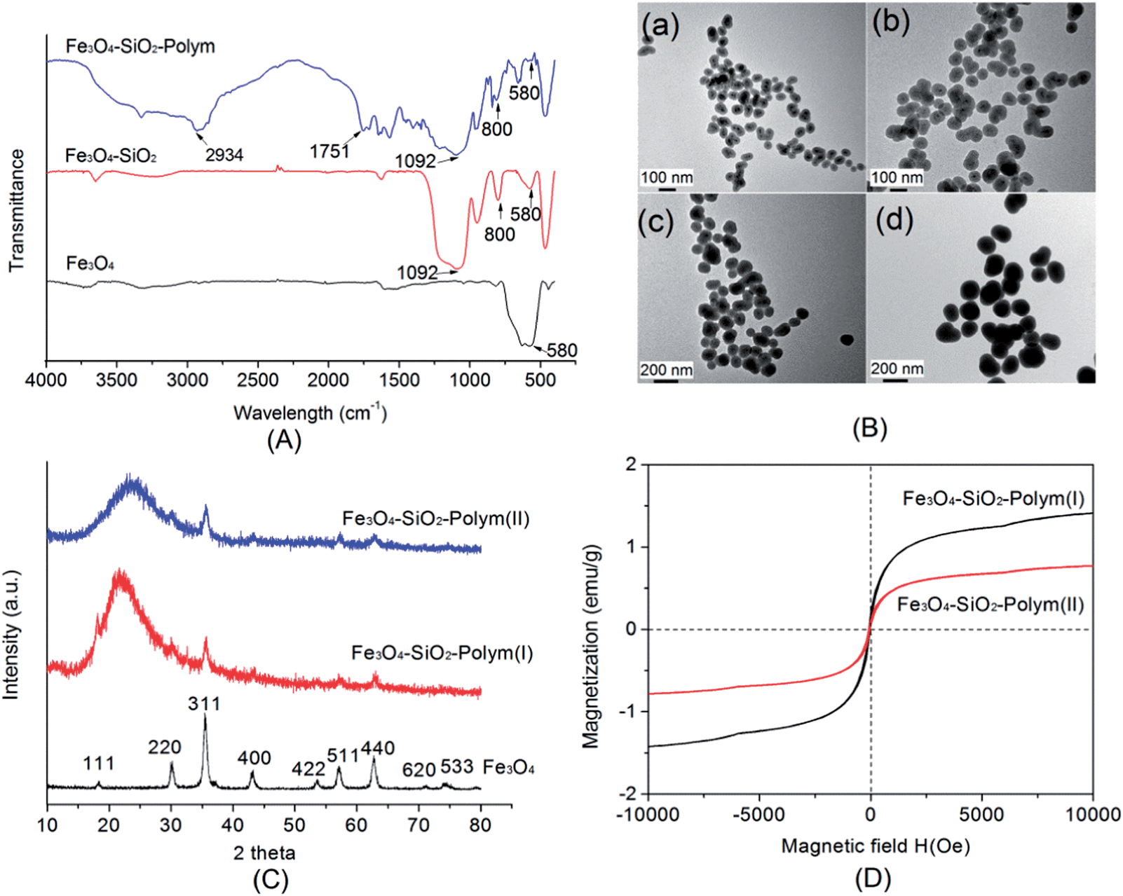 | ||
| Fig. 2 The characterization of dual layer-coated SPIONs: (A) FTIR (Fourier transform infrared spectroscopy) spectra of Fe3O4, Fe3O4–SiO2, and Fe3O4–SiO2–Polym; (B) TEM (transmission electron microscopy) images of Fe3O4–SiO2 (I), Fe3O4–SiO2 (II) (a and b), and Fe3O4–SiO2–Polym (I), Fe3O4–SiO2–Polym (II) (c and d); (C) XRD (X-ray diffraction) spectra of Fe3O4, Fe3O4–SiO2–Polym (I), and Fe3O4–SiO2–Polym (II); (D) magnetization curves of Fe3O4–SiO2–Polym (I) and Fe3O4–SiO2–Polym (II). Polym represents mPEG–PLA polymer and the thickness of SPIONs coating increases from type I to type II, correspondingly. | ||
The XRD spectra demonstrated that the Fe3O4 core of dual layer-coated SPIONs maintained a face-centered cubic crystal structure (Fig. 2C). The broad peak at ca. 22 degree (2θ) for Fe3O4–SiO2–Polym (I and II) was due to the contribution of the silica layer. This can be explained by the low rotational energy for the Si–O bond and the open Si–O–Si bond angle, which led to a flexible and mobile molecular structure of silica.15 As a result of the shielding effect of dual coating, the intensity of the characteristic peaks for Fe3O4 decreased markedly. However, two peaks (311 and 440) remained clear after silica and polymer coating, while another peak (511) was also noticeable. The obtained SPIONs with tailored dual layers exhibited superparamagnetic behavior without magnetic hysteresis (Fig. 2D). Despite being much lower than the control Fe3O4 (at about 60 emu g−1), the degree of the saturation magnetization of Fe3O4–SiO2–Polym decreased when increasing the coating thickness from ca. 50 nm (type I) to 70 nm (type II). With the help of inductively coupled plasma atomic emission spectroscopy (ICP-AES), the mass of the coating layer was subtracted and the weight-normalized saturation magnetization values for both types of Fe3O4–SiO2–Polym were consistent with that of the control Fe3O4. In terms of the threshold of saturation magnetization, it was reported that the value greater than 7 emu g−1 was sufficient for biomedical applications.16 Although the Fe3O4–SiO2–Polym nanoparticles obtained in the current study exhibited a lower saturation magnetization compared to the threshold, the level of the saturation magnetization could be finely tuned and optimized to suit in vivo tumor theranostic application via the manipulation of coating material and thickness.17 Moreover, the amphiphilic nature of mPEG–PLA would extend the systemic circulation of SPIONs, thus avoiding elimination prior to reaching the target tissue.
The paclitaxel loading in Fe3O4–Polym, Fe3O4–SiO2–Polym (I), and Fe3O4–SiO2–Polym (II) was 44 ± 2, 3092 ± 10, and 8369 ± 33 μg per mg Fe, respectively (Fig. 3). The employment of the dual coating strategy considerably increased the loading of paclitaxel in Fe3O4–SiO2–Polym up to nearly 190 times compared to that with only single layer of coating (i.e. Fe3O4–Polym). In addition, the thicker polymer coating in Fe3O4–SiO2–Polym (II) in comparison to Fe3O4–SiO2–Polym (I) gave rise to a relatively higher loading. It was postulated that the polymer layer that held the majority of the encapsulated drug mainly via the hydrophobic interaction considering the hydrophobicity of both PLA and paclitaxel. Based on the TEM particle size analysis, the volume ratio of the outer polymer layer was calculated to be ca. 2.6 times between Fe3O4–SiO2–Polym (II) and Fe3O4–SiO2–Polym (I), which agreed well with the ratio of paclitaxel loading (i.e. 2.7) in these two SPIONs. It is the increased particle surface area and PLA content that contribute to the enhanced drug loading. A previous study employed poly(beta-amino ester) to generate single-layer SPIONs (ca. 80 nm by DLS) and the loading of the active agent (doxorubicin) and polymer was ca. 700 and 7000 μg per mg Fe, respectively.18 In spite of the divergence of the utilized polymer and model drug, the dual layer method employed in the current study efficiently enlarged the “apparent” core of SPIONs without losing their superparamagnetic feature. This leads to a drug loading capability 10 times higher than the above reported value at the cost of increased mPEG–PLA packing (ca. 77![[thin space (1/6-em)]](https://www.rsc.org/images/entities/char_2009.gif) 824 μg per mg Fe) for Fe3O4–SiO2–Polym (II). The SPIONs with the highest loading in the current study were chosen for the drug release experiment that was carried out in sink conditions. The profile exhibited a rapid drug release at the beginning and the curve started getting plateaued after 8 h; over 24 h nearly 80% of the total drug was released. Although PLA is biodegradable, the release was thought to be primarily controlled by diffusion as the SIPONs's disintegration induced by PLA degradation is negligible within a short time at a neutral condition.
824 μg per mg Fe) for Fe3O4–SiO2–Polym (II). The SPIONs with the highest loading in the current study were chosen for the drug release experiment that was carried out in sink conditions. The profile exhibited a rapid drug release at the beginning and the curve started getting plateaued after 8 h; over 24 h nearly 80% of the total drug was released. Although PLA is biodegradable, the release was thought to be primarily controlled by diffusion as the SIPONs's disintegration induced by PLA degradation is negligible within a short time at a neutral condition.
 | ||
| Fig. 3 The drug (paclitaxel) loading of Fe3O4–Polym and Fe3O4–SiO2–Polym (top); the drug release profile from Fe3O4–SiO2–Polym (II) (bottom). Fe3O4–Polym was coated only by the mPEG–PLA polymer with APS as the linker. Fe3O4–SiO2–Polym was coated by both silica and polymer, and the coating thickness of Fe3O4–SiO2–Polym (II) was higher than that of Fe3O4–SiO2–Polym (I); the polymer volume in single-layer Fe3O4–Polym nanoparticles was two orders of magnitude smaller compared to that in Fe3O4–SiO2–Polym (I and II). | ||
In summary, both silica and polymer coated SPIONs successfully addressed the poor drug loading issue of conventional SPIONs. The precise manipulation of the thickness of dual layers would balance the requirement of drug loading and saturation magnetization, which is particularly true for potent active agents. Such an approach could be further manipulated to engineer multifunctional nanotheranostics with imaging, therapeutics loading and targeting for improved tumor therapy.
We gratefully acknowledge the funding support from Tianjin Research Program of Application Foundation and Advanced Technology (11JCZDJC20600; 13JCQNJC13300) and National Natural Science Foundation of China (11001197). Prof. Tonglei Li from Purdue University kindly went through this manuscript and made valuable suggestions.
Notes and references
- Z. L. Cheng, A. Al Zaki, J. Z. Hui, V. R. Muzykantov and A. Tsourkas, Science, 2012, 338, 903 CrossRef CAS PubMed.
- M. Ferrari, Nat. Rev. Cancer, 2005, 5, 161 CrossRef CAS PubMed.
- Y. Lalatonne, C. Paris, J. M. Serfaty, P. Weinmann, M. Lecouvey and L. Motte, Chem. Commun., 2008, 2553 RSC; Y. Pan, X. Du, F. Zhao and B. Xu, Chem. Soc. Rev., 2012, 41, 2912 RSC.
- C. Rumenapp, B. Gleich and A. Haase, Pharm. Res., 2012, 29, 1165 CrossRef PubMed.
- O. Veiseh, J. W. Gunn and M. Zhang, Adv. Drug Delivery Rev., 2010, 62, 284 CrossRef CAS PubMed.
- M. V. Yigit, A. Moore and Z. Medarova, Pharm. Res., 2012, 29, 1180 CrossRef CAS PubMed; S. A. Corr, A. O'Byrne, Y. K. Gun'ko, S. Ghosh, D. F. Brougham, S. Mitchell, Y. Volkov and A. Prina-Mello, Chem. Commun., 2006, 4474 RSC.
- Q. M. Quan, J. Xie, H. K. Gao, M. Yang, F. Zhang, G. Liu, X. Lin, A. Wang, H. S. Eden, S. Lee, G. X. Zhang and X. Y. Chen, Mol. Pharmaceutics, 2011, 8, 1669 CrossRef CAS PubMed; M. H. El-Dakdouki, D. C. Zhu, K. El-Boubbou, M. Kamat, J. J. Chen, W. Li and X. F. Huang, Biomacromolecules, 2012, 13, 1144 CrossRef PubMed.
- K. M. Krishnan, IEEE Trans. Magn., 2010, 46, 2523 CrossRef CAS PubMed.
- P. Zou, Y. Yu, Y. Wang, Y. Zhong, A. Welton, C. Galban, S. Wang and D. Sun, Mol. Pharmaceutics, 2010, 7, 1974 CrossRef CAS PubMed.
- N. Schleich, P. Sibret, P. Danhier, B. Ucakar, S. Laurent, R. Muller, C. Jerome, B. Gallez, V. Preat and F. Danhier, Int. J. Pharm., 2013, 447, 94 CrossRef CAS PubMed.
- Q. Liu, J. A. Finch and R. Egerton, Chem. Mater., 1998, 10, 3936 CrossRef CAS.
- R. Al-Oweini and H. El-Rassy, J. Mol. Struct., 2009, 919, 140 CrossRef CAS PubMed; S. K. Choi and D. Kim, J. Appl. Polym. Sci., 2002, 83, 435 CrossRef.
- H. Maeda, J. Wu, T. Sawa, Y. Matsumura and K. Hori, J. Controlled Release, 2000, 65, 271 CrossRef CAS.
- I. J. Majoros, T. P. Thomas, C. B. Mehta and J. R. Baker, J. Med. Chem., 2005, 48, 5892 CrossRef CAS PubMed.
- J. Zhang, M. Sun, A. Fan, Z. Wang and Y. Zhao, Int. J. Pharm., 2013, 441, 389 CrossRef CAS PubMed.
- X. He, X. Wu, X. Cai, S. Lin, M. Xie, X. Zhu and D. Yan, Langmuir, 2012, 28, 11929 CrossRef CAS PubMed.
- F. M. Kievit and M. Zhang, Acc. Chem. Res., 2011, 44, 853 CrossRef CAS PubMed.
- F. Chen, F. M. Kievit, O. Veiseh, Z. R. Stephen, T. Wang, D. Lee, R. G. Ellenbogen and M. Zhang, J. Controlled Release, 2012, 162, 233 CrossRef PubMed.
Footnote |
| † Electronic supplementary information (ESI) available: Details of SPIONs generation and characterization. See DOI: 10.1039/c3ra42861c |
| This journal is © The Royal Society of Chemistry 2014 |