
 Open Access Article
 This Open Access Article is licensed under a Creative Commons Attribution-Non Commercial 3.0 Unported Licence
Oligonucleotide-capped mesoporous silica nanoparticles as DNA-responsive dye delivery systems for genomic DNA detection†
Lluís
Pascual
abc,
Isabel
Baroja
abc,
Elena
Aznar
ac,
Félix
Sancenón
abc,
M. Dolores
Marcos
abc,
Jose Ramón
Murguía
abc,
Pedro
Amorós
d,
Knut
Rurack
e and
Ramón
Martínez-Máñez
*abc
aCentro de Reconocimiento Molecular y Desarrollo Tecnológico (IDM), Unidad Mixta Universidad Politécnica de Valencia-Universidad de Valencia, Spain
bDepartamento de Química, Universidad Politécnica de Valencia, Camino de Vera s/n, 46022, Valencia, Spain. E-mail: rmaez@qim.upv.es
cCIBER de Bioingeniería, Biomateriales y Nanomedicina (CIBER-BBN), Spain
dInstitut de Ciència dels Materials (ICMUV), Universitat de València, P.O. Box 2085, 46071 Valencia, Spain
eFachbereich 1.9 Sensormaterialien, Bundesanstalt für Materialforschung und -prüfung (BAM), Richard-Willstätter-Straße 11, 12489 Berlin, Germany
First published on 19th November 2014
Abstract
New hybrid oligonucleotide-capped mesoporous silica nanoparticles able to detect genomic DNA were designed.
Nanodevices equipped with molecular gates based on mesoporous silica nanoparticles have proven their potential for use in biological and medical applications.1 So far, capped materials have been mainly used in drug delivery and their use in sensing protocols is still scarce.2 For sensing, the mesoporous support is loaded with an indicator before the material is capped with a responsive chemistry. Uncapping and cargo delivery is then selectively achieved only in the presence of a target analyte.3 In this context we are interested in the use of DNA sequences for the design of gated materials and others and we have recently reported the use of oligonucleotides as caps on mesoporous supports in the context of the detection of biomolecules via delivery of optical or electro-active probes.4 However, in this context, the detection of genomic DNA using capped materials is still very rare.
Inspired by these preliminary studies we report herein a new hybrid gated material with a covalently attached double-stranded DNA (dsDNA) sequence that selectively delivers a dye in the presence of genomic DNA. As a proof of concept the detection of Mycoplasma genomic DNA was pursued in this work. Detection of bacteria of genus Mycoplasma is of interest in diverse areas. Mycoplasma contamination is a common problem in cell culture laboratories of research institutions and hospitals.5 Moreover, several Mycoplasma species are related to the development of certain pathologies such as pneumonia, rheumatoid arthritis and non-gonococcal urogenital diseases.6
The approach we followed is depicted in Scheme 1. MCM-41 mesoporous silica nanoparticles of ca. 100 nm diameter were selected as inorganic supports because of their unique and advantageous properties for the design of capped materials. The MCM-41 support was first loaded with the dye rhodamine B and then the external surface was functionalized with (3-isocyanatopropyl)triethoxysilane, yielding solid S1-I. For the gating mechanism two single stranded oligonucleotides were selected; i.e. (i) a short DNA sequence, functionalized with an aminohexyl moiety at the 5′-end position (NH2-(CH2)6-5′-GAC TAC CAG GGT ATC-3′, O1) that could be covalently attached to solid S1-Ivia the formation of urea bonds and (ii) a single stranded oligonucleotide (5′-AAG CGT GGG GAG CAA ACA GGA TTA GAT ACC CTG GTA GTC-3′, O2) which is a highly conserved sequence of the 16S ribosomal subunit of the Mycoplasma species genome.
 | ||
| Scheme 1 Representation of the gated material S1-O1/O2 capped with a dsDNA. Delivery of the entrapped dye (rhodamine B) is selectively accomplished in the presence of a single strand of Mycoplasma genomic DNA. | ||
In a typical experiment the two single stranded DNA sequences (O1 and O2) were first hybridized and then covalently attached to the surface of the S1-I solid, yielding the final material S1-O1/O2. The dsDNA, anchored on the mesoporous support, was expected to be bulky enough to block the pores and inhibit dye delivery. Moreover it was anticipated that the presence of a single strand of Mycoplasma genomic DNA would displace O2 from the solid with the subsequent pore opening and dye release.
MCM-41 nanoparticles were obtained according to reported procedures. Solid S1-I was first loaded with rhodamine B in acetonitrile and then an excess of (3-isocyanatopropyl)triethoxysilane was added to the suspension. Solid S1-I was collected by centrifugation, washed with acetonitrile and dried (see ESI† for details). For the preparation of the final capped material (i.e.S1-O1/O2) O1 and O2 were first mixed, heated at 90 °C and then gradually cooled to achieve the hybridization of both oligonucleotides. Then the hybridized O1/O2 sequences were anchored onto the outer surface of the S1-I solid through the formation of a urea bond by reaction of the amine group in O1/O2 and the isocyanatopropyl groups attached on S1-I (see ESI† for further details).
The starting MCM-41-like calcined nanoparticles and S1-I were characterized by powder X-ray diffraction, TEM, porosimetry, thermogravimetry and elemental analysis (see ESI† for details). Table 1 summarizes the particle diameter, BET specific surface area, pore volumes and pore sizes obtained for the starting nanoparticles and for solid S1-I. Moreover, from elemental and thermogravimetric analyses contents of 0.469 and 0.358 mmol g−1 for isocyanatopropyl moieties and rhodamine B, respectively, were determined in solid S1-I. Finally, dynamic light scattering studies in water gave diameters for the MCM-41, S1-I and S1-O1/O2 nanoparticles of 88 ± 2, 114 ± 3 and 154 ± 2 nm, respectively.
The response of S1-O1/O2 was tested in the presence of Mycoplasma fermentans genomic DNA by measuring the emission of rhodamine B dye delivered from the pores of the solid. In a typical experiment 0.2 mg of S1-O1/O2 were suspended in 400 μL of hybridization buffer (20 mM tris-HCl, 37.5 MgCl2, pH 7.5) and separated into 2 aliquots of 200 μL. Both samples were filled to a final volume of 900 μL with hybridization buffer. At the same time a solution of 4000 copies μL−1 of Mycoplasma fermentans quantification standard in water was heated to 95 °C for 5 min in order to dehybridize the double helix structure and then cooled in an ice bath for 3 min. After cooling, 100 μL of the dehybridized genomic solution was added to one of the aliquots whereas 100 μL of water (that was subjected to the same thermal treatment) were added to the other aliquot. Both suspensions were maintained at 25 °C and, at certain times, fractions were taken and centrifuged to eliminate the solid. Cargo release to the solution was then measured by the rhodamine B fluorescence at 575 nm (λexc = 555 nm). Fig. 1 shows the delivery profile of rhodamine B from solid S1-O1/O2 in the presence and in the absence of Mycoplasma fermentans genomic DNA. As it can be seen, in the absence of the target DNA (Fig. 1, curve a) a poor rhodamine B delivery was observed (less than 15% after 1 h) indicative of a remarkable pore closure. In contrast, in the presence of Mycoplasma genomic DNA, the pores were opened due to the displacement of oligonucleotide O2 from the solid S1-O1/O2 with the subsequent delivery of the dye (Fig. 1, curve b). Under these conditions ca. 90% of the maximum delivery of rhodamine B was observed after 30 min.
 | ||
| Fig. 1 Release of rhodamine B from solid S1-O1/O2 in (a) the absence and (b) in the presence of Mycoplasma fermentans genomic DNA (4000 copies μL−1) in Tris-HCl buffer at pH 7.5. The inset shows the emission changes (under UV lamp, excitation at 254 nm) of the rhodamine B released from solid S1-O1/O2 in the absence (left) and in the presence (right) of Mycoplasma fermentans genomic DNA (4000 copies μL−1). | ||
The high performance of solid S1-O1/O2 pointed to the effective anchoring of the O1/O2 dsDNA on the external surface of the inorganic scaffold through the formation of urea bonds. Additional experimental evidence of the correct grafting arose from the fact that solid S1-O1/O2 was able to deliver rhodamine B in the presence of urease, due to the enzyme-induced hydrolysis of this bond with the subsequent detachment of the O1/O2 double strand (see ESI†). Moreover, in the presence of DNAse I, a remarkable dye release was also observed indicating the crucial role played by the O1/O2 sequence in the capping protocol (see ESI†).
The next step was to assess the sensitivity of S1-O1/O2 nanoparticles towards genomic DNA with the determination of the limit of detection. Following a procedure similar to that described above various solutions with different concentrations of Mycoplasma fermentans genomic DNA were added to several suspensions of S1-O1/O2 and the rhodamine B released after 30 min was measured (see ESI†). A correlation between the number of copies of Mycoplasma genomic DNA added and the rhodamine B delivered was found in agreement with an uncapping process related to the displacement of O2 from the S1-O1/O2 solid. A limit of detection of ca. 70 DNA copies μL−1 was determined (see ESI†). The limit of detection following this simple approach is not far from those reported using commercially available PCR Mycoplasma detection kits (around ca. 10 copies μL−1),7 suggesting that this procedure for genomic DNA sensing possesses the potential of finding application in medical diagnosis protocols.
In further studies the selectivity in the detection of Mycoplasma genomic DNA by the S1-O1/O2 solid was investigated by carrying out similar experiments in the presence of genomic DNA of the pathogens Candida albicans and Legionella pneumophila in amounts of 1000 copies μL−1. The results obtained are shown in Fig. 2. As can be seen there were no significant differences between the blank (dye release in Tris-HCl buffer) and the potentially interfering genomic DNA of Candida albicans and Legionella pneumophila. In addition, when a mixture of Candida albicans–Legionella pneumophila (each one at a concentration of 1000 copies μL−1) was used no delivery of rhodamine B was observed. Furthermore, when genomic DNA of Mycoplasma was present in the mixture a clear delivery of rhodamine B was found pointing to a high degree of selectivity of the S1-O1/O2 material.
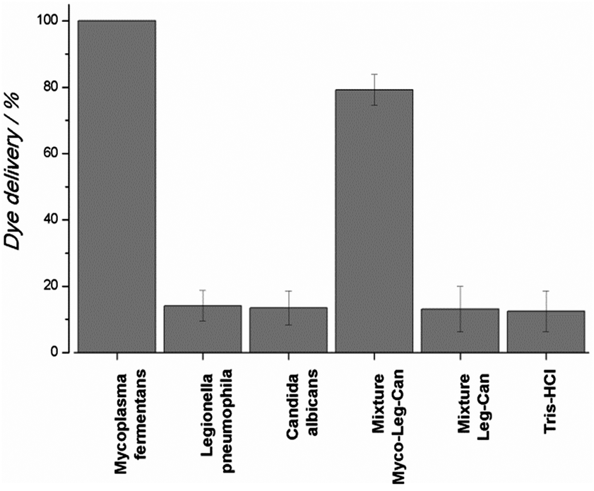 | ||
| Fig. 2 Release of rhodamine B from solid S1-O1/O2 in the presence of (from right to left): Tris-HCl buffer, a mixture of Legionella pneumophila–Candida albicans (Leg–Can) genomic DNA, a mixture of Leg–Can and Mycoplasma fermentans (Myco) genomic DNA, Can genomic DNA, Leg genomic DNA and Myco genomic DNA. All DNA were at a concentration of 1000 copies μL−1. | ||
In summary, we have prepared mesoporous silica nanoparticles loaded with rhodamine B and capped with a covalently linked double stranded DNA containing a highly conserved sequence of the 16S ribosomal subunit of the Mycoplasma species genome. The nanoparticles were able to deliver the entrapped dye in the presence of Mycoplasma fermentans genomic DNA. A remarkable detection limit of ca. 70 DNA copies μL−1 was found. Moreover the selectivity of the system was assessed, for instance, addition of Candida albicans and Legionella pneumophila genomic DNA was unable to induce dye release. We believe that DNA-based gated materials could be suitable alternatives to the standard bacteria detection techniques such as PCR and can find application as alternative rapid point-of-care diagnostic systems or in situations and places where sophisticated techniques or equipment are not available.
Financial support from the Spanish Government (Project MAT2012-38429-C04) and the Generalitat Valencia (Project PROMETEOII/2014/047) is gratefully acknowledged. Ll. P. is grateful to the Universidad Politécnica de Valencia for his grant.
Notes and references
- (a) A. Agostini, L. Mondragón, A. Bernardos, R. Martínez-Máñez, M. D. Marcos, F. Sancenón, J. Soto, A. Costero, C. Manguan-García, R. Perona, M. Moreno-Torres, R. Aparicio-Sanchis and J. R. Murguía, Angew. Chem., Int. Ed., 2012, 51, 10556–10560 CrossRef CAS PubMed; (b) Q. Zhang, X. Wang, P.-Z. Li, K. T. Nguyen, X.-J. Wang, Z. Luo, H. Zhang, N. S. Tan and Y. Zhao, Adv. Funct. Mater., 2014, 24, 2450–2461 CrossRef CAS; (c) C. Chen, J. Geng, F. Pu, X. Yang, J. Ren and X. Qu, Angew. Chem., Int. Ed., 2011, 50, 882–886 CrossRef CAS PubMed; (d) L. Zhou, Z. Chen, K. Dong, M. Yin, J. Ren and X. Qu, Adv. Mater., 2014, 26, 2424–2430 CrossRef CAS PubMed.
- (a) A. Agostini, L. Mondragón, L. Pascual, E. Aznar, C. Coll, R. Martínez-Máñez, F. Sancenón, J. Soto, M. D. Marcos, P. Amorós, A. M. Costero, M. Parra and S. Gil, Langmuir, 2012, 28, 14766–14776 CrossRef CAS PubMed; (b) C. Coll, A. Bernardos, R. Martínez-Máñez and F. Sancenón, Acc. Chem. Res., 2013, 46, 339–349 CrossRef CAS PubMed; (c) E. Climent, R. Martínez-Máñez, A. Maquieira, F. Sancenón, M. D. Marcos, E. M. Brun, J. Soto and P. Amorós, ChemistryOpen, 2012, 1, 251–259 CrossRef CAS PubMed.
- (a) M. Oroval, E. Climent, C. Coll, R. Eritja, A. Avino, M. D. Marcos, F. Sancenon, R. Martinez-Manez and P. Amoros, Chem. Commun., 2013, 49, 5480–5482 RSC; (b) M. Chen, C. Huang, C. He, W. Zhu, Y. Xu and Y. Lu, Chem. Commun., 2012, 48, 9522–9524 RSC.
- (a) E. Climent, R. Martínez-Máñez, F. Sancenón, M. D. Marcos, J. Soto, A. Maquieira and P. Amorós, Angew. Chem., Int. Ed., 2010, 49, 7281–7283 CrossRef CAS PubMed; (b) E. Climent, L. Mondragón, R. Martínez-Máñez, F. Sancenón, M. D. Marcos, J. R. Murguía, P. Amorós, K. Rurack and E. Pérez-Payá, Angew. Chem., Int. Ed., 2013, 52, 8938–8942 CrossRef CAS PubMed; (c) Z. Zhang, D. Balogh, F. Wang, S. Y. Sung, R. Nechushtai and I. Willner, ACS Nano, 2013, 7, 8455–8468 CrossRef CAS PubMed; (d) L. Wu, J. Ren and Q. Xiaogang, Nucleic Acids Res., 2014 DOI:10.1093/nar/gku858.
- H. Drexler and C. Uphoff, Cytotechnology, 2002, 39, 75–90 CrossRef CAS.
- (a) L. Matas Andreu, S. Molinos Abós, G. Fernández Rivas, V. González Soler and V. Ausina Ruiz, Enferm. Infecc. Microbiol. Clin., 2006, 24, 19–23 CrossRef PubMed; (b) V. Ausina, Infecciones causadas por micoplasmas, Medicina Interna, Elsevier España, 2004, 15th edn, pp. 2363–2365 Search PubMed.
- (a) http://www.lifetechnologies.com ; (b) http://www.sigmaaldrich.com ; (c) http://www.lgcstandards-atcc.org (all websites accessed on 01/10/2014).
Footnote |
| † Electronic supplementary information (ESI) available: Synthesis of S1-I and S1-O1/O2 materials, characterization of the prepared solids, and release experiments with solid S1-O1/O2. See DOI: 10.1039/c4cc08306g |
| This journal is © The Royal Society of Chemistry 2015 |