Biocompatibility, biodistribution and efficacy of magnetic nanohydrogels in inhibiting growth of tumors in experimental mice models†
Manish K.
Jaiswal
a,
Manashjit
Gogoi
b,
Haladhar
Dev Sarma
c,
Rinti
Banerjee
b and
D.
Bahadur
*a
aDepartment of Metallurgical Engineering & Materials Science, Indian Institute of Technology Bombay, Mumbai, 400076, India
bDepartment of Biosciences & Bioengineering, Indian Institute of Technology Bombay, Mumbai, 400076, India
cRadiation Biology and Health Sciences Division, Bhabha Atomic Research Centre, Mumbai, 400085, India. E-mail: dhirenb@iitb.ac.in; Fax: +91-22-2572-3480; Tel: +91-22-2576-7632
First published on 20th November 2013
Abstract
We report in vivo evaluation of a thermo-responsive poly(N-isopropylacrylamide)-chitosan based magnetic nanohydrogel (MNHG) incorporated with Fe3O4 nanoparticles (NPs) in mice models with expandible scope for use in localized delivery of chemotherapeutics. Biocompatibility and biodistribution of the MNHG are studied in normal Swiss mice while efficacy in tumor growth inhibition is studied in a subcutaneous fibrosarcoma tumor. The ex vivo time-dependent pattern of accumulated MNHG into vital organs; lung, liver, spleen, kidney and brain collected at 1 h, 48 h, 7 d and 14 d post intravenous administration are investigated using both a vibrating sample magnetometer (VSM) and inductively coupled plasma-atomic emission spectroscopy (ICP-AES) method. The doses of MNHG (dose I ∼ 650 and dose II ∼ 325 μg g−1 body wt) used in the study are determined based on induced thermal activation of MNHG under an AC magnetic field (AMF). Fibrosarcoma tumor bearing mice are subjected to hyperthermia with a field of 325 Oe and 265 kHz for 30 min following intratumoral administration of dose I. Tumor size measured at an interval of 72 h for a period of 2 weeks reveals that the NPs mediated therapy decelerated the growth of the transplanted tumor by about three-fold (size, 1545 ± 720 mm3) as compared to the exponential growth of the tumor (size, 4510 ± 735 mm3) in control mice. These results suggest the feasibility of using poly(NIPAAm)-chitosan hydrogels loaded with NPs for combined thermo-chemotherapy where the efficacy may further be improved by temperature dependent release of the drugs from the magneto hydrogels.
1. Introduction
Thermo-responsive hydrogels are three dimensional cross-linked polymeric submicron size structures which are hydrophilic below a specific temperature, defined as the lower critical solution temperature (LCST). Above the LCST, the polymeric hydrogels manifest conformational changes from ‘expanded coil’ to ‘compact globule’ form by expelling aqueous content as it transforms to the hydrophobic state. These stimuli-responsive hydrogels have received attention in drug delivery because of their improved aqueous solubility below the LCST, which facilitates easy encapsulation of therapeutics that may be released upon a temperature trigger.1,2 Poly(N-isopropylacrylamide, poly(NIPAAm)) is one of the most widely used thermo-responsive polymers (LCST ∼ 32–34 °C), which can be easily used for hyperthermia and drug delivery applications above 42 °C.3–5Fe3O4 nanoparticle (NPs) based systems are proven to have many biomedical applications such as tumor-specific cell targeting,6 lymph node imaging,7 magnetic resonance imaging and visualization of RNA interference,8–11 target-cell-specific labeling,12 inhibition of tumor cell invasion,13 stem cell tracking14 and magnetic hyperthermia among many others.15–17
Biocompatible dual pH/temperature stimuli-responsive poly(NIPAAm)-chitosan (CS) based nanohydrogels with incorporated Fe3O4 magnetic nanoparticles (MNHG) have not been explored much for magnetic hyperthermia, and their fate in the body after being administered through blood vessels is also unknown. Our preliminary studies have shown the feasibility of causing a shift in the LCST of poly(NIPAAm)-CS when loaded with NPs. Poly(NIPAAm) based MNHG (250 nm) with CS (by 2 wt%) and Fe3O4 NPs (by 25 wt%) have been found to have their LCST above 42 °C which is suitable for magnetic thermal therapy.4 The presence of CS may provide the additional advantage of releasing therapeutics in the tumor tissues where the pH of the environment is mild acidic in nature. It may also be worthwhile evaluating the efficacy of the MNHG in treating tumors under mild hyperthermia conditions with an optimal interval period between two bouts of hyperthermia as compared to existing literatures.18–22
We present an account of biodistribution, biocompatibility and efficacy studies using NPs incorporated within poly(NIPAAm)-chitosan hydrogels. Further, we have utilized the magnetic properties of the various tissues to record the distribution of magnetic hydrogels. This is expected to be a more sensitive and accurate method of detecting even trace amounts of magnetic nanoparticle accumulation. The results were further corroborated by direct quantification of iron accumulated into organs. Spectroscopic techniques based on inductively coupled plasma atomic emission spectroscopy (ICP-AES) yield reliable information about iron concentrations and are therefore used in this work. It fares better over other available methods e.g. isotope tagging of NPs followed by the counting of radiolabeled nuclides method, which has certain practical limitations.23,24
The scheme shown in Fig. 1 presents the proposed idea of treating tumor-bearing animals through nanohydrogels incorporated with Fe3O4 NPs and an aqueous soluble anti-cancer therapeutic agent. Upon switching the AMF ‘ON’ the NPs heat up and stimulate the release of loaded agent due to structural collapse of the hydrogels at the site of interest.
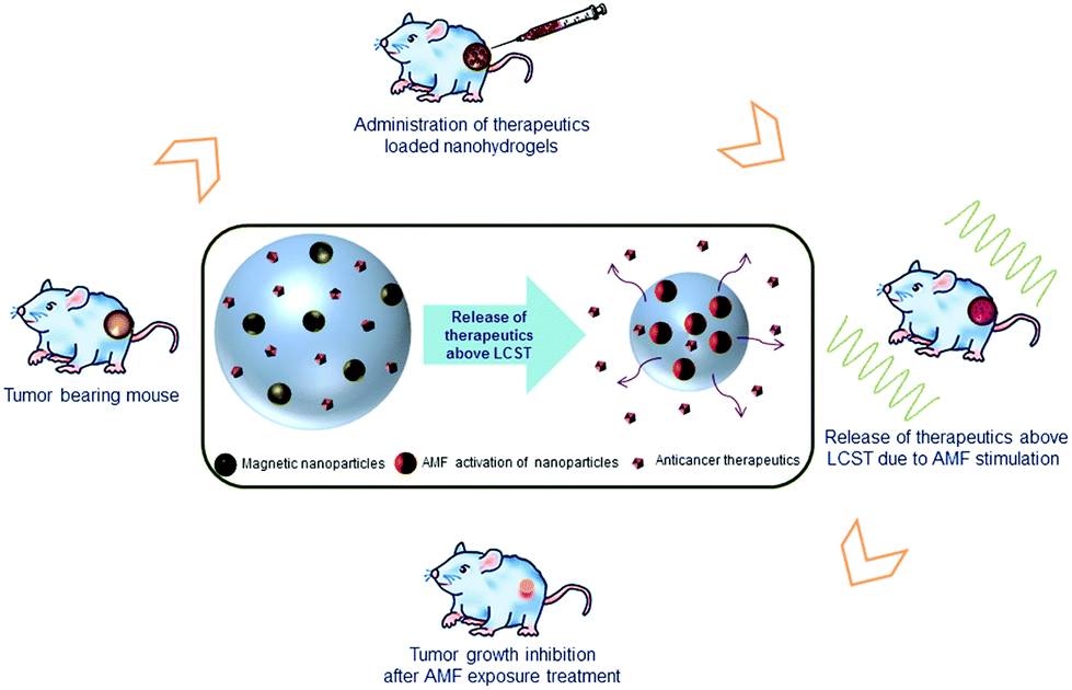 | ||
| Fig. 1 Schematic illustration of localized therapy of tumor grown around the flank of a Swiss mouse. Magnetic nanohydrogels incorporated with Fe3O4 nanoparticles (NPs) carrying anti-cancer therapeutics can be stimulated by an AC magnetic field (AMF) to deliver the drug and hyperthermia at the tumor site. The central scheme shows the behavior of hydrogel on administration at the target site, it undergoes a conformational change upon a rise in temperature above the LCST (lower critical solution temperature) and thereby releases the drug. | ||
2. Experimental methods
2.1 Materials
N-Isopropylacrylamide (NIPAAm, 97%), ammonium persulphate (APS, 98%), sodium metabisulphite (SBS, 99%), ferric chloride hexahydrate (FeCl3·6H2O, ≥98%), ferrous chloride tetrahydrate (FeCl2·4H2O, 99%), crosslinker N,N′-methylene-bis-acrylamide (MBAAm), EDTA (ethylenediaminetetraacetic acid) and low molecular wt (Mw ∼ 10 kg mol−1) chitosan (CS) with 85% deacetylation degree of reagent grade were purchased from Sigma-Aldrich USA. Acetic acid (glacial, 99%) was purchased from Merck Chemicals. All chemicals were directly used without further purification.2.2 Synthesis of magnetic nanohydrogels (MNHG)
The LCST optimized magnetic nanohydrogels (MNHG) for hyperthermia application is synthesized using a reported method.4 In brief, aqueous NIPAAm (0.036 M, 80 mL) and freshly prepared chitosan (0.004 M, 20 mL, 20% acetic acid glacial) stabilized Fe3O4 NPs (200 mg) were mixed in a 250 mL 4-neck flask under vigorous stirring. The crosslinker MBAAm (methylene-bis-acrylamide, 0.1 M, 8 mL) in water was added to the mixture and temperature was slowly raised to 70 °C. Subsequently, to initiate the polymerization, aqueous APS (ammonium persulphate, 0.01 M, 4 mL) and catalyst SBS (sodium metabisulphite, 0.005 M, 4 mL) were added and the system was refluxed for the next 5 h. The system was cooled down to room temperature (ca. 25 °C) thereafter and the obtained latex was dialyzed against ultrapure water (resistivity 18.2 MΩ cm) for one week before further application.2.3 Animal experiment
Experimental animals were procured from the animal house facility of Bhabha Atomic Research Centre (BARC), Mumbai, India and the experiments were carried out in the experimental animal house facility of the center. All the animal experiments were done in compliance with the national laws governing control and supervision of experiments on animals. Swiss mice (6–8 weeks age) were used for the biocompatibility and biodistribution study of the MNHG. In vivo hyperthermia studies were carried out on the fibrosarcoma tumor model raised in the same species of mice.| Grouping of animals | Number of animalsb | Time of collection of sample (post-administration) | ||
|---|---|---|---|---|
| Group (MNHG) | Sub-group | For histopath and haematology studies | For Fe3O4/iron accumulation studies | |
| a h = hour, d = day. b Total number of animals in each group (except XII) was 8, out of which 4 animals were used for VSM and ICP-AES studies for dose I treated mice groups. | ||||
| Dose I (650 μg g−1) | I | 8 | 4 | 1 h |
| II | 8 | 4 | 24 h | |
| III | 8 | 4 | 48 h | |
| IV | 8 | 4 | 7 d | |
| V | 8 | 4 | 14 d | |
| Dose II (325 μg g−1) | VI | 8 | — | 1 h |
| VII | 8 | — | 24 h | |
| VIII | 8 | — | 48 h | |
| IX | 8 | — | 7 d | |
| X | 8 | — | 14 d | |
| Control | XI | 8 | 4 | 1 h |
| XII | 4 | — | 14 d | |
The doses of MNHG for biocompatibility and in vivo biodistribution studies are determined based on AMF induction heating results. MNHG at 650 μg g−1 body wt was taken as dose I and half of the dose (i.e. 325 μg g−1 body wt) was taken as dose II for this study. For the sake of readers’ knowledge, MNHG concentrations were 65 and 32.5 mg mL−1 in dose I and dose II respectively, out of which 200 μL was administered to the animals of respective groups for biocompatibility and biodistribution studies (Table 1).
Four to eight mice from the sub groups were sacrificed at the various time points; 1 h, 24 h, 48 h, 7 d and 14 d for collection of blood and the vital tissue organs; lung, liver spleen, kidney and brain. The detail protocol for CBC and serum biochemistry is described in the ESI.†
Tumor bearing animals (on 12th day of transplantation) were randomly divided into six (I, II, III, IV, V and control) groups consisting of three animals each. Details of grouping of the experimental animals, applied field strength, duration of hyperthermia and frequency of dose of MNHG are listed in Table 2.
| Group (n = 3) | Field (Oe) | Dose | Exposure time (min) |
|---|---|---|---|
| a n = number of animals; SD = single dose; DD = double dose. | |||
| I | 325 | DD | 30 |
| II | 325 | SD | 30 |
| III | 290 | DD | 30 |
| IV | 290 | SD | 30 |
| V | 290 | SD | 20 |
| Control | — | — | — |
Tumor diameters of all the animals were measured with the help of a digital vernier caliper prior to hyperthermia treatment (day 1). Subsequent measurements of the tumors were done at 72 h intervals on at least four occasions. Tumor size (or volume) was calculated according to the following formula;25
| Volume = (π/6) × (major dimension) × (minor dimension)2 |
Animals were anesthetized with intra-peritoneal injections of ketamine–HCl (50–80 μg g−1) and xylazine–HCl (5–10 μg g−1) before administration of the MNHG. A dose of 150 μL of MNHG (65 mg mL−1) based on an induction heating experiment was administered via multiple point injections directly into the tumor region of each mouse using a hypodermic syringe with a 27 gauge needle before application of an external field.
All the animals were then subjected to AMF at 290 or 325 Oe for 30 or 20 min as mentioned in Table 2. The magnetic field was generated by a water-cooled cylindrical copper coil (diameter 6.5 cm, 4 turns) connected to a radio frequency generator (Ameritherm, USA). Mice were placed inside the coil to maximize field exposure. The rise in temperature at different points on the surface of the tumor was measured with an optical probe (model, OEM Fiber-optic thermosensor, LumaSense USA) at 10 min intervals. Animals of the experimental groups were subjected to AMF three times for hyperthermia treatment at an interval of 72 h. Animals of group I and III received a 2nd dose of MNHG 72 h after the first dose.
3. Materials characterization
The synthesized magnetic hydrogels were fully characterized for structural, morphological, thermal and magnetic properties. The morphological and surface topographical images of MNHG were captured through scanning electron microscopy (SEM, model Hitachi 3400, Japan) and atomic force microscopy (AFM, model Veeco Instruments, Nanoscope IV, USA). The TEM images of the sample were taken at 200 keV using a Philips model CM 200 to confirm the presence of NPs in the MNHG.To determine the LCST of the MNHG, a change in enthalpy method was used where a small portion of the dried sample was dissolved in water and kept overnight at 4 °C to allow it to swell fully. Subsequently, the sample was centrifuged and excess water was decanted. The obtained slurry was subjected to DSC (differential scanning calorimetry, TA Instruments, USA) under a nitrogen environment. The change in enthalpy was recorded with temperature from 20 to 60 °C at ramp rate of 2 °C min−1 at constant pressure. The onset of a sudden decrease in the enthalpy was considered as the hydrophilic to hydrophobic transition. For magnetization, M–H data was recorded of the dried powder of the sample up to an applied field of 20 kOe through a vibrating sample magnetometer (VSM).
Statistical analysis
All the data are expressed as mean ± s.d. (standard deviation). Statistical significance levels are computed using paired one-tailed student's t-test and p ≤ 0.05 was used as the criteria to determine the confidence intervals.4. Results and discussion
4.1 Microstructural characterization
Fig. 2 shows (a) SEM and (b) AFM microstructures of the sample confirming the regular shape and size (approx. 250 nm) of the hydrogels. Fig. 2(c and d) show the visual photographs of an aqueous solution of the hydrogel below and above the LCST. The hydrogels are hydrophilic and appear transparent below the phase transition temperature, while above it they precipitate out in the medium and thereby the solution turns turbid. Fig. 3 presents the (a) TEM picture of the MNHG and its (b) magnetization data at room temperature measured up to 20 kOe. The inset image on the right upper corner of Fig. 3(a) shows the magnified picture of the hydrogel where the arrow indicates the presence of Fe3O4 NPs inside it. | ||
| Fig. 2 Surface morphology and topography observed through (a) SEM and (b) AFM confirming the regular shape of the hydrogels. Visual photographs of aqueous hydrogel taken (c) below and (d) above its LCST (lower critical solution temperature) display hydrophilic to hydrophobic transition of the sample which turns a transparent solution into a turbid one due to precipitation in the medium. | ||
 | ||
| Fig. 3 (a) TEM image of the magnetic nanohydrogel (MNHG) and its (b) magnetization at room temperature. The inset picture in (a) is a magnified image of the hydrogel where arrow indicates the presence of Fe3O4 NPs. | ||
The hydrophilic to hydrophobic transition of the sample characterized by LCST is shown in Fig. 4(a). It is expected to be enhanced due to the presence of NPs in it. For the given sample it is found to be 42.5 °C which is otherwise only 32 °C. Fig. 4(b) exhibits the rise in temperature due to the AMF induction of the aqueous MNHG (65 mg mL−1) suspension. As is evident from the figure, the temperature of 200 and 150 μL of the MNHG sample was raised to 42 °C within 15 and 20 min, respectively, on the application of AMF (325 Oe at 265 kHz).
 | ||
| Fig. 4 (a) DSC plot of the magnetic nanohydrogel confirming its enhanced lower critical solution temperature (LCST), 42 °C suitable for cancer applications. (b) The rise in temperature of the medium by MNHG under a given field of 325 Oe and frequency of 265 kHz. The experiment was carried out by taking an aliquot of 200 and 150 μL from the chosen concentration of 65 mg mL−1, which were administered for biodistribution and hyperthermic studies respectively. | ||
The FT-IR spectra of the composite along with that of poly(NIPAAm) and CS was recorded to confirm the grafting into the hydrogel, and the X-ray diffraction pattern to establish the presence of Fe3O4 NPs in the hydrogel was also recorded for these samples. The results are illustrated in Fig. S1 of the ESI.† The aqueous hydrodynamic diameter (dH) and zeta potential (ζ) of MNHG were found to be ∼275 nm with s.d. <10% and ζ, 9.3 ± 1.2 mV respectively (measured through Zeta PALS, Brookhaven Instruments Corp., USA; using 632 nm wavelength of He–Ne laser).
4.2 Biocompatibility and biodistribution studies on MNHG treated mice
Fig. 5(a,b) shows the glutamic oxaloacetic transaminase (GOT) and glutamic pyruvic transaminase (GPT) enzyme levels which basically govern liver functions. There was an instant rise in activity (p < 0.05) of both the enzymes at dose I in a 1 h study, which subsequently decreased over time. This transient enzyme surge may be attributed to the inflammatory response of the body towards the samples. Although dose II has little impression on both the enzyme levels over a period of 7 days, both the parameters returned to their normal control levels of 173.15 ± 20.27 and 30.2 ± 6.55 units litre−1, respectively, suggesting that there was no toxicity due to the materials.
 | ||
| Fig. 5 Variations in serum biochemistry parameters. Histograms showing (a) GOT, (b) GPT, (c) BUN and (d) creatinine activities in plasma of mice at 1 h, 24 h, 48 h, and 7 d of intravenous (i.v.) administration of magnetic nanohydrogel (MNHG) at dosage 650 μg g−1 (dose I) and 325 μg g−1 (dose II). Results are expressed as mean ± s.d. (n = 8). * p < 0.05 with respect to control. h = hour, d = day, dL = decilitre. | ||
Changes in blood urea nitrogen (BUN) levels were found to be significant (p < 0.05) at 1 h of treatment for both the doses (Fig. 5c). The level showed a gradual decline with the time elapsed. Creatinine, on the other hand, showed little variation on the 7th day of the observation period (Fig. 5d); however both parameters governing renal function have been found to be below their critical levels in this study.26,27
Fig. 6 shows minor variations in RBC count and Hb level at 1 h and 24 h, which returned to the control level at 48 h. In brief, there was no significant change in RBC and Hb 24 h after administration of MNHG at both the doses. The WBC count was marginally increased after 24 h, which decreased thereafter and on the 7th d of observation the count was found to be normal.
 | ||
| Fig. 6 Variations in blood parameters. Histogram showing (a) RBC, (b) WBC, (c) haemoglobin (Hb) and (d) platelets (PLT) count in mice at 1 h, 24 h, 48 h, and 7 d of i.v. administration of magnetic nanohydrogel (MNHG) at dosage 650 μg g−1 (dose I) and 325 μg g−1 (dose II). Results are expressed as mean ± s.d. (n = 8). * p < 0.05 and ** p < 0.005 with respect to control. h = hour, d = day, cmm = cubic millimeter. | ||
Platelets circulate in the blood and play an important role in hemostasis. The present study demonstrated significant (p < 0.05) changes in the platelet count up to 7 d post-administration of MNHG (Fig. 6d). However, extension of the observation period revealed a normal platelet count on 14 d of MNHG administration at dose I (Fig. S2, ESI†).
WBC typically constitutes 1% of total blood by volume in a healthy body. Administration of MNHG did not cause any significant variation in differential leukocyte counts (DLC), mean corpuscular volume (MCV) and mean corpuscular haemoglobin (MCH), as shown in Fig. S3 and S4 respectively of the ESI†). Likewise, no change in the mean corpuscular haemoglobin concentration (MCHC) was detected during the study.
Further, histological sections of vital organs were examined for any lesions or abnormality. Images of routine hematoxylin and eosin (H & E) stained sections of lung, spleen, liver, brain and kidney are shown in Fig. S5 of the ESI†. The overall microscopic impression indicates a normal histophysiological status of the organs in the treated animals.
 | ||
| Fig. 7 Biodistribution of magnetic nanohydrogel (MNHG) through quantification of materials accumulated in organs. (a) Highest magnetizations obtained at 20 kOe and (b) iron levels of vital organ tissues collected at different time points post i.v. administration of magnetic nanohydrogel (MNHG) at 650 μg g−1 (dose I). Results are expressed as mean ± s.d. (n = 4). The basal (control) level is already deducted from the plots. h = hour, d = day. | ||
Amongst all the samples, the maximum magnetization was obtained for lung tissue at all the time points of measurement (Fig. 7a and b). The magnetization was 0.7 ± 0.2 emu g−1 after 1 h of dose I administration, which gradually declined over a period attaining its lowest value, 0.23 ± 0.14 emu g−1, over 14 d. The corresponding iron concentration was estimated to be 6968 ± 1417 μg g−1 at 1 h, which reached 1817 ± 981 μg g−1 on 14th day. Further, spleen tissue showed the second highest accumulation of MNHG while liver and kidney tissue showed nearly 10 and 100 fold less accumulation than lung at any of the time points.
The trend of localization of MNHG in spleen and liver tissue was interesting and provided a better understanding of its distribution with the passage of time. The magnetization for spleen tissue was found to be minimal (0.04 ± 0.01 emu g−1) after 1 h, at which it reached its maximum (0.44 ± 0.07 emu g−1 or 4395 ± 1327 μg g−1 iron) after 48 h of dose administration and then gradually declined over 14 days of analysis indicating its clearance from the organ. The localization of MNHG in liver tissue followed a similar trend. It was lowest (0.04 ± 0.004 emu g−1) at 1 h and reached a maximum (0.08 emu g−1 or 1087 ± 102 μg g−1 iron) at 48 h of dose administration, which further declined over 14 days of observation. The accumulation in kidney tissue at 1 h and 48 h post administration also appear to be very low as was indicated by the value of magnetization 21 ± 5 × 10⁻⁴ emu g−1. However, it becomes diamagnetic after 7 through to 14 days, suggesting complete clearance from the organs. The pattern of insignificant iron accumulation in brain tissue as confirmed by both ICP-AES and VSM techniques indicates that foreign materials were not able to cross the blood brain barrier (BBB). Fig. 8(a–e) shows the complete magnetization profile of all vital organ tissues collected at different time intervals up to 20 kOe.
 | ||
| Fig. 8 Magnetization profiles of (a) lung, (b) liver, (c) spleen, (d) kidney and (e) brain tissues of mice collected at 1 h, 48 h, 7 d and 14 d post i.v. administration of magnetic nanohydrogel (MNHG) at 650 μg g−1 (dose I). Results are expressed as mean ± s.d. (n = 4). (f) Shows the Prussian blue stained histological tissue section of lung indicating the presence of iron into the treated section. h = hour, d = day. | ||
Distribution of Fe3O4 NPs: Prussian blue staining. Upon i.v. administration of MNHG, the incorporated Fe3O4 NPs are distributed throughout the body and are taken up by cellular matrices. To observe the distribution of NPs, the tissue sections were stained with potassium ferrocyanide. The underlying principle of the staining is the formation of a bright blue pigment called Prussian blue due to chemical interaction of ferric (3+) ions present in the tissue sections with ferrocyanide. The representative image of Prussian blue stained tissue section of lung taken at the 7th day of MNHG administration clearly confirms the presence of Fe3O4 NPs, as displayed in Fig. 8(f).
4.3 In vivo hyperthermia and tumor growth inhibition
After intratumoral dose administration, mice were placed inside an AMF coil and the rise in the temperature was monitored at several points on the tumor, as mentioned in section 2. Fig. S6 (ESI†) shows the representative mean temperature on the surface of the tumor during the hyperthermia treatment and after MNHG administration at fields of 325 and 290 Oe, respectively. The mean temperature recorded at multiple points on the tumor surface showed a gradual increase from nearly 33 to 38.5 °C at 10 min, which ultimately reached 40.4 °C after 20 min of AMF exposure at the higher field (Fig. S6a†). The tumor surface temperature was nearly 38 °C after 20 min of AMF exposure at 290 Oe (Fig. S6b†).It is important to note that at a field strength of 325 Oe, a single dose (650 μg g−1) of MNHG could raise the surface temperature of the tumor to 40 °C in 20 min. A second dose of MNHG was given after 72 h and the application of AMF (325 Oe) for 20 min raised the temperature to 40 °C. The subsequent AMF exposure at 72 h interval was continued. Administration of a second dose of MNHG has the additional advantage of raising and maintaining the temperature after further interval of 72 h, during subsequent exposure of field.
AMF exposure caused significant reduction in the growth of transplanted tumors (Fig. 9). The effects of a single dose (SD) and a double dose (DD) of MNHG on the reduction of tumor size growth following hyperthermia treatment carried out at field strengths of 325 or 290 Oe are shown in Fig. 9(a) and (b).
 | ||
| Fig. 9 Growth kinetics of tumor after intratumoral administration of MNHG (150 μL of 65 mg mL−1). Change in tumor size of mice exposed to hyperthermia for 30 min at field strength (a) 325 Oe and (b) 290 Oe following administration of a single dose (SD) or a double dose (DD) of MNHG. Comparative effect of (c) field strengths; 290 and 325 Oe following administration of DD and (d) duration of AMF exposure for 20 and 30 min following administration of SD of MNHG. The circle mark in the figures indicates when the second dose was administered in the animal while asterisks marks indicate the day on which the animal was exposed to AMF for the mentioned duration. | ||
The average tumor size on the 13th day of treatment was 4510 ± 735 mm3 in control animals. The tumors were markedly reduced to 3420 ± 680 mm3 and 1545 ± 720 mm3 in the animals exposed to hyperthermia at a field of 325 Oe for 30 min following intratumoral administration of SD and DD of MNHG respectively (Fig. 9a). However, there was no significant difference in the inhibition pattern of the tumor growth when the field strength was reduced to 290 Oe, the other parameters remained unaltered (Fig. 9b). Fig. 9(c) shows a comparison of the effect of field strength on tumor regression. Tumor bearing animals subjected to the field strength of 325 Oe for 30 min following DD of MNHG treatment revealed significant inhibition in tumor growth compared to the control.
Fig. 9(d) shows the effect of duration of hyperthermia treatment on tumor growth inhibition. Animals of group IV and V were subjected to hyperthermia (SD, 290 Oe) for 30 and 20 min, respectively. A longer duration (30 min) of hyperthermia treatment caused significant inhibition in tumor growth (observed up to 10 days) compared to the treatment for a shorter (20 min) duration.
Fig. 10 shows the representative visual photographs of tumor bearing mice before and after treatment along with the control. The treated mouse (325 Oe, DD, 30 min exposure, 3 stints of hyperthermia at 72 h interval each) clearly showed tumor growth suppression up to the 14th day of observation, whilst the control tumor has grown almost 3 to 4 times in size. It is worth mentioning here that no tumor growth rate reduction was observed after a group of control mice were subjected to AMF without MNHG administration (data not shown).
 | ||
| Fig. 10 Representative photographs of a transplanted fibrosarcoma tumor in the right thigh region of treated and untreated mice as indicated by arrows. Control animal on (a) day 1 of starting the experiment and (b) treated animal after 13 days, indicating reduced tumor size, and (c) untreated animal with a tumor over three-times the size with respect to (a), demonstrating the potential efficacy of the MNHG in treating cancer. | ||
This study was designed to investigate the feasibility of using MNHG for treatment of fibrosarcoma bearing mice. Biodistribution and biocompatibility studies were carried out following i.v. administration of MNHG. This resulted in systemic exposure of MNHG formulation of which the toxicity level was evaluated in terms of changes in CBC, and certain enzymes and metabolites associated with liver and kidney functions. The amounts of NPs localized and their further clearance from vital organs over a period of two weeks were determined using VSM and ICP-AES techniques. Here dose I (650 μg g−1) was chosen for this study. It is interesting to note that the pluronic coated Fe3O4 based NPs (11 ± 2 nm) exhibited the tendency to localize more in liver and spleen tissue sections in the 6 h of observation.24 However, in the present work, their localization was found to be much larger in lung than spleen, liver, brain and kidney tissue, especially in the first hour post administration. Further, the preferential accumulation of the hydrogels in the lungs may be exploited for passive targeting of the nanohydrogels in primary or metastatic lung tumors. After biodistribution and biocompatibility studies, the same formulation was studied for hyperthermia therapy on subcutaneous tumor bearing mice. The formulation was administered intratumorally to maintain the desired level of concentration within the tumor mass. During the experimental period, the maximum temperature achieved on the surface of the tumor was 39–40.5 °C for higher field strengths. So, practically, the temperature at the core of the tumor was expected to be around 42 °C. The MNHG administered mice were subjected to AMF exposure thrice at an interval of 72 h each. Unlike the earlier studies18–20 where tumor surface temperature raised to 45 °C and mice were exposed to an external field repeatedly within a 24 h interval, we treated them under milder conditions. In a recent study, Hou et al.21 also reported exposure of mice to AMF nine times following intratumoral administration of magnetic hydroxyapatite over a period of 15 days and while maintaining the tumor surface temperature at 45–46 °C, which seems high if the anatomical and physiological features of the species are considered. In another study,22 aimed at investigating the efficacy of core–shell ferrite nanoparticles for treating subcutaneous tumors (U87MG) in mice, the researchers used 500 kHz and 470 Oe for thermal treatment in order to suppress the tumor growth. In contrast we attempted a far gentler approach by giving reasonable breaks between the two treatments and under relatively low field (265 kHz, 290 to 325 Oe) exposure.
The results of the present study revealed the efficacy of the magnetic nanohydrogels in suppressing the tumor growth via magnetic hyperthermia. The higher strength of external AMF has better effect on tumor growth inhibition if applied for a longer duration of time. A double dose of the MNHG had an added advantage over single dose treatment in regressing the growth of the tumor, which was more obvious in higher fields and longer periods of exposure. An initial time based study revealed that 20 min of field exposure was required to raise the tumor surface temperature of the tumor to 40 °C. Therefore, an additional 10 min exposure was given to the tumor bearing animals in the subsequent experiments to help kill more tumor cells. However, the growth inhibition pattern observed for a single dose treatment group revealed that the difference in exposure time was more effective only in the first week of treatment. The tumor growth thereafter was relatively high compared to that of the double dose treatment group at the same field exposures. Here, we recognize that double dose treatment would be much more effective.
Further, in view of the observed data for tumor growth inhibition through AMF, we perceive that the system loaded with anti-cancer agent would provide additional benefits in suppression of cancer by triggering the release of anti-cancer agents under the influence of AMF, which will be our next plan of study. It involves doxorubicin loaded hydrogels (both poly(NIPAAm) and a composite of poly(NIPAAm)-CS) to be administered into the tumor where the drug would be released by AMF stimulation. Being a pH sensitive formulation, it is expected that drug release from this MNHG would be more prolific within the tumor tissue at low pH.28 It is noteworthy that while earlier studies related to magnetic nanohydrogels were limited to in vitro drug release and cytotoxicity analysis,29,30 this was the first attempt of its kind using poly(NIPAAm)-CS based thermo-sensitive hydrogels for the thermal treatment of cancer, which provides an account of biodistribution and localization of magnetic carriers in vital organs along with results of tumor growth inhibition using mild hyperthermia parameters successfully.
5. Conclusions
The biocompatibility and biodistribution studies of Fe3O4 NPs incorporated thermo-responsive poly(N-isopropylacrylamide)-chitosan based nanohydrogels as well as their potential application for in vivo cancer hyperthermia have been investigated. No significant change in biochemical parameters were observed over a period of 14 days, even at a dose of 650 μg g−1, suggesting the biocompatibility of the magnetic nanohydrogels. The biodistribution of magnetic nanohydrogels in various vital organs were determined using both VSM and ICP-AES techniques. Interestingly, the two sets of data obtained by VSM and ICP-AEC methods for accumulation of magnetic materials in the form of Fe3O4 NPs and iron concentrations, respectively, substantiated each other and established the time-dependent localization pattern of the materials through different organs. The preferential accumulation of magnetic materials more in the lung sections than in the liver may be due to the size and surface hydrophilicity of poly(NIPAAm)-chitosan hydrogels.In vivo hyperthermia experiments were carried out on fibrosarcoma tumor bearing mice following intratumoral administration of the composite magnetic nanohydrogels. The results showed that NPs mediated hyperthermia was effective in arresting the tumor growth. Significant tumor growth inhibition was observed upon application of double doses of magnetic nanohydrogel in comparison to the exponential tumor growth in the control. The effect of varying magnetic fields suggests that even a field strength of 290 Oe is effective in suppressing the tumor growth rate if dose and exposure time are optimal. This study has shown the potential of Fe3O4 NPs loaded thermo-responsive poly(N-isopropylacrylamide)-chitosan based MNHG as a modality for cancer treatment.
Acknowledgements
Financial support from Nanomission DST and the nanotechnology division of DIT, Govt. of India are gratefully acknowledged. The authors are thankful to Mr Arpan Pradhan, IIT Bombay for assisting in the synthesis of materials and Dr R. S. Ningthoujam, Chemistry Division, BARC, Mumbai for his generous offer of using the hyperthermia machine to carry out the animal experiments during this project. One of the authors MKJ is thankful to UGC, India for providing senior research fellowship during this work.Notes and references
- L. A. Lyon, Z. Meng, N. Singh, C. D. Sorrell and A. St. John, Chem. Soc. Rev., 2009, 38, 865–874 RSC.
- N. Satarkar, D. Biswaland and J. Z. Hilt, Soft Matter, 2010, 6, 2364–2371 RSC.
- R. Rubio-Retama, N. E. Zafeiropoulos, C. Serafinelli, R. Rojas-Reyna, B. Voit, E. L. Cabarcos and M. Stamm, Langmuir, 2007, 23, 10280–10285 CrossRef PubMed.
- M. K. Jaiswal, R. Banerjee, P. Pradhan and D. Bahadur, Colloids Surf., B, 2010, 81, 185–194 CrossRef CAS PubMed.
- M. K. Jaiswal, S. Mehta, R. Banerjee and D. Bahadur, Colloid Polym. Sci., 2012, 290, 607–617 CAS.
- S. P. Foy, R. L. Manthe, S. T. Foy, S. Dimitrijevic, N. Krishnamurthy and V. Labhasetwar, ACS Nano, 2010, 4, 5217–5224 CrossRef CAS PubMed.
- A. Bumb, C. A. Regino, J. G. Egen, M. Bernardo, P. J. Dobson and R. N. Germain, Mol. Imaging Biol., 2010, 13, 1163–1172 CrossRef PubMed.
- K. C. Barick, M. Aslam, Y. P. Lin, P. V. Prasad, D. Bahadur and V. P. Dravid, J. Mater. Chem., 2009, 19, 7023–7029 RSC.
- D. Bhattacharya, M. Das, D. Mishra, I. Banerjee, S. K. Sahu, T. K. Maiti and P. Pramanik, Nanoscale, 2011, 3, 1653–1662 RSC.
- Y. L. Wu, Q. Ye, L. M. Foley, T. K. Hitchens, K. Sato, J. B. Williams and C. Ho, Proc. Natl. Acad. Sci. U. S. A., 2006, 103, 1852–1857 CrossRef CAS PubMed.
- A. Moore, E. Marecos, A. Bogdanov and R. Weissleder, Radiology, 2000, 214, 568–574 CrossRef CAS PubMed.
- P. Pradhan, J. Giri, F. Rieken, C. Koch, O. Mykhaylyk, M. Döblinger, R. Banerjee, D. Bahadur and C. Plank, J. Controlled Release, 2010, 142, 108–121 CrossRef CAS PubMed.
- C. Alexiou, W. Arnold, R. J. Klein, F. G. Parak, P. Hulin, C. Bergemann, W. Erhardt, S. Wagenpfeil and A. S. Lubbe, Cancer Res., 2000, 60, 6641–6648 CAS.
- R. Guzman, N. Uchida, T. M. Bliss, D. He, K. K. Christopherson and D. Stellwagen, Proc. Natl. Acad. Sci. U. S. A., 2007, 104, 10211–10216 CrossRef CAS PubMed.
- C. V. Durgadas, C. P. Sharma and K. Sreenivasan, Nanoscale, 2011, 3, 4780–4787 RSC.
- P. Pradhan, J. Giri, R. Banerjee, J. Bellare and D. Bahadur, J. Magn. Magn. Mater., 2007, 311, 208–215 CrossRef CAS PubMed.
- P. Pradhan, J. Giri, G. Samanta, H. D. Sarma, K. P. Mishra, J. Bellare, R. Banerjee and D. Bahadur, J. Biomed. Mater. Res., Part B, 2007, 8, 12–22 CrossRef PubMed.
- M. Yanase, M. Shinkai, H. Honda, T. Wakabayashi, J. Yoshida and T. Kobayashi, Cancer Sci., 1998, 89, 463–470 CrossRef CAS.
- Y. Shido, Y. Nishida, Y. Suzuki, T. Kobayashi and N. Ishiguro, J. Bone Jt. Surg., Br. Vol., 2010, 92(B), 580–585 CAS.
- A. Ito, K. Tanaka, H. Honda, S. Abe, H. Yamaguchi and T. Kobayashi, J. Biosci. Bioeng., 2003, 96(4), 364–369 CAS.
- C. H. Hou, S. M. Hou, Y. S. Hsueh, J. Lin, H. C. Wu and F. H. Lin, Biomaterials, 2009, 30, 3956–3960 CrossRef CAS PubMed.
- J.-H. Lee, J.-T. Jang, J.-s. Choi, S. H. Moon, S.-h. Noh, J.-w. Kim, J.-G. Kim, I.-S. Kim, K. I. Park and J. Cheon, Nat. Nanotechnol., 2011, 6, 418–422 CrossRef CAS PubMed.
- B. Chertok, A. J. Cole, A. E. David and V. C. Yang, Mol. Pharmaceutics, 2010, 7, 375–385 CrossRef CAS PubMed.
- T. K. Jain, M. K. Reddy, M. A. Morales, D. L. Leslie-Pelecky and V. Labhasetwar, Mol. Pharmaceutics, 2008, 5, 316–327 CrossRef CAS PubMed.
- K. C. Lee, D. A. Hamstra, S. Bullarayasamudram, M. S. Bhojani and B. A. Moffat, et al. , Gene Ther., 2006, 13, 127–137 CrossRef CAS PubMed.
- L. E. Ta, P. A. Low and A. J. Windebank, Mol. Pain, 2009, 5 Search PubMed.
- I. Szwarc, S. Soullier, N. Gayrard, C. Mejean, G. Mourad and A. Argiles, Transplant Proc., 2007, 39, 2554–2556 CrossRef CAS PubMed.
- M. K. Jaiswal, A. Pradhan, R. Banerjee and D. Bahadur, J. Nanosci. Nanotechnol., 2013, 13, 1–8 CrossRef PubMed.
- N. S. Satarkar and J. Z. Hilt, J. Controlled Release, 2008, 130, 246–251 CrossRef CAS PubMed.
- Y. Deng, C. Wang, X. Shen, W. Yang, L. Jin, H. Gao and H. S. Fu, Chem.–Eur. J., 2005, 11, 6006–6013 CrossRef CAS PubMed.
Footnote |
| † Electronic supplementary information (ESI) available: ESI appended with the manuscript contains FT-IR and XRD of the materials in detail. It further discusses the protocol for CBC and serum biochemistry analysis; the extended study for platelet count; differential leukocyte count, MCH and MCV as a part of CBC. H & E stained histological tissue sections of vital organs and tumor surface temperature recorded at multiple points following administration of MNHG and AMF exposure. See DOI: 10.1039/c3bm60225g |
| This journal is © The Royal Society of Chemistry 2014 |