History of phototherapy in dermatology†
Herbert
Hönigsmann
Department of Dermatology, Medical University of Vienna, Vienna, Austria. Tel: +43-1-40400-7702; Fax: +43-1-40400-7699; E-mail: herbert.hoenigsmann@meduniwien.ac.at
First published on 28th May 2012
Abstract
Over many centuries, treatment with sunlight or “heliotherapy” was used in the treatment of skin diseases. More than 3500 years ago, ancient Egyptian and Indian healers used the ingestion of plant extracts or seeds in addition to sunlight for treating “leucoderma”. Modern phototherapy began with Nobel Prize winner Niels Finsen who developed a “chemical rays” lamp with which he treated patients with skin tuberculosis. However, it took several decades until phototherapy was introduced anew into the dermatological armamentarium. It was the development of photochemotherapy (PUVA) in 1974 that marked the beginning of a huge upsurge in photodermatology. The subsequent development of high intensity UV sources with defined spectra facilitated an optimized therapy for psoriasis and led to an expansion of indications for photo(chemo)therapy also in combination with topical and systemic agents. The introduction of extracorporeal photopheresis in 1987 for cutaneous T-cell lymphoma and of topical photodynamic therapy widely expanded the therapeutic possibilities in dermato-oncology.
Ancient history
Over many centuries, treatment with sunlight or “heliotherapy” was used in the treatment of skin diseases. One form, more than 3500 years ago, consisted of ingestion of a boiled extract derived from a weed growing in the Nile Delta, Ammi majus L. followed by exposure to the Egyptian sun for the treatment of vitiligo (mentioned in the Papyrus Ebers), which was mistakenly thought to be leprosy. Independent of the Egyptian invention, the Atharva-Veda in India reported about healers who used a treatment consisting of ingestion of seeds of the Bavachee plant (Psoralea corylifolia) and sunlight for treating “leucoderma”. Around 1100 AD the Arab physician Ibn al-Bitar in his book “Mofradat El-Adwiya” mentioned a remedy for vitiligo with oral extracts from Ammi majus and sunlight. These crude treatments were the very earliest form of what is now called PUVA photochemotherapy, a treatment for psoriasis, vitiligo and other diseases and that uses the same chemical, psoralen, derived from the same plant source, Ammi majus L. and followed by exposure to specially designed computerized UVA units. Herodotus in 525 BC reporting on heliotherapy correlated the strength of the human skull to the degree of sun exposure (Herodotus, The Histories: “…I noticed that the skulls of the Persians are so thin that the merest touch with a pebble will pierce them, but those of the Egyptians on the other hand [are] so tough that it is hardly possible to break them with a stone. I was told…that the reason was that the Egyptians shave their heads from childhood, so that the bone of the skull is indurated by the sun…”.) In the Middle Ages followed the dark period of phototherapy.1Modern phototherapy
In the 19th century, observations were made that sunlight may be beneficial for medical purposes. In 1877 Downes and Blunt showed that sunlight exerts a bactericidal action and could kill anthrax bacilli.2 In 1890 Palm from Edinburgh suggested that the sun could play a therapeutic role in rickets.3However, real modern phototherapy began with Niels Ryberg Finsen, the father of ultraviolet therapy. In 1896, Finsen, aware of the bacteria-destroying effects of sunlight, developed a “chemical rays” lamp with which he treated a friend who had lupus vulgaris; within a few months the lesions were completely resolved4,5 (Fig. 1). Finsen then assembled a focusable carbon-arc torch. With this “Finsen Lamp” he treated over 800 patients with lupus vulgaris in his Phototherapy Institute in Copenhagen (Fig. 2). In total, 80% were cured.6 At a time when no antibiotics or anti-inflammatory drugs were available, Finsen's phototherapy was a major breakthrough. In 1903, Finsen was awarded the Nobel Prize in medicine “in recognition of treatment of lupus vulgaris, with concentrated light rays”. So far, the only Nobel Prize ever awarded for dermatology or photomedicine.7 Unfortunately, he was too ill to travel to Stockholm to receive this Prize.
 | ||
| Fig. 1 Nils Ryberg Finsen (1860–1904) (Courtesy of the Clendening History of Medicine Library, University of Kansas Medical Center. | ||
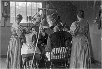 | ||
| Fig. 2 Lupus vulgaris treatment at the Finsen Institute in Copenhagen (Courtesy of Prof. Hans Christian Wulf, Copenhagen). | ||
Treatment of lupus vulgaris was not the only example of the use of phototherapy in dermatology. In 1923, William Henry Goeckerman introduced his regime (artificial broadband UVB from a high pressure mercury lamp plus topical coal tar) for psoriasis. In 1925 he published his first results8 (Fig. 3). This treatment became very popular, particularly in the U.S.A, and was used for decades to treat psoriasis. In 1953, John Ingram, in the United Kingdom, combined this treatment with anthralin.9 Surprisingly, Goeckerman’s or Ingram’s regimens have never been popular in central Europe.
 | ||
| Fig. 3 William H. Goeckerman (1884–1954) from: http://www.psoriasiscouncil.org/publications/slide_library/66. | ||
Subsequently, it was found that broadband UVB on its own, given in slightly erythematous doses, could clear mild forms of psoriasis, mainly seborrheic and guttate psoriasis (see below). By the beginning of the 1960s, Wiskemann in Hamburg, Germany, had constructed a phototherapy system with Osram Ultravitalux lamps and another with fluorescent UVB tubes.10
Photochemotherapy and phototherapy
The revival of photochemotherapy started in 1947 when the active ingredients of Ammi majus, 8-methoxypsoralen (8-MOP) and 5-methoxypsoralen (5-MOP) were isolated11,12 (Fig. 4), and the first trials with 8-MOP and sun exposure were performed in vitiligo patients by El-Mofty in Egypt.13 Later studies used topical 8-MOP in combination with UV irradiation to treat psoriasis.14 The first oral use of 8-MOP to treat psoriasis was reported in 1967.15 In 1972 Mortazawi used so-called blacklight UVA tubes in a total-body-irradiation unit with topical 8-MOP to treat psoriasis.16 However, the UVA output of these tubes was insufficient when 8-MOP was administered orally.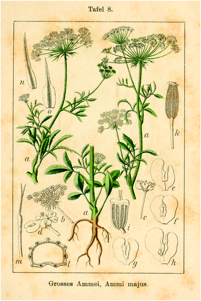 | ||
| Fig. 4 Ammi Majus (Deutschlands Flora in Abbildungen (1796). From: http://www.BioLib.de. | ||
In the 1970s, lighting engineers, photophysicists, and dermatologists worked together to develop ultraviolet irradiators emitting high intensity UVA. These UVA irradiators were designed for oral psoralen photochemotherapy to deliver uniform high dose UVA irradiation. The seminal publication of Parrish, Fitzpatrick, Tanenbaum, and Pathak reported the use of this new type of UVA tube in combination with oral 8-MOP in the treatment of psoriasis.17 This approach was much more effective than the blacklight method and represented the real start of PUVA therapy. The term PUVA was coined by Fitzpatrick as an acronym for psoralen and UVA (Fig. 5). The effectiveness of PUVA was confirmed by well controlled clinical trials in thousands of patients, both in the USA and in European countries18 (Fig. 6). Combination therapy with oral retinoids and PUVA contributed to greater effectiveness and long-term safety of psoralen photochemotherapy.19 The use of psoralen baths and subsequent UVA exposure (bath PUVA) originated in Scandinavia and is still in use as it avoids 8-MOP side effects such as nausea and dizziness.20
 | ||
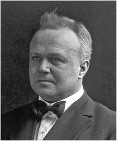
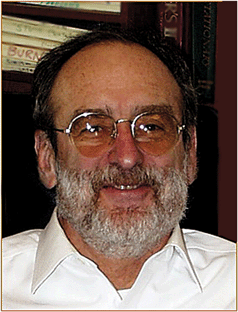
| Fig. 5 Thomas B. Fitzpatrick, Edward Wigglesworth Professor of Dermatology, (1919–2003) (Courtesy of Klaus Wolff MD). | ||
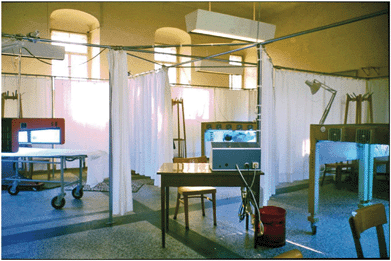 | ||
| Fig. 6 First PUVA unit in Vienna 1975 (Courtesy of Herbert Hönigsmann MD). | ||
PUVA revolutionized dermatological therapy and it became a standard treatment for many skin diseases. PUVA in fact was the driving force in the mid-70s that sparked a whole new series of discoveries during the next two decades i.e. newly created high intensity ultraviolet sources such as narrowband UVB, and later UVA1 (340–400 nm).
As mentioned above, it was the development of PUVA that led to the discovery of narrowband UVB (311–313 nm) irradiation, which replaced broadband UVB, as the first line therapy for psoriasis due to being more efficacious. In 1976, Parrish & Jaenicke defined the action spectrum for psoriasis with a peak at 313 nm21 (Fig. 7). However, it took almost a decade for commercially produced artificial lamps at this wavelength to be available as narrowband UVB. Van Weelden, Baart de la Faille, Young and van der Leun in 198422 demonstrated the clinical efficacy of narrowband UVB (Fig. 8) which was confirmed a few months later by Green et al.23 Since then, it has proven to be more effective than broadband UVB and is increasingly used in various parts of the world. It is also beneficial for a variety of other dermatoses that were previously treated with PUVA. The use of PUVA nowadays has declined with the emergence of narrowband UVB because of its easier handling. Another reason may be the increased risk of skin carcinoma after excessive exposures. Exposure to more than 350 PUVA treatments greatly increases the risk of squamous cell carcinoma (SCC), whereas exposure to fewer than 150 PUVA treatments has, at most, modest effects on SCC risk.24 So far this risk has not been shown with narrowband UVB.25 PUVA, however, still remained the gold standard for comparing with other phototherapeutic modalities and serves an important therapeutic role in cases of dermatoses recalcitrant to conventional phototherapy and in dermatoses that penetrate deeper into the skin such as plaque stage mycosis fungoides.
 | ||
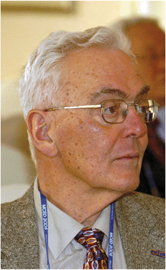
| Fig. 7 John A. Parrish, Professor of Dermatology emeritus (Courtesy of Herbert Hönigsmann MD). | ||
 | ||
| Fig. 8 Jan C. van der Leun (Photo taken at the Fifth Ministerial Conference on Environment and Development in Asia and the Pacific, 24 March 2005). | ||
These effective therapies—PUVA and narrowband UVB—for psoriasis have been a boon for the patients with generalized psoriasis, providing efficacious ambulatory treatments and avoiding the systemic problems of methotrexate and cyclosporine.
In 1992, UVA1 (340–400 nm) was introduced first for the treatment of atopic dermatitis. UVA1 penetrates deeper than UVA2 (320–340 nm) and thus is able to reach deep dermal components of the skin.26 A few studies with small numbers of patients showed efficacy in some other dermatoses such as localized scleroderma, urticaria pigmentosa and mycosis fungoides. The published evidence on how best to use UVA1 has still remained limited and of variable quality. UVA1 is effective in various diseases, but appears to be the first-line phototherapy only for some types of sclerosing diseases such as morphea.27
In the early 1980s a new form of phototherapy, namely, extracorporeal photochemotherapy (photopheresis, ECP) was introduced for the palliative treatment of erythrodermic cutaneous T-cell lymphoma (CTCL).28 Its efficacy was confirmed later by several uncontrolled clinical trials and approved as a device in 1988 by the FDA for the treatment of this disease. In 1994, at the International Consensus Conference on Staging and Treatment Recommendations for CTCL, ECP was recommended as the first-line of treatment for patients with erythrodermic CTCL.29
Meanwhile, besides CTCL, ECP also plays an important role in the treatment of chronic GVHD after allogeneic bone marrow transplantation with excellent response rates.30 In addition, positive results have also been published for acute GVHD.31 ECP has also been used in several other autoimmune diseases including acute allograft rejection among cardiac, lung and renal transplant recipients and Crohn's disease, with some success.32
Photodynamic therapy
At the beginning of the 20th century, the first experiments were performed with photosensitizers and visible light in the treatment of skin cancer, which is now known as photodynamic therapy (PDT). While studying the effects of acridine on paramecia cultures, Oscar Raab, a student of Hermann von Tappeiner in Munich observed a toxic effect. Fortuitously, Raab also observed that light was required for the killing of paramecia cultures to take place.33 Subsequent work in the laboratory of von Tappeiner showed that oxygen was essential for the “photodynamic action”—a term coined by von Tappeiner34 (Fig. 9).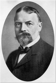 | ||
| Fig. 9 Hermann Tappeiner, Edler von Tappein (1847–1927). © Ludwig-Maximilian University, Munich. | ||
With the discovery of photodynamic effects, von Tappeiner and colleagues went on to perform the first PDT trial in patients with skin cancer using the photosensitizers eosin and Magdala-red solution. Out of 6 patients with a facial basal cell carcinoma treated with the dyes and exposure either to sunlight or to arc-lamp light, 4 patients showed total tumor resolution and a relapse-free period of 12 months35 (Fig. 10).
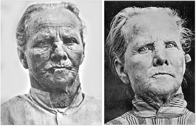 | ||
| Fig. 10 Patient with “rodent ulcer” treated by Jesionek & Tappeiner (a) before, and (b) after PDT with topical 5% Magdala-red solution and sun exposure. From: Dtsch. Arch. Klin. Med., 1905, 82, 223–226. | ||
Although they reported this success, it would take most of the 20th century to verify the utility of “photodynamic therapies” until Thomas Dougherty and co-workers at Roswell Park Cancer Institute, Buffalo NY, clinically started PDT again. In 1978, they published impressive results in which they treated 113 cutaneous or subcutaneous malignant tumors and observed a total or partial resolution of 111 tumors36 (Fig. 11). The photosensitizer used in the clinical PDT trials by Dougherty was an agent called hematoporphyrin derivative (HpD) which was applied intravenously. However, the problem with this route of administration was that the whole skin became photosensitized for up to 6 weeks.
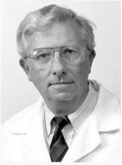 | ||
| Fig. 11 Thomas J. Dougherty, Professor of Oncology emeritus, Roswell Park Cancer Institute. From: http://www.roswellpark.edu/thomas-dougherty. | ||
The milestone for PDT in dermatology was the observation of Kennedy, Pottier and Pross that topically applied 5-aminolevulinic acid would induce tissue accumulation of protoporphyrin IX (PPIX) in skin tumors. PPIX acted as an endogenous photosensitizer and upon illumination with red light destroyed tumor tissue.37 The major advantage of this regimen was that the patients’ skin was sensitized only in the desired area.
Besides 5-aminolevulinic acid, methyl-aminolevulinate (MAL) produced commercially under the name Metvix® is now quite commonly used with similar success. PDT has become a routine treatment for widespread actinic keratosis, superficial basal cell carcinoma and Bowen's disease.38 New sensitizers are now tested for other dermatological conditions such as psoriasis, however no major breakthrough has been reported so far.
The successful use of the new ultraviolet and light techniques for the treatment of disease was a stimulus for the development of a new sub-specialty called photodermatology, which encompasses all of the applications of the diagnosis and treatment of light-induced disorders as well as the use of the new modalities for therapy of diseases. There now exists both national and international Photomedicine Societies as well as specialized journals of photodermatology. According to a quote by Kendric C. Smith,39 one of the founding fathers of the American Society for Photobiology, I would like to close: “Photodermatology—The Future is Bright”.
References
- M. A. Pathak and T. B. Fitzpatrick, The evolution of photochemotherapy with psoralens and UVA (PUVA): 2000 BC to 1992 AD, J. Photochem. Photobiol., B, 1992, 14, 3–22 CrossRef CAS.
- A. H. Downes and T. P. Blunt, Researches on the effect of light upon bacteria and other organisms, Proc. R. Soc. London, 1877, 26, 488–500 Search PubMed.
- T. A. Palm, The geographical distribution and aetiology of rickets. The Practitioner, October and November, 1890.
- N. R. Finsen, Om Lysets Indvirkninger paa Huden [On the effects of light on the skin], Hospitalstidende, 1893, 36, 721–728 Search PubMed.
- N. R. Finsen, Om Anvendelse i Medicinen af koncentrerede kemiske Lysstraaler. [On the application in medicine of concentrated chemical rays of light], Gyldendalske Boghandels Forlag, Copenhagen, 1896 Search PubMed.
- N. R. Finsen and H. Forchhammer, Resultate der Lichtbehandlung bei unseren ersten 800 Fällen von Lupus vulgaris. [Results of light therapy in our first 800 cases of lupus vulgaris], Mitt. Fins. Med. Lichtinst., 1904, 5/6, 1–48 Search PubMed.
- R. Roelandts, A new light on Niels Finsen, a century after his nobel prize, Photodermatol., Photoimmunol. Photomed., 2005, 21, 115–117 CrossRef.
- W. H. Goeckerman, Treatment of psoriasis, Northwest Med., 1925, 24, 229–231 Search PubMed.
- J. T. Ingram, The approach to psoriasis, Br. Med. J., 1953, 12(2), 591–594 CrossRef.
- A. Wiskemann, Die neuere Entwicklung der Lichttherapie. [Recent developments in light therapy] , Dermatol. Wochenschr., 1963, 20(147), 377–383 Search PubMed.
- I. R. Fahmy, H. Abu-Shady and A. A. Schönberg, Crystalline principle from Ammi majus L., Nature, 1947, 160(4066), 468 CrossRef CAS.
- I. R. Fahmy and H. Abu-Shady, Ammi majus Linn: The isolation and properties of ammoidin, ammidin and majudin, and their effect in the treatment of leukoderma, Q. J. Pharm. Pharmacol., 1948, 21(4), 499–503 Search PubMed.
- A. M. El-Mofty, A preliminary clinical report on the treatment of leukoderma with Ammi majus Linn, J. Egypt. Med. Assoc., 1948, 31, 651–665 Search PubMed.
- B. Allyn, Studies on phototoxicity in man and laboratory animals. Paper presented at the Twenty-First Annual Meeting of the American Academy of Dermatology, Chicago, December 1962 Search PubMed.
- L. Oddoze, P. Témime, J. P. Marchand and M. Benne, L'association “meladinine” per os et rayons U.V. dans le traitement du psoriasis. [Combined oral meladinine and ultraviolet rays in the treatment of psoriasis], Bull. Soc. Fr. Dermatol. Syphiligr., 1967, 74(5), 609–610 Search PubMed.
- S. M. Mortazawi, Meladinine und UVA bei Vitiligo, Psoriasis, Parapsoriasis und Akne vulgaris. [Meladinine and UVA in vitiligo, psoriasis, parapsoriasis and acne vulgaris], Dermatol. Monatsschr., 1972, 158, 908–909 Search PubMed.
- J. A. Parrish, T. B. Fitzpatrick, L. Tanenbaum and M. A. Pathak, Photochemotherapy of psoriasis with oral methoxsalen and longwave ultraviolet light, N. Engl. J. Med., 1974, 291(23), 1207–1211 CAS.
- K. W. Wolff, T. B. Fitzpatrick, J. A. Parrish, F. Gschnait, B. Gilchrest, H. Hönigsmann, M. A. Pathak and L. Tanenbaum, Photochemotherapy for psoriasis with orally administered methoxsalen, Arch. Dermatol., 1976, 112(7), 943–950 CAS.
- H. Hönigsmann and K. Wolff, Results of therapy for psoriasis using retinoid and photochemotherapy (RePUVA), Pharmacol. Ther., 1989, 40(1), 67–73 CrossRef CAS.
- T. Fischer and J. Alsins, Treatment of psoriasis with trioxsalen baths and dysprosium lamps, Acta Derm. Venereol., 1976, 56(5), 383–390 CAS.
- J. A. Parrish and K. F. Jaenicke, Action spectrum for phototherapy of psoriasis, J. Invest. Dermatol., 1981, 76(5), 359–362 CrossRef CAS.
- H. van Weelden, H. Baart de la Faille, E. Young and J. C. van der Leun, A new development in UVB phototherapy of psoriasis, Br. J. Dermatol., 1988, 119(1), 11–9 CrossRef CAS.
- C. Green, J. Ferguson, T. Lakshmipathi and B. E. Johnson, 311 nm UVB phototherapy – an effective treatment for psoriasis, Br. J. Dermatol., 1988, 119(6), 691–696 CrossRef CAS.
- R. S. Stern, PUVA follow-up study. The risk of squamous cell and basal cell cancer associated with psoralen and ultraviolet A therapy: a 30-year prospective study, J. Am. Acad. Dermatol., 2012, 66(4), 553–562 CrossRef CAS.
- R. M. Hearn, A. C. Kerr, K. F. Rahim, J. Ferguson and R. S. Dawe, Incidence of skin cancers in 3867 patients treated with narrow-band ultraviolet B phototherapy, Br. J. Dermatol., 2008, 159(4), 931–935 CrossRef CAS.
- J. Krutmann, W. Czech, T. Diepgen, R. Niedner, A. Kapp and E. Schöpf, High-dose UVA1 therapy in the treatment of patients with atopic dermatitis, J. Am. Acad. Dermatol., 1992, 26(2 Pt 1), 225–230 CrossRef CAS.
- E. B. Kroft, N. J. Berkhof, P. C. van de Kerkhof, R. M. Gerritsen and E. M. de Jong, Ultraviolet A phototherapy for sclerotic skin diseases: a systematic review, J. Am. Acad. Dermatol., 2008, 59(6), 1017–1030 CrossRef.
- R. Edelson, C. Berger and F. Gasparro, et al., Treatment of cutaneous T-cell lymphoma by extracorporeal photochemotherapy. Preliminary results, N. Engl. J. Med., 1987, 316, 297–303 CAS.
- F. Trautinger, R. Knobler and R. Willemze, et al., EORTC consensus recommendations for the treatment of mycosis fungoides/Sézary syndrome, Eur. J. Cancer, 2006, 42, 1014–1030 CrossRef.
- H. T. Greinix, B. Volc-Platzer and W. Rabitsch, et al., Successful use of extracorporeal photochemotherapy in the treatment of severe acute and chronic graft-versus-host disease, Blood, 1998, 92, 3098–3104 CAS.
- H. T. Greinix, R. M. Knobler and N. Worel, et al., The effect of intensified extracorporeal photochemotherapy on long-term survival in patients with severe acute graft versus-host disease, Haematologica, 2006, 91, 405–408.
- R. Dall'Amico and L. Murer, Extracorporeal photochemotherapy: a new therapeutic approach for allograft rejection, Transfus. Apheresis Sci., 2002, 26, 197–204 Search PubMed.
- O. Raab, Über die Wirkung Fluorescierenden Stoffe auf Infusorien. [On the efffect of fluorescent substances on infusoria], Z. Biol., 1904, 39, 524–546 Search PubMed.
- H. von Tappeiner and A. Jodlbauer, Über die Wirkung der photodynamischen (fluorescierenden) Stoffe auf Protozoen und Enzyme. [On the effect of photodynamic (fluorescent) substances on protozoa and enzymes], Dtsch. Arch. Klin. Med., 1904, 80, 427–487 Search PubMed.
- H. Jesionek and H. von Tappeiner, Zur Behandlung der Hautcarcinome mit fluoreszierenden Stoffen. [On the treatment of skin carcinomas with fluorescent substances], Dtsch. Arch. Klin. Med., 1905, 82, 223–226 Search PubMed.
- T. J. Dougherty, J. E. Kaufman, A. Goldfarb, K. R. Weishaupt, D. Boyle and A. Mittleman, Photoradiation therapy for the treatment of malignant tumors, Cancer Res., 1978, 38(8), 2628–2635 CAS.
- J. C. Kennedy, R. H. Pottier and D. C. Pross, Photodynamic therapy with endogenous protoporphyrin IX: basic principles and present clinical experience, J. Photochem. Photobiol., B, 1990, 6, 143–148 CrossRef CAS.
- P. Babilas, S. Karrer, A. Sidoroff, M. Landthaler and R. M. Szeimies, Photodynamic therapy in dermatology—an update, Photodermatol., Photoimmunol. Photomed., 2005, 21(3), 142–149 CrossRef CAS.
- K. C. Smith, Photobiology and photomedicine: the future is bright, J. Invest. Dermatol., 1981, 77(1), 2–7 CrossRef CAS.
Footnote |
| † This article is published as part of a themed issue on current topics in photodermatology. |
| This journal is © The Royal Society of Chemistry and Owner Societies 2013 |