Encapsulation of bio-based PCM with coaxial electrospun ultrafine fibers
Wen
Hu
and
Xun
Yu
*
Department of Mechanical and Energy Engineering, University of North Texas, TX 76203, USA. E-mail: Xun.Yu@unt.edu; Fax: +1940-565-8675; Tel: +1940-565-2742
First published on 18th April 2012
Abstract
In this study, bio-based phase change material (bio-PCM) was successfully encapsulated in ultrafine fibers via coaxial electrospinning technique. Natural soy wax was used as the bio-PCM for thermal storage and Polyurethane (PU) was used as the shell material for encapsulation. The bio-PCM fibers were characterized by environmental scanning electron microscopy (ESEM), transmission electron microscopy (TEM), differential scanning calorimetry (DSC), and X-ray Diffraction (XRD). The results indicate that coaxial electrospinning resulted in a uniform fiber morphology with a core-shell structure, and a homogeneous wax distribution throughout the core of the fibers. Thermal analysis results show that the enthalpy increases with wax content. The fibrous structures exhibited balanced thermal storage and releasing properties for thermo-regulating functions. The thermal properties were unaltered after 100 heating-cooling test cycles, demonstrating that the composite fibers have good thermal stability and reliability.
1. Introduction
With the growing energy crisis, more and more interest has been focused on the research of renewable sources and materials. Phase change materials (PCMs) have received great attention in recent years for their great capacity in thermal energy storage, as they will absorb and slowly release the latent heat involved in a phase change process.1 PCMs can be used in many applications, such as energy storage and thermal protection systems as well as in active and passive cooling of electronic devices,2,3 of which thermal energy storage is one of the most important. Heat is stored through the phase transition within the material for later use. The most popular phase change selected for thermal energy storage is the solid–liquid transition as there is a large melting enthalpy associated with this phase change while volumetric and temperature variation during the transition is small. Common PCMs include inorganic compounds such as salt hydrates, salts, metals and alloys as well as organic compounds such as paraffin, non-paraffin organics and poly-alcohols depending on the specific application. Paraffin waxes are the most commonly used PCM in the room temperature range applications due to their high latent heat of fusion, negligible super-cooling, low vapor pressure in the melt, chemical inertness and stability, lack of phase segregation and commercial availability at relatively low cost. PCMs bridge the time gap between energy requirements and energy use and contribute to the effective use of energy. However, they also have some inherent limitations, such as low thermal conductivity and the need for containers. More importantly, paraffin is a petroleum-based material, so using paraffin as a PCM is indirect use of fossil fuels, which in reality is not renewable and not environmental friendly. On the other hand, natural wax, processed from fruits, nuts and seeds, has its environmental advantages and has attracted a lot of interest. Soybean wax is one of the most available natural waxes. It comprises a large number of identified chemicals as well as unidentified ones, which are all environmentally safe and non-toxic. Soybeans are also very low cost.4 In recent years, there has been a big effort to produce environmentally friendly compositions, biopolymers and biodiesels from soybeans.5–7 In this study, natural soy wax, a partially hydrogenated form of soybean oil, which possesses similar properties to paraffin wax but is friendly to the environment and human beings, is used as a new bio-based PCM.Encapsulation of PCMs is an important step for their application as liquid migration and reactivity with other substances in the environment could be an issue during phase changes. Most current research focusses on microencapsulating PCMs with convenient polymers.8–10 A spherical polymeric shell will encapsulate the PCMs and provide its own specific properties.11–13 As well as the traditional sphere encapsulation, the melt coaxial electrospining technique is also reported for the fabrication of PCMs.14,15 Electrospinning as a simple and versatile method has been established for producing continuous fibers with diameters ranging from nanometer to submicrometer scale.16 Co-axial electrospinning using concentrically aligned spinnerettes consisting of tubes or syringes has attracted the greatest attention in the past decade, as it provides a straightforward method for generating long (centimeter-scale) nanoscale core/sheath or hollow fibers.17 As coaxial electrospinning utilizes two separate channels for the different solutions, it is beneficial for maintaining the functional activity of PCMs. Melt electrospinning is designed to melt the polymer to a suitable viscosity, which is easily electrospun. Some underlying difficulties in melt electrospinning include a high temperature setup, high viscosity and low conductivity of polymer melts.18 In this study, solution coaxial electrospinning of encapsulated PCMs is proposed. Instead of heating soy wax over its melting temperature, soy wax was dissolved in chloroform. PCMs with a series of soy wax contents were prepared, and had a homogeneous wax distribution throughout the core of the nanofibers.
The aim of the present study is to generate shape-stabilized bio-PCM. The coaxial electrospinning method was used to facilitate the fabrication of double layer PU/wax composite nanofibers. The morphology and thermal properties of PU/wax composite PCMs were investigated. The fibrous encapsulated bio-PCM can potentially be used for thermal storage and thermal protection,19–21 which has appealing environmental advantages.
2. Experimental
Polyurethane (PU) was purchased from BASF, natural soy wax was obtained from Candlewic Co and chloroform, tetrahydrofuran (THF) and dimethylformamide (DMF) were obtained from Aldrich Corp. All the chemicals were used without further purification. In this study, the PU solution was prepared by dissolving in 1![[thin space (1/6-em)]](https://www.rsc.org/images/entities/char_2009.gif) :1 v/v DMF:THF for 2–3 h at the ambient temperature. The wax was dissolved in chloroform. The PU solution was made in a weight ratio of 10 wt% and the wax solution was made with different concentrations varying from 10 wt% to 60 wt%.
:1 v/v DMF:THF for 2–3 h at the ambient temperature. The wax was dissolved in chloroform. The PU solution was made in a weight ratio of 10 wt% and the wax solution was made with different concentrations varying from 10 wt% to 60 wt%.
Fig. 1 shows a diagram of the coaxial electrospinning setup. Different from normal electrospinning, the coaxial electrospinning setup has an inner needle coaxially placed inside of an outer one. Core material solution (soy wax) and shell material solution (PU) were fed into the capillary, and their speeds were controlled by a variable speed syringe pump (NE-300 Pump Systems Inc.). A DC voltage was applied via a high voltage power supply (PO20HP 1.5M, Acopian, USA). The applied voltage makes the pendent drop of solution highly electrified and the induced charges evenly distributed over the surface. Once the strength of the electric field is high enough to allow the electrostatic forces to overcome the surface tension of the solution, a liquid jet forms and moves towards the counter electrode. During the movement of the liquid jet, the solvent evaporates (or solidifies), and solid fibers are precipitated on the counter electrode. In the experiments, the outer and inner syringe pumps were set to a flow rate of 2 ml h−1 and 0.5 ml h−1 respectively to provide a continuous flow of solution to the tip. Both needles were connected to the same electrical potential with an applied voltage of 20 kV DC. The spinning distance was set at 13–15 cm.
 | ||
| Fig. 1 Illustration of the coaxial electrospinning setup. | ||
The surface morphology of the electrospun fibers were examined by an environmental scanning electron microscope (ESEM) (Quanta 200, Jeol). The electrospun membranes were sputter coated with a thin layer of gold. The morphological features were observed at an accelerating voltage of 20 kV under high vacuum. Fiber diameters of nanofibers were analyzed with image visualization software (ImageJ 1.38 e/Java 1.5.0_09). Average 100 fibers were used to calculate the fibers diameter. The core-shell structure of the PCMs was examined using a Tecnai G2 F20 S-Twin 200 keV field-emission Scanning Transmission Electron Microscope (STEM). The samples for TEM were prepared by direct deposition of the electrospun fibers onto copper grids. Differential scanning calorimetry (DSC) (PerkinElmer DSC 4000) was carried out in flowing nitrogen. Samples were heated from 0 °C to 80 °C at a heating rate of 10 °C min−1 and then cooled at the same rate. Thermal properties, namely melting temperatures and enthalpies of melting, were determined from the 100th heating run.
3. Results and discussion
PCM fibrous membranes with a core-shell structure were obtained by coaxial electrospinning. Different concentrations of PU solutions were prepared in this study to figure out the perfect outer layer concentration, varying from 7 wt%–15 wt%. At concentrations below 10 wt%, nanofibers with beads on string morphology were often observed. The solutions with the concentrations above 12 wt% were too viscous to form steady jets during electrospinning. Continuous fibers with bead-free morphologies could be formed between 10 wt% and 12 wt% solutions. Meanwhile, the fiber diameter increased with the solution concentration. A concentration of 10 wt% was chosen as the optimal outer layer solution concentration in this work. The voltage was set to 20 kV and the feeding rate was set as 2 ml h−1.The compositions of the core-shell fibers were modulated simply by varying the feed rate and the concentration of the inner wax solution. PU served as the outer liquid and wax was chosen for the inner liquid. The effect of the concentration of inner solution on fiber morphology is shown in Fig. 2. It can be seen that a fairly homogeneous surface and a relatively tight fiber diameter distribution was obtained at wax concentrations below 30 wt%. No indication of wax separation was observed. Bead formation increased as the wax concentration increased as shown in Fig. 2e and 2f, and the beads became elongated at the edges forming a spindle-like shape. Furthermore, these beads are found to fuse together and solidify into larger molten structures that are randomly dispersed throughout the membrane when the wax concentration is 60 wt% (see Fig. 2g). There was an increase in the average diameter and fiber diameter distribution of the composite fibers. Pure PU nanofibers showed a tight fiber diameter distribution and an average fiber diameter of 650.2 nm. An increase in the concentration of the core solution resulted in an increase in both the wax content and fibrous diameter (see Fig. 2h).
 | ||
| Fig. 2 The morphology of core-shell (PU/wax) fibrous membranes as a function of wax concentration (Fig. 2A, B, C, D, E, F, G represent 10%, 20%, 30%,40%, 50% and 60% wax in the inner layer, respectively, with the fixed outer layer concentration of 10wt%. The graphs of a, b, c, d, e, f, g show the distribution diameter of nanofibers corresponding to A, B, C, D, E, F, G, respectively. The curve of h shows the trend of averaged diameters corresponding to A, B, C, D, E, F, G, respectively.) | ||
The morphology and structure of the PU/wax composite membrane was further investigated by TEM. Fig. 3 demonstrates that the coaxially electrospun PCMs exhibited an obvious core-shell structure, indicated by the differences in electron density between the inner core and outer shell of the fibers. The sharp boundary between PU shell and wax core phase demonstrated that the wax was completely encapsulated by the PU nanofibers along the axis. Since the wax cannot be electrospun into fibers itself, the inner wax fluid was compressed by the outer PU fluid to form a thin thread. Thereby, the soy wax was encapsulated by PU fibers. The coaxial nanofibers exhibited a relatively homogeneous wax distribution under the low wax concentration (see Fig. 3A). When the shell concentration increased to 50 wt%, increasing the wax concentration lead to axial anisotropy of core/shell fluids, which formed bi-component nanofibers side by side.
 | ||
| Fig. 3 TEM micrographs of core-shell structured PU/wax electrospun nanofibers (A for 10% wax and B for 50% wax). | ||
DSC was used to investigate the effect of natural soy wax on the thermal properties of nanocomposites and its capacity to store thermal energy. Both the melting and cooling behaviors of PU/wax were recorded. The DSC heating curves for both pure components and composite membranes are shown in Fig. 4. All the parameters, as well as the heating curves, were obtained from the 100th scan to eliminate the effect of thermal history and more accurately measure thermal stability and reliability. The DSC curve of pure wax displays two well defined separated peaks at 27 °C and 50 °C, which correspond to the solid–solid transition and melting/crystallization of natural soy wax. The DSC heating curves of the PU/wax composite show one slight peak at around 27 °C and one endothermic event (Fig. 4) over a broad temperature range (30–60 °C). This event is in the same temperature region as the melting peak of pure wax. Increasing the wax concentration from 10 wt% to 60 wt% leads to a large increase in the total enthalpy of composite membranes. The wax content with wax concentrations from 10% to 60% in the composite are 14 wt%, 25 wt%, 33 wt%, 40 wt%, 45 wt% and 50 wt% respectively, which are in correspondence with total melting enthalpies (see Table 1), indicating that no wax leakage occurred during sample preparation. By increasing the wax content, the melting temperature (Tm) is slightly increased, which means that the wax elevates the thermal stability of PU/wax composites. Similar behavior was observed for cooling. The cooling curves of the composites depict two exothermic peaks, related to the crystallization of wax (the solid–solid transition in the wax structure). It can be seen that the Tm for both peaks gradually decreases with an increase of wax content. DSC results show that the fibrous structures exhibited balanced thermal storage and releasing properties for thermo-regulating functions.
 | ||
| Fig. 4 DSC melting thermograms of electrospun composite membranes. | ||
| Index | PU/wax 10% | PU/wax 20% | PU/wax 30% | PU/wax 40% | PU/wax 50% | PU/wax 60% | Wax |
|---|---|---|---|---|---|---|---|
| T 1,o,m(°C) | 20.61 | 22.78 | 22.43 | 22.09 | 22.43 | 23.04 | 22.37 |
| T 1,m (°C) | 26.15 | 26.74 | 26.56 | 27.42 | 27.87 | 28.12 | 26.89 |
| ΔH1,m(J g−1) | 0.86 | 0.92 | 0.93 | 1.16 | 1.65 | 1.88 | 3.17 |
| T 2,o,m(°C) | 32.58 | 33.12 | 34.09 | 34.27 | 35.88 | 36.72 | 33.92 |
| T 2,m (°C) | 45.04 | 47.41 | 48.27 | 48.81 | 50.87 | 51.38 | 50.87 |
| ΔH2,m(J g−1) | 9.24 | 19.12 | 20.71 | 25.89 | 34.30 | 36.47 | 69.97 |
In order to illustrate the differences in crystalline content, X-ray diffraction patterns were studied. Fig. 5 shows the XRD analysis of PU/wax composites. It can be seen that the neat PU appears to have an amorphous structure. The sharp peaks appearing at 5.79°, 19.13°, 22.64°, 23.31° are the characteristic of wax. Comparing the spectra of pure PU membrane, the peaks related to the wax crystal appear in the XRD patterns of the coaxial nanofibers, showing the existence of the same wax crystalline structure in the electrospun nanofibers. Incorporation of natural wax with 10 wt% PU increased its crystallinity which is in line with the observed strengthening of the sharp peaks present at 5.79°, 20.88°and 22.90°. An increase of wax content, results in the same trend as observed in DSC analysis, which may explain the improvement of thermal stability.
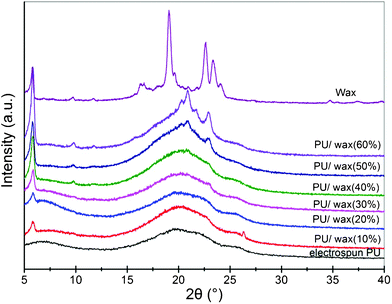 | ||
| Fig. 5 X-ray diffraction patterns of natural wax and its composites with PU. | ||
Conclusions
In this study, a core-shell structured PU/bio-based wax PCM was successfully fabricated by using a coaxial electrospining technique. SEM, TEM, DSC, and XRD were used to characterize the morphology, thermal properties and crystallinity of PCMs. A smooth morphology could be obtained when the PU concentration was set as 10 wt% and the flow rate of outer and inner liquids was 2 ml h−1 and 0.5 ml h−1, respectively. The wax concentration was varied from 10 wt% to 60 wt% to evaluate the thermal effect of wax on composite membranes. The results indicate that the wax was completely incorporated into the PU layer. The wax increased the total enthalpy of the composite nanofibers and consequently increased their crystallinities. This new kind of bio-PCM can be used in thermal storage and thermal shielding applications.References
- A. Abhat, Low temperature latent heat thermal energy storage, Heat storage materials, Sol. Energy, 1983, 30(4), 313–32 CrossRef CAS.
- M. Kenisarin and K. Mahkamov, Solar energy storage using phase change materials, Renewable Sustainable Energy Rev., 2007, 11(9), 1913–1965 CrossRef CAS.
- B. Zalba, J. Marin, L. Cabeza and H. Mehling, Review on thermal energy storage with phase change: materials, heat transfer analysis and applications, Appl. Therm. Eng., 2003, 23, 251–283 CrossRef CAS.
- S. Tascıoglu, B. Inem and N. Akar, Conversion of an investment casting sprue wax to a pattern wax by the modification of its properties, Mater. Des., 2004, 25, 499–505 CrossRef.
- S. G. Rogers, Biotechnology and the soybean, Am. J. Clin. Nutr., 1998, 68, 1330–2 Search PubMed.
- Y. T-P. Ly and L. A. Johnson, J. Jane Soy protein as biopolymer, Biopolym. Renewable Resour., 1998, 144–76 Search PubMed.
- R. Alcantara, J. Amores, L. Canoira, E. Fidalgo, M. J. Franco and A. Navarro, Catalytic production of biodiesel from soybean oil, used frying oil and tallow, Biomass Bioenergy, 2000, 18(6), 515–27 CrossRef CAS.
- Y. Hong and G. Xin-Shi, Preparation of polyethylene-paraffin compound as a form-stable solid-liquid phase change material, Sol. Energy Mater. Sol. Cells, 2000, 64(1), 37–44 CrossRef CAS.
- Mónica Delgado, Ana Lázaro, Javier Mazo and Belén Zalba, Review on phase change material emulsions and microencapsulated phase change material slurries: Materials, heat transfer studies and applications, Renewable Sustainable Energy Rev., 2012, 16(1), 253–73 CrossRef CAS.
- D. Hale, V. Hoover, M. J. O'Neill, Phase Change Materials Handbook, NASA Technical Report 72N19956, National Aeronautics and Space Administration, Washington, DC, 1971 Search PubMed.
- Y. B. Cai, Y. Hu and L. Song, Flammability and thermal properties of high density polyethylene/paraffin hybrid as a form-stable phase change material, J. Appl. Polym. Sci., 2006, 99(4), 1320–27 CrossRef CAS.
- M. Xiao, B. Feng and K. C. Gong, Preparation and performance of shape stabilized phase change thermal storage materials with high thermal conductivity, Energy Convers. Manage., 2002, 43(1), 103–08 CrossRef CAS.
- I. Krupa and A. S. Luyt, Thermal properties of uncross-linked and cross-linked LLDPE/wax blends, Polym. Degrad. Stab., 2000, 70(1), 111–17 CrossRef CAS.
- T. Jesse McCann, Manuel Marquez and Younan Xia, A Versatile Method for the Encapsulation of Solid Materials and Fabrication of Phase Change Nanofibers, Nano Lett., 2006, 6(12), 2868–2872 CrossRef.
- Fengyu Li, Yong Zhao and Sen Wang, Thermochromic core-shell nanofibers fabricated by melt coaxial electrospinning, J. Appl. Polym. Sci., 2009, 112(1), 269–274 CrossRef CAS.
- T. N. Mtshali, I. Krupa and A. S. Luyt, The effect of cross-linking on the thermal properties of LDPE/wax blends, Thermochim. Acta, 2001, 380(1), 47–54 CrossRef CAS.
- A. S. Luyt and I. Krupa, PE/wax blends: interesting observations, Macromol. Symp., 2002, 178(1), 109–16 CrossRef CAS.
- Huajun Zhou, B. Thomas Green and Yong Lak Joo, The thermal effects on electrospinning of polylactic acid melts, Polymer, 2006, 47, 7497–7505 CrossRef CAS.
- S. Wang, Y. P. Li and X. L. Fei, Preparation of a durable superhydrophobic membrane by electrospinning poly(vinylidene fluoride) (PVDF) mixed with epoxy–siloxane modified SiO2 nanoparticles: A possible route to superhydrophobic surfaces with low water sliding angle and high water contact angle, J. Colloid Interface Sci., 2011, 359, 380–88 CrossRef CAS.
- C. Z. Chen, L. G. Wang and Y. Huang, Electrospun phase change fibers based on polyethylene glycol/cellulose acetate blends, Appl. Energy, 2011, 88(9), 3133–39 CrossRef CAS.
- D. Hale, V. Hoover, M. J. O'Neill. Phase Change Materials Handbook; NASA Technical Report 72N19956; National Aeronautics and Space Administration: Washington, DC ( 1971) Search PubMed.
| This journal is © The Royal Society of Chemistry 2012 |