Aluminium, iron and copper in human brain tissues donated to the medical research council's cognitive function and ageing study
Emily
House
a,
Margaret
Esiri
b,
Gill
Forster
c,
Paul G
Ince
c and
Christopher
Exley
*a
aThe Birchall Centre, Lennard-Jones Laboratories, Keele University, Stoke-on-Trent, Staffordshire, ST5 5BG, UK. E-mail: c.exley@chem.keele.ac.uk; Fax: + 44(0)1782 712378; Tel: + 44(0)1782 734080
bDepartment of Clinical Neurology, University of Oxford, Department of Neuropathology, Oxford Radcliffe NHS Trust, Oxford, UK
cSheffield Institute for Translational Neuroscience, Department of Neuroscience, University of Sheffield, Sheffield, UK
First published on 1st November 2011
Abstract
Aluminium, iron and copper are all implicated in the aetiology of neurodegenerative diseases including Alzheimer's disease. However, there are very few large cohort studies of the content of these metals in aged human brains. We have used microwave digestion and TH GFAAS to measure aluminium, iron and copper in the temporal, frontal, occipital and parietal lobes of 60 brains donated to the Cognitive Function and Ageing Study. Every precaution was taken to reduce contamination of samples and acid digests to a minimum. Actual contamination was estimated by preparing a large number of (170+) method blanks which were interspersed within the full set of 700+ tissue digests. Subtraction of method blank values (MBV) from tissue digest values resulted in metal contents in all tissues in the range, MBV to 33 μg g−1 dry wt. for aluminium, 112 to 8305 μg g−1 dry wt. for iron and MBV to 384 μg g−1 dry wt. for copper. While the median aluminium content for all tissues was 1.02 μg g−1 dry wt. it was informative that 41 brains out of 60 included at least one tissue with an aluminium content which could be considered as potentially pathological (> 3.50 μg g−1 dry wt.). The median content for iron was 286.16 μg g−1 dry wt. and overall tissue iron contents were generally high which possibly reflected increased brain iron in ageing and in neurodegenerative disease. The median content for copper was 17.41 μg g−1 dry wt. and overall tissue copper contents were lower than expected for aged brains but they were commensurate with aged brains showing signs of neurodegenerative disease. In this study we have shown, in particular, the value of carrying out significant numbers of method blanks to identify unknown sources of contamination. When these values are subtracted from tissue digest values the absolute metal contents could be considered as conservative and yet they may still reflect aspects of ageing and neurodegenerative disease in individual brains.
Introduction
There are very few large cohort studies of the metal content of aged human brains. Such data are of burgeoning significance as they will contribute towards understanding of the role of metals in neurodegenerative disease.1,2 The aluminium content of human brain tissue has recently been reviewed3 and it was concluded that the normal range is 0.1–4.5 μg g−1 dry wt. with the higher values being measured in brains taken from non-demented elderly. Higher values still were recorded for diseased states such as Alzheimer's disease (AD) (up to 11.5 μg g−1 dry wt.), dialysis encephalopathy (up to 14.1 μg g−1 dry wt.), congophilic amyloid angiopathy (CAA) (up to 23.0 μg g−1 dry wt.) and other aluminium-related encephalopathies (up to 47.4 μg g−1 dry wt.). There have been several recent reviews concerning the iron and copper content of human brain tissue.4,5 The normal range for iron is ca 150–300 μg g−1 dry wt. with higher values being associated with the substantia nigra and/or ageing.6 There is some debate as to whether brain iron content is different in diseased states such as AD and CAA.7 A normal range for brain copper is 15–50 μg g−1 dry wt. with higher values in the hippocampus and, possibly, with ageing.6,8 Recent research has suggested that the copper content of brain tissue might be lower in AD and CAA.7Herein we have gained access to the Medical Research Council Cognitive Function and Ageing Neuropathology Study (MRC CFAS) brain donor resource which currently includes more than 500 brains9 and we have measured the content of aluminium (Al), iron (Fe) and copper (Cu) in 60 brains taken from the Cambridge cohort of CFAS.
Experimental
General
Significant precautions were taken throughout the study to minimise contamination. These included storage of all plastic-based laboratory-ware in 5% v/v conc. HCl and, before use, rinsing of all such apparatus in several volumes of ultrapure water (cond. < 0.067 μS cm−1). Where required, rinsed apparatus was air-dried in a dedicated incubator at 37 °C.Tissue collection
Tissues were obtained post-mortem from brains which had been donated to MRC CFAS (donors aged 70–103 years) and were stored at −80 °C at Sheffield Brain Tissue Bank, UK. One of us (EH) travelled to Sheffield and personally obtained 1–2 cm3 samples of frontal (F), parietal (P), temporal (T) and occipital (O) lobe using a stainless steel knife and cutting from Brodman areas, 8/9, 40, 20/21 and 18/19 respectively. Samples were then transported to Keele on dry ice and stored at −18 °C.Tissue digestion
Tissues were thawed and each sample of lobe was sub-divided into 3 sub-samples using either a stainless steel knife (first 20 brains, each sub-sample ca 100 mg wet weight) or a ceramic knife (final 40 brains, each sub-sample ca 250 mg). All tissue samples were dried to a constant weight in an incubator at 37 °C. The median dry/wet weight ratio was ca 0.19. Dried tissues were digested in a microwave (MARS Xpress CEM Microwave Technology Ltd) in a mixture of 1 mL 15.8 M HNO3 (Fischer Analytical Grade) and 1 mL of 30% w/vH2O2 (BDH Aristar Grade). Digests were clear and colourless or light yellow with no visible precipitate or fatty residue. Upon cooling each digest was diluted to a total volume of 5 mL with ultrapure water.TH GFAAS determination of aluminium, iron and copper
Total Al, Fe and Cu were measured using an AAnalyst 600 atomic absorption spectrometer with a transversely heated graphite atomizer (THGA) and longitudinal Zeeman-effect background corrector and an AS-800 autosampler with WinLab32 software (Perkin Elmer, UK). Standard THGA pyrolitically-coated graphite tubes with integrated L'Vov platform (Perkin Elmer, UK) were used. The Zeeman background-corrected peak area of the atomic absorption signal was used for the determinations. Each experimental extract datum was the arithmetic mean of three injections with a relative standard deviation <10%. The TH GFAAS was calibrated by automated serial dilution of 40, 60 and 100 μg L−1 solutions of Fe, Al and Cu respectively with 1% HNO3. Non-linear zero intercept WinLab 32-generated fits were applied. Al was measured immediately post digestion while Fe and Cu were measured over the following 2 days during which time the digests were stored in FEP bottles (Nalgene, USA). Furnace programmes for Al, Fe and Cu are given in Table 1. Optimisation data for pretreatment and atomisation temperatures are shown in Fig. 1. Instrumental drift was monitored by measurement of initial calibration verification standards (ICVS) immediately following calibration and continuing calibration verification standards (CCVS) after every 10 samples.| Instrument Parameters: | Iron | Copper | Aluminium |
|---|---|---|---|
| Lamp Current (mA) | 30 | 15 | 25 |
| Wavelength (nm) | 248.3 | 324.8 | 309.3 |
| Bandwidth (nm) | 0.2 | 0.7 | 0.7 |
| Injection volume (μL) | 20 | 20 | 30 |
| Injection T/°C | 20 | 20 | 20 |
| Pipette speed (%) | 100 | 100 | 100 |
| Read time (s) | 3 | 3 | 2 |
| Delay time (s) | 0 | 0 | 0 |
| BOC time (s) | 2 | 2 | 3 |
| Step | T/°C | Ramp (s) | Hold (s) | Internal Ar flow (mL min−1) |
|---|---|---|---|---|
| Iron | ||||
| Drying | 110 | 10 | 40 | 250 |
| Drying | 130 | 15 | 40 | 250 |
| Pyrolysis | 1300 | 10 | 20 | 250 |
| Atomisation | 2100 | 0 | 5 | 0 |
| Clean out | 2500 | 1 | 5 | 250 |
| Copper | ||||
| Drying | 110 | 10 | 40 | 250 |
| Drying | 130 | 15 | 40 | 250 |
| Pyrolysis | 1100 | 10 | 20 | 250 |
| Atomisation | 1900 | 0 | 5 | 0 |
| Clean out | 2500 | 1 | 5 | 250 |
| Aluminium | ||||
| Drying | 110 | 10 | 40 | 250 |
| Drying | 130 | 15 | 40 | 250 |
| Pyrolysis | 500 | 10 | 10 | 250 |
| 1200 | 10 | 15 | 250 | |
| Atomisation | 2300 | 0 | 5 | 0 |
| Clean out | 2500 | 1 | 5 | 250 |
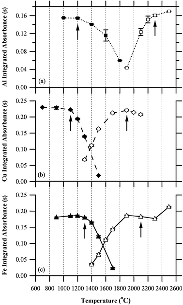 | ||
| Fig. 1 Pyrolysis (filled symbols) and atomisation (open symbols) profiles of (a) Al, (b) Cu and (c) Fe in brain tissue digests. Values are the means of 3 injections, error bars are standard deviations. Arrows indicate the optimum temperatures applied. | ||
Instrument performance
Characteristic masses were calculated from the absorbances of each calibration standard (6 for Cu, 4 for Al and Fe) for all calibrations (62 for Al, 60 for Cu and 59 for Fe). Mean characteristic masses for Al, Cu and Fe were 34.8 pg per 0.0044 s (standard deviation, SD, 4.4 pg per 0.0044 s, n = 248), 16.7 pg per 0.0044 s (SD 1.4 pg per 0.0044 s, n = 360) and 13.1 pg per 0.0044 s (SD 1.1 pg per 0.0044 s, n = 236) respectively. Values were in good agreement with manufacturer's cited values of 31 pg Al per 0.0044 s (±20%), 17 pg Cu per 0.0044 s (±20%) and 12 pg Fe per 0.0044 s (±20%).Mean absorbances of all initial and continuing calibration verification standards of 30 μg L−1Al, 25 μg L−1Cu and 20 μg L−1Fe were 0.13 A-s (SD 0.02 A-s, n = 181), 0.15 A-s (SD 0.02 A-s, n = 177) and 0.16 A-s (SD 0.02 A-s, n = 173) respectively. Values compared well with manufacturer's sensitivity checks for Al, Cu and Fe of 0.09 A-s, 0.15 A-s and 0.15 A-s respectively. Slopes of calibration curves suggested similar sensitivity to manufacturer's cited values. Means of Winlab 32-generated non-linear through zero slopes of Al, Cu and Fe calibrations were 0.0043 A-s per μg L−1 (SD 0.0006 A-s per μg L−1, n = 62), 0.0050 A-s per μg L−1 (SD 0.0005 A-s per μg L−1, n = 60) and 0.0069 A-s per μg L−1 (SD 0.0007 A-s per μg L−1, n = 59) respectively. Means of slopes of linear (method of least squares) non-zero fits of the same calibrations for Al, Cu and Fe were 0.0034 A-s per μg L−1 (SD 0.0003 A-s per μg L−1), 0.0056 A-s per μg L−1 (SD 0.0004 A-s per μg L−1) and 0.0066 A-s per μg L−1 (SD 0.0005 A-s per μg L−1) respectively.
Quality assurance
Each batch of digests included brain tissue samples, method blanks, standard reference materials and reagent spikes for each element. Examples of these data are summarised in Table 2. Instrument detection limits (IDL) were estimated from three times the standard deviation on the 1% HNO3 calibration blank absorbance (n = 3 injections) divided by the Winlab32 generated calibration slope. Mean IDLs for Al, Cu and Fe were 0.13 μg L−1 (SD 0.13 μg L−1, n = 62), 0.10 μg L−1 (SD 0.10 μg L−1, n = 60) and 0.16 μg L−1 (SD 0.14 μg L−1, n = 59) respectively.| SRM1566B Concentration (μg g−1 dry mass): | |||||||
|---|---|---|---|---|---|---|---|
| Metal | n | Median | IQR a | Mean | SD | AD (P)b | Certified |
| a IQR denotes interquartile range. b AD denotes the Anderson-Darling test statistic, a corresponding P of less than 0.05 indicates rejection of the null hypothesis that the data was normally distributed. | |||||||
| Al | 32 | 121.21 | 19.21 | 123.82 | 19.05 | 0.48 (0.21) | 197.2 ± 6 |
| Cu | 32 | 72.27 | 9.64 | 71.48 | 8.20 | 0.34 (0.48) | 71.6 ± 16 |
| Fe | 32 | 225.13 | 12.26 | 227.05 | 14.97 | 1.79 (<0.005) | 205.8 ± 6.8 |
Statistics
Statistical analyses were performed with Microsoft Excel 2007 and Minitab 15.Results
Method blanks
The mass of Al in method blanks ranged from ca 5–150 ng/vessel (Fig. 2c). The median Al content for 174 method blanks was ca 22 ng/vessel. The data were not normally distributed and a log transformation was used to determine a contaminant level of 54 ng/vessel (mean + 1.654SD). This value was subtracted from all Al tissue digests.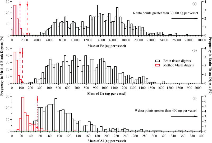 | ||
| Fig. 2 Mass of (a) Fe, (b) Cu and (c) Al in vessels containing ca 5 mL method blank digests and brain tissue digests uncorrected for contamination. Arrows and dashed lines show the contaminant level which was subtracted from tissue digest values. | ||
The mass of Fe in method blanks ranged from ca 40–>2000 ng/vessel (Fig. 2a). The median Fe content for all 175 method blanks was ca 390 ng/vessel. Method blanks were treated as per tissue digests and contaminant levels of 1286 and 2424 ng/vessel (mean + 1.654SD) were determined for digest dilutions of 125 and 200-fold respectively. These values were subtracted from the respective Fe tissue digests.
The mass of Cu in method blanks ranged from ca 10–100 ng/vessel (Fig. 2b). The median Cu content for all 176 method blanks was ca 22 ng/vessel. Method blanks were treated as per tissue digests and contaminant levels of 115 and 87 ng/vessel (mean + 1.654SD) were determined for digest dilutions of 4 and 5-fold respectively. These values were subtracted from the respective Cu tissue digests.
Tissue digests
The Al content of 713 tissue digests following subtraction of the method blank were not normally distributed and ranged from ca 0 (MBV) to 33 μg g−1 dry wt. (Table 3). The median value was 1.02 μg g−1 dry wt. and 75% of all values were <2.01 μg g−1 dry wt. (Fig. 3c).| Brain | Contamination Corrected Aluminium Concentration (μg g−1 dry mass tissue) | |||||||||||
|---|---|---|---|---|---|---|---|---|---|---|---|---|
| Frontal | Occipital | Parietal | Temporal | |||||||||
| Sample 1 | Sample 2 | Sample 3 | Sample 1 | Sample 2 | Sample 3 | Sample 1 | Sample 2 | Sample 3 | Sample 1 | Sample 2 | Sample 3 | |
| 1 | 1.10 | −0.99 | 0.46 | −1.15 | 0.99 | 0.92 | −0.06 | −1.05 | 0.04 | 1.70 | −0.52 | 1.56 |
| 2 | −0.95 | −0.68 | −0.31 | 3.80 | −0.03 | −0.17 | 0.71 | −0.12 | 0.47 | 0.31 | −0.61 | 3.23 |
| 3 | 1.17 | 0.07 | −0.08 | 2.23 | 4.66 | 2.36 | −1.12 | 1.50 | 0.53 | 1.56 | −0.96 | 3.48 |
| 4 | 0.48 | −0.32 | −0.56 | 5.41 | −0.84 | 0.15 | 2.79 | 0.65 | −0.68 | 3.22 | −0.32 | 1.47 |
| 5 | 0.14 | 0.16 | 1.77 | 2.12 | −0.44 | −1.18 | 0.62 | −0.72 | 5.95 | 0.71 | 0.69 | 0.24 |
| 6 | 2.34 | 0.89 | 3.07 | 0.03 | 1.69 | 2.48 | 4.28 | 0.39 | 5.20 | 0.61 | −0.06 | 7.07 |
| 7 | 1.37 | −0.52 | 1.83 | −0.13 | 0.08 | 0.37 | 0.71 | −0.94 | 9.18 | −0.37 | 0.75 | 1.68 |
| 8 | −0.09 | −0.49 | −0.16 | 1.21 | 4.28 | 1.78 | 0.26 | 2.57 | 0.61 | 2.80 | 6.82 | 2.65 |
| 9 | 0.00 | 1.67 | 1.53 | 1.83 | 0.70 | 0.01 | −0.26 | 0.26 | 2.11 | 1.29 | 1.49 | −0.38 |
| 10 | 2.95 | −0.95 | −0.26 | 0.20 | −0.47 | 0.76 | 2.42 | 0.06 | 0.81 | 1.76 | 0.19 | 2.77 |
| 11 | 2.95 | 1.88 | 1.69 | 1.35 | 0.66 | 2.33 | −0.95 | 4.46 | 0.56 | 0.66 | 31.96 | 1.64 |
| 12 | −3.99 | 3.07 | −0.51 | 2.59 | 1.86 | 0.78 | −0.29 | 3.56 | 1.23 | −2.27 | 3.70 | 3.87 |
| 13 | −1.10 | 20.79 | 3.82 | −0.58 | 1.45 | 1.17 | −0.07 | 3.61 | 0.08 | 0.18 | 1.81 | 1.53 |
| 14 | 2.71 | 0.42 | 0.17 | −0.53 | 1.67 | 28.93 | 0.09 | 2.21 | 1.70 | 1.62 | 6.14 | 3.34 |
| 15 | −2.11 | 4.72 | 0.90 | 0.37 | 32.99 | 1.42 | 1.03 | 0.86 | 0.04 | −0.02 | 11.50 | 5.19 |
| 16 | 1.92 | −0.93 | 5.46 | 2.19 | 0.11 | −2.02 | −0.33 | 2.18 | −1.31 | 6.30 | 2.57 | −0.18 |
| 17 | 13.90 | −0.12 | −0.80 | 1.62 | 12.07 | 1.97 | −0.56 | 0.19 | 1.74 | 3.75 | 0.93 | −0.77 |
| 18 | −4.91 | −0.20 | 0.99 | 1.21 | 0.29 | 4.39 | 2.51 | 1.83 | 0.85 | 1.90 | 1.47 | 1.25 |
| 19 | 5.36 | 0.02 | 3.12 | 0.79 | 8.53 | 0.29 | 5.82 | 3.58 | 6.47 | 8.58 | 1.04 | 1.75 |
| 20 | 9.53 | 2.33 | 0.71 | 2.56 | 3.74 | 1.99 | 3.02 | 4.95 | 0.95 | 2.11 | 0.45 | 0.21 |
| 21 | 1.76 | 0.40 | 1.61 | 0.29 | 1.06 | 1.12 | 4.50 | 1.75 | 0.15 | 1.30 | 0.88 | 3.94 |
| 22 | 2.59 | 0.12 | 1.48 | 1.82 | 0.74 | 1.86 | 0.33 | 1.73 | 0.09 | 2.29 | 18.48 | −0.09 |
| 23 | 1.60 | 0.40 | 1.04 | 1.11 | 0.19 | 0.54 | 2.22 | 0.96 | 1.48 | 1.59 | 1.21 | 0.65 |
| 24 | 0.41 | 1.45 | 0.59 | 1.00 | 0.11 | 0.25 | 1.69 | 4.63 | 0.60 | 1.90 | 0.83 | 1.98 |
| 25 | 4.56 | 1.09 | 2.64 | 2.02 | 0.76 | 3.74 | 0.19 | 0.46 | 0.62 | 1.89 | 0.62 | 1.54 |
| 26 | 0.31 | 0.48 | 1.01 | 0.58 | 1.67 | 1.08 | 1.89 | 0.07 | 1.06 | 0.46 | 0.75 | 1.03 |
| 27 | 1.29 | 1.90 | 1.68 | 0.59 | 0.99 | 0.92 | 0.65 | 0.33 | 1.31 | 1.30 | 1.76 | 2.09 |
| 28 | 0.25 | 0.26 | 0.22 | 0.99 | 0.53 | 0.29 | 1.07 | 0.62 | 1.85 | 1.02 | 1.19 | 2.17 |
| 29 | 0.37 | 0.54 | 0.69 | 0.98 | −0.02 | 0.54 | 0.63 | 0.80 | 1.60 | 1.16 | 1.13 | 1.33 |
| 30 | 0.70 | 1.01 | 0.95 | 0.78 | 0.92 | 1.80 | 0.48 | 0.79 | 0.60 | 1.09 | 0.87 | 1.31 |
| 31 | 0.74 | 1.01 | 0.22 | 0.82 | 2.20 | 3.63 | 2.61 | 2.25 | nm | 0.43 | 0.73 | 0.54 |
| 32 | 0.07 | 0.49 | 0.19 | 0.55 | 2.09 | 0.93 | −0.02 | 1.37 | 0.69 | 0.02 | 1.17 | 1.20 |
| 33 | 0.15 | 1.76 | 0.71 | 0.07 | 0.41 | 1.07 | 0.94 | 0.52 | 1.58 | 1.45 | 1.92 | 0.26 |
| 34 | 0.46 | 1.14 | −0.10 | 1.06 | 0.39 | 0.96 | 0.79 | 2.65 | −0.24 | 0.00 | 1.17 | 4.08 |
| 35 | −0.05 | 2.23 | 0.33 | 0.38 | 1.44 | 0.72 | 1.44 | 2.68 | 1.20 | 1.04 | 1.64 | 0.91 |
| 36 | 0.67 | nm | 0.26 | 0.60 | 0.77 | 0.72 | 3.96 | 1.71 | 0.55 | 3.30 | 0.25 | 0.07 |
| 37 | 0.78 | 0.32 | 1.73 | 0.24 | 0.07 | 1.34 | 0.52 | 0.26 | 1.64 | 0.19 | 1.43 | 1.56 |
| 38 | 1.76 | 1.10 | −0.06 | 1.01 | 0.67 | 2.56 | 0.31 | 0.56 | 0.28 | 0.98 | 1.13 | 1.31 |
| 39 | 0.30 | 1.19 | 0.59 | 3.71 | 0.27 | 2.01 | 2.41 | 1.28 | 1.19 | 1.33 | 0.27 | 3.12 |
| 40 | 0.49 | 0.60 | 1.14 | 0.21 | 0.75 | 2.84 | 0.18 | 0.99 | 2.83 | 0.36 | 0.64 | 0.32 |
| 41 | −0.05 | −0.13 | 0.34 | 1.53 | −0.22 | 0.56 | 0.57 | −0.41 | 0.61 | 1.11 | 0.91 | 1.26 |
| 42 | 0.51 | 0.76 | 0.41 | 0.98 | 0.32 | 0.45 | 0.28 | 1.05 | 0.79 | 0.64 | 2.91 | 3.92 |
| 43 | 1.86 | 0.02 | 0.60 | 1.69 | 2.60 | 0.92 | 2.17 | 2.13 | 4.27 | 0.98 | 1.77 | 0.85 |
| 44 | 3.32 | 0.60 | 0.56 | 1.25 | 2.17 | 4.58 | 2.12 | 1.45 | 0.13 | 2.12 | 1.02 | 1.96 |
| 45 | 1.11 | 1.30 | 1.37 | 2.01 | −0.06 | 0.06 | 0.73 | 0.82 | 0.48 | 2.65 | 1.10 | 0.93 |
| 46 | 1.78 | 2.79 | −0.30 | 2.22 | 0.30 | 1.10 | 3.66 | 2.48 | 1.81 | 2.14 | 1.96 | −0.07 |
| 47 | 0.60 | 4.73 | 1.15 | 0.42 | 2.49 | 1.04 | 1.28 | 0.51 | 1.54 | 0.95 | 1.22 | 1.55 |
| 48 | 1.73 | 1.34 | 1.24 | −0.23 | 1.10 | 1.32 | 2.78 | 1.81 | 0.98 | 6.41 | 1.38 | 0.54 |
| 49 | −0.48 | 0.71 | 0.08 | 0.62 | 0.01 | 0.29 | 0.07 | 0.77 | 0.81 | 0.56 | 1.14 | 0.57 |
| 50 | 4.06 | 0.83 | 0.44 | 0.11 | 0.80 | 0.00 | 0.34 | 0.64 | 0.75 | 2.05 | 0.31 | 0.81 |
| 51 | 0.41 | 2.43 | 1.17 | 1.04 | 0.79 | 2.62 | 1.85 | 1.91 | 1.40 | 0.53 | 0.33 | 2.64 |
| 52 | 0.76 | 3.78 | 2.68 | 0.91 | 1.01 | 2.23 | 0.94 | 1.68 | 2.29 | 0.26 | 3.31 | 5.86 |
| 53 | 1.34 | 1.56 | 3.17 | 1.04 | 1.93 | 3.41 | 0.92 | 3.47 | 4.45 | 0.43 | 3.34 | 16.94 |
| 54 | 0.40 | 2.93 | 3.30 | 0.24 | 7.47 | 2.16 | 1.30 | 1.43 | nm | 0.72 | 2.86 | 2.36 |
| 55 | 1.01 | 2.22 | 0.77 | −0.01 | 1.49 | 0.42 | 0.25 | 1.79 | 1.03 | 2.50 | 2.31 | 3.53 |
| 56 | 0.66 | 2.07 | 7.63 | 1.24 | 0.23 | 0.63 | 2.52 | 0.08 | nm | 1.64 | −0.02 | 5.04 |
| 57 | 4.27 | 3.28 | 2.83 | 1.14 | 0.30 | 2.25 | 3.35 | 0.00 | 11.18 | −0.01 | 0.15 | 2.67 |
| 58 | nm | 0.65 | 1.87 | 17.76 | 0.31 | 0.58 | 0.18 | 0.76 | 5.31 | 2.43 | 0.65 | 1.07 |
| 59 | 3.77 | 1.00 | 7.45 | 1.63 | 0.79 | 0.43 | 5.27 | 0.09 | 4.13 | 6.60 | 2.26 | nm |
| 60 | 5.04 | 12.14 | nm | 6.22 | 7.03 | 1.00 | 1.71 | 0.18 | 2.67 | 2.54 | 8.23 | 1.51 |
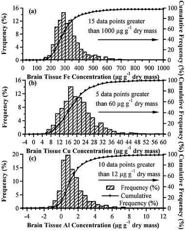 | ||
| Fig. 3 Percentage frequency (bars) and cumulative frequency (line and marker) distributions of (a) Fe, (b) Cu and (c) Al concentrations in (n = 719, 720 and 713 respectively) brain tissues after subtraction of contamination. | ||
The median content of Al in each lobe were 0.83 Frontal (F), 0.98 Occipital (O), 0.95 Parietal (P) and 1.30 Temporal (T) μg g−1 dry wt. and significant differences (P < 0.05; Sign test) between lobes were found for T-F, T-O, T-P and F-P (Fig. 4a).
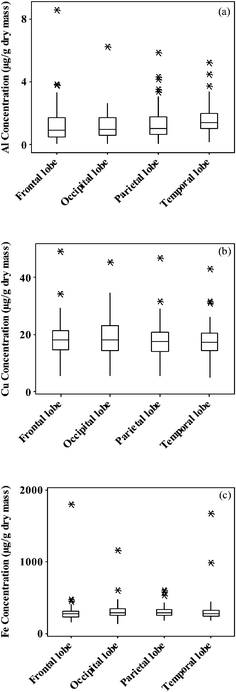 | ||
| Fig. 4 Boxplots of the concentrations of (a) Al, (b) Cu and (c) Fe by lobe in 60 brains. The medians of concentrations in the three tissue samples of a given lobe were used, to give one value for each lobe for each brain. | ||
The Fe content of 719 tissue digests following subtraction of the method blank were not normally distributed and ranged from ca 100 to 8300 μg g−1 dry wt. (Table 5). The median value was 286.16 μg g−1 dry wt. and 75% of all values were < 345.36 μg g−1 dry wt. (Fig. 3a). The median content of Fe in each lobe were ca 271 (F), 304 (O), 295 (P) and 280 (T) μg g−1 dry wt. and significant differences (P < 0.05; Sign test) between lobes were found for F-O and F-P (Fig. 4b).
The Cu content of 720 tissue digests following subtraction of the method blank were not normally distributed and ranged from ca 0 (MBV) to 380 μg g−1 dry wt. (Table 4). The median value was 17.41 μg g−1 dry wt. and 75% of all values were <22.19 μg g−1 dry wt. (Fig. 3b). The median content of Cu in each lobe were 17.5 (F), 17.9 (O), 17.4 (P) and 16.6 (T) μg g−1 dry wt. and there were no significant differences (P < 0.05; Sign test) between any of the lobes (Fig. 4c).
| Brain | Contamination Corrected Copper Concentration (μg g−1 dry mass tissue) | |||||||||||
|---|---|---|---|---|---|---|---|---|---|---|---|---|
| Frontal | Occipital | Parietal | Temporal | |||||||||
| Sample 1 | Sample 2 | Sample 3 | Sample 1 | Sample 2 | Sample 3 | Sample 1 | Sample 2 | Sample 3 | Sample 1 | Sample 2 | Sample 3 | |
| 1 | 29.50 | 23.82 | 19.53 | 22.72 | 26.13 | 18.97 | 28.53 | 21.56 | 19.30 | 20.26 | 18.79 | 17.43 |
| 2 | 14.57 | 15.06 | 11.01 | 13.37 | 8.94 | 12.88 | 15.27 | 21.68 | 11.91 | 11.18 | 15.83 | 15.94 |
| 3 | 18.09 | 19.61 | 24.60 | 25.80 | 11.93 | 16.69 | 13.49 | 23.81 | 23.92 | 16.55 | 26.77 | 20.65 |
| 4 | 49.00 | 67.04 | −1.74 | 43.23 | 45.25 | 55.78 | 46.58 | 54.91 | 32.44 | 42.84 | 45.18 | 33.37 |
| 5 | 20.52 | 28.34 | 21.04 | 25.79 | 17.35 | 25.74 | 18.97 | 20.20 | 21.56 | 24.14 | 11.42 | 23.51 |
| 6 | 21.90 | 24.60 | 26.70 | 26.89 | 21.19 | 22.88 | 21.46 | 25.87 | 23.37 | 16.03 | 22.02 | 19.82 |
| 7 | 16.86 | 8.43 | 15.12 | 19.52 | 29.56 | 18.33 | 27.11 | 10.23 | 15.32 | 16.08 | 9.55 | 13.80 |
| 8 | 19.12 | 17.76 | 19.20 | 21.78 | 30.07 | 37.74 | 20.03 | 20.43 | 19.87 | 25.53 | 26.94 | 14.12 |
| 9 | 19.49 | 17.19 | 29.86 | 25.13 | 23.76 | 22.59 | 19.25 | 14.37 | 28.09 | 25.88 | 25.58 | 25.67 |
| 10 | 22.22 | 12.65 | 16.75 | 20.78 | 17.42 | 20.06 | 18.37 | 13.53 | 15.39 | 20.68 | 20.23 | 14.72 |
| 11 | 10.68 | 26.48 | 7.79 | 15.49 | 8.61 | 17.34 | 15.43 | 16.87 | 9.19 | 21.31 | 30.14 | 4.34 |
| 12 | 21.94 | 14.04 | 20.29 | 16.87 | 10.93 | 3.99 | 19.73 | 10.55 | 13.82 | 15.02 | 13.05 | 13.04 |
| 13 | 34.22 | 384.44 | 28.28 | 28.48 | 35.76 | 34.55 | 95.20 | 31.63 | 24.42 | 34.58 | 30.52 | 31.44 |
| 14 | 21.99 | 19.81 | 25.44 | 93.44 | 26.01 | 23.62 | 15.12 | 19.43 | 20.40 | 25.91 | 34.74 | 20.76 |
| 15 | 13.53 | 20.13 | 15.35 | 17.92 | 39.08 | 17.90 | 15.30 | 15.13 | 18.96 | 19.45 | 21.40 | 15.38 |
| 16 | 29.24 | 19.98 | 16.92 | 24.67 | 20.42 | 39.38 | 21.41 | 19.16 | 22.62 | 20.17 | 28.73 | 17.50 |
| 17 | 25.05 | 29.22 | 32.76 | 47.98 | 33.20 | 33.67 | 28.67 | 28.78 | 32.46 | 42.18 | 18.35 | 25.34 |
| 18 | 73.53 | 19.25 | 11.55 | 24.35 | 23.45 | 25.72 | 37.05 | 21.07 | 16.31 | 19.78 | 12.86 | 14.19 |
| 19 | 33.73 | 12.59 | 19.44 | 29.07 | 14.22 | 12.23 | 31.24 | 17.69 | 14.36 | 26.61 | 13.16 | 15.86 |
| 20 | 41.78 | 21.31 | 19.82 | 15.79 | 17.89 | 15.53 | 48.37 | 19.89 | 17.01 | 20.23 | 18.07 | 12.45 |
| 21 | 15.67 | 22.18 | 18.37 | 13.59 | 18.74 | 26.74 | 18.11 | 25.10 | 12.27 | 16.93 | 14.78 | 34.73 |
| 22 | 20.99 | 18.56 | 22.97 | 17.40 | 23.16 | 27.14 | 12.71 | 18.85 | 19.86 | 13.63 | 15.64 | 14.70 |
| 23 | 17.58 | 21.53 | 21.79 | 22.99 | 20.78 | 23.92 | 26.59 | 20.21 | 32.21 | 21.87 | 24.04 | 20.20 |
| 24 | 11.71 | 18.71 | 14.93 | 16.19 | 18.09 | 19.05 | 13.02 | 20.35 | 15.06 | 16.42 | 15.81 | 16.67 |
| 25 | 23.58 | 28.21 | 33.30 | 14.77 | 22.13 | 25.47 | 17.48 | 21.68 | 23.28 | 17.37 | 25.53 | 27.15 |
| 26 | 11.45 | 14.67 | 15.90 | 10.18 | 17.67 | 15.04 | 13.63 | 20.46 | 18.19 | 12.87 | 16.25 | 14.92 |
| 27 | 20.38 | 18.07 | 31.79 | 18.44 | 19.18 | 14.47 | 11.47 | 18.54 | 22.45 | 19.75 | 22.78 | 14.57 |
| 28 | 6.97 | 5.67 | 4.44 | 16.19 | 5.56 | 5.46 | 5.68 | 5.44 | 9.71 | 5.41 | 5.78 | 7.13 |
| 29 | 12.23 | 8.64 | 10.08 | 12.01 | 11.50 | 12.15 | 10.72 | 8.13 | 13.90 | 14.96 | 8.75 | 13.19 |
| 30 | 18.05 | 16.20 | 24.60 | 12.37 | 15.32 | 25.82 | 16.06 | 16.36 | 13.86 | 17.83 | 18.55 | 25.02 |
| 31 | 16.12 | 14.46 | 12.97 | 14.15 | 14.43 | 14.16 | 15.40 | 17.61 | 21.31 | 15.06 | 14.47 | 13.91 |
| 32 | 28.60 | 25.96 | 24.22 | 33.47 | 26.14 | 22.45 | 22.97 | 24.85 | 31.54 | 27.79 | 20.27 | 22.90 |
| 33 | 16.85 | 13.50 | 14.49 | 11.35 | 8.65 | 16.97 | 15.81 | 11.44 | 13.40 | 17.70 | 14.32 | 18.08 |
| 34 | 9.00 | 8.28 | 9.36 | 9.79 | 10.08 | 11.17 | 10.39 | 8.40 | 9.36 | 7.87 | 6.78 | 11.45 |
| 35 | 7.09 | 8.19 | 13.41 | 8.09 | 10.89 | 12.17 | 9.39 | 12.85 | 14.76 | 7.19 | 9.78 | 11.61 |
| 36 | 11.22 | 10.52 | 13.71 | 7.82 | 9.72 | 11.90 | 13.24 | 15.68 | 15.58 | 11.80 | 10.32 | 12.85 |
| 37 | 11.48 | 11.38 | 15.56 | 7.60 | 11.95 | 17.62 | 11.27 | 18.65 | 14.71 | 10.41 | 15.64 | 19.38 |
| 38 | 13.84 | 18.78 | 14.84 | 14.56 | 19.15 | 17.57 | 12.24 | 20.08 | 15.57 | 12.51 | 18.47 | 16.91 |
| 39 | 14.79 | 15.94 | 17.26 | 24.94 | 23.22 | 16.56 | 18.47 | 17.28 | 14.89 | 19.79 | 10.54 | 23.59 |
| 40 | 10.40 | 10.23 | 7.66 | 8.20 | 7.06 | 9.23 | 9.99 | 9.54 | 13.17 | 10.16 | 12.59 | 8.69 |
| 41 | 15.68 | 13.10 | 25.09 | 21.89 | 20.52 | 17.12 | 23.85 | 16.50 | 20.34 | 15.67 | 15.80 | 14.45 |
| 42 | 9.04 | 9.43 | 9.65 | 9.29 | 11.10 | 7.68 | 9.14 | 7.53 | 7.21 | 2.31 | 9.24 | 5.09 |
| 43 | 15.75 | 14.39 | 16.56 | 14.82 | 17.60 | 14.66 | 13.02 | 14.08 | 15.49 | 14.18 | 14.23 | 10.25 |
| 44 | 16.31 | 17.49 | 13.84 | 29.74 | 19.00 | 15.46 | 16.33 | 15.88 | 13.56 | 18.03 | 17.35 | 14.92 |
| 45 | 28.62 | 15.42 | 22.58 | 18.39 | 17.88 | 14.60 | 23.37 | 16.70 | 16.60 | 15.00 | 15.45 | 12.36 |
| 46 | 22.36 | 13.29 | 19.35 | 18.68 | 12.52 | 15.88 | 12.44 | 11.59 | 17.74 | 15.80 | 17.01 | 15.48 |
| 47 | 17.67 | 16.76 | 20.72 | 15.43 | 17.77 | 16.97 | 19.35 | 18.66 | 17.79 | 16.99 | 15.03 | 21.02 |
| 48 | 6.45 | 11.20 | 6.31 | 11.45 | 12.71 | 9.90 | 7.41 | 9.25 | 5.81 | 8.81 | 9.66 | 7.06 |
| 49 | 6.52 | 18.88 | 13.52 | 15.11 | 14.48 | 21.43 | 8.92 | 17.85 | 12.48 | 8.40 | 13.24 | 11.86 |
| 50 | 20.82 | 28.83 | 14.55 | 18.23 | 27.76 | 20.13 | 18.38 | 16.34 | 20.50 | 18.67 | 19.23 | 21.05 |
| 51 | 17.25 | 25.68 | 21.94 | 30.53 | 14.91 | 22.77 | 35.89 | 23.64 | 19.03 | 19.35 | 15.55 | 31.00 |
| 52 | 27.56 | 29.82 | 22.99 | 31.25 | 23.54 | 21.35 | 23.12 | 21.02 | 22.95 | 22.48 | 19.99 | 28.75 |
| 53 | 27.97 | 38.26 | 28.25 | 42.14 | 31.87 | 29.97 | 31.41 | 28.81 | 38.31 | 33.24 | 30.90 | 28.45 |
| 54 | 24.45 | 20.30 | 22.76 | 22.58 | 21.29 | 23.72 | 21.10 | 17.76 | 18.26 | 15.94 | 18.07 | 24.81 |
| 55 | 21.12 | 16.02 | 13.18 | 21.84 | 21.29 | 12.22 | 11.89 | 11.19 | 12.68 | 16.56 | 11.78 | 19.28 |
| 56 | 18.42 | 13.07 | 12.89 | 14.28 | 13.71 | 15.54 | 17.66 | 9.66 | 9.10 | 16.35 | 14.21 | 13.70 |
| 57 | 15.93 | 15.46 | 12.24 | 20.05 | 12.55 | 9.06 | 11.55 | 13.88 | 10.04 | 16.76 | 10.90 | 12.94 |
| 58 | 18.34 | 22.82 | 16.88 | 14.18 | 19.54 | 12.81 | 20.98 | 22.66 | 11.43 | 17.43 | 17.83 | 23.97 |
| 59 | 44.51 | 19.07 | 17.51 | 15.01 | 21.61 | 18.04 | 15.55 | 24.20 | 16.29 | 37.95 | 20.43 | 19.12 |
| 60 | 10.68 | 12.53 | 9.65 | 13.96 | 9.79 | 6.76 | 13.49 | 9.46 | 10.63 | 10.40 | 11.20 | 8.62 |
The Al content was positively correlated (P = 0.05; Spearman's rank) with both Fe and Cu content across all lobes. Al was positively correlated with Cu in the frontal lobe though there were no other significant correlations between Al and Fe or Cu for the individual lobes. Fe and Cu content were positively correlated across all lobes and in each individual lobe (Fig. 5).
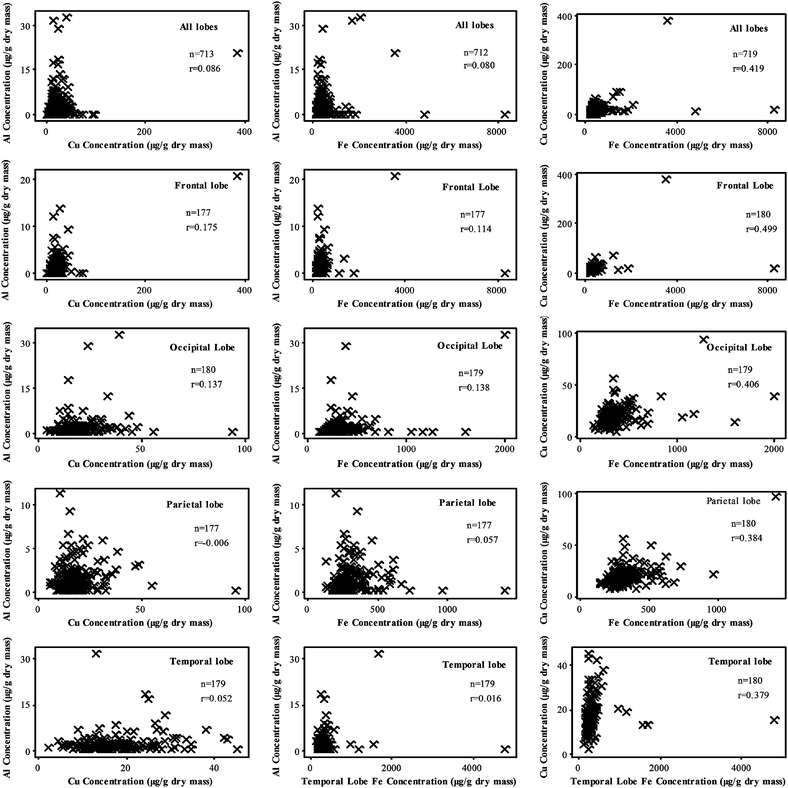 | ||
| Fig. 5 Scatter plots to show the correlation between Al, Cu and Fe concentrations using all data and data for specific lobes. Concentrations are values after subtraction of contamination with negative values assigned a value of zero. Data were not normally distributed and r is the Spearman's rank coefficient. | ||
Discussion
Instrument performance and quality assurance data demonstrated a high level of precision and reproducibility in measuring Al, Fe and Cu in acid digests by TH GFAAS. Contamination of samples and of procedures is the major constraint in determining absolute amounts of these metals in brain tissues and every precaution was taken to minimise this issue. To better understand and quantify the potential contribution of contamination to absolute measurements we carried out a large number of method blanks. These data revealed non-normal distributions of contaminants and showed a wider variance for Al than either Fe or Cu (Fig. 2).It was of interest that while there was little overlap between the method blank and tissue digest distributions for Fe and Cu the overlap was significant for Al and this was simply a reflection of the significantly lower amounts of the non-essential Al in both method blanks and tissue digests. It should be noted that absolute amounts of contamination by the so-called ‘ubiquitous contaminant’ Al were significantly lower than for Fe or Cu. As an upper estimate of the method blank contamination we used the mean + tα0.05,1-tail SD10,11, to determine a level of contamination per vessel and subtracted this from each tissue digest. For Al this resulted in a significant number of ‘zero’ values which are shown in Table 3 as negative values. All negative values were treated as zero in subsequent statistical analyses. A significant observation for Al were the high variances for each set of 3 tissue samples taken from the same lobe. This showed that Al was not evenly distributed within the brain tissue samples and supported focal accumulations of Al which reflected possible associations between Al and particular compartments and structures.3 These differences in metal content between sub-samples of a single tissue sample were not as consistently evident for Fe (Table 5) or Cu (Table 4). Overall there were few significant differences between the metal content of individual lobes with the main exception being the higher content of Al in the temporal lobe.
| Brain | Contamination Corrected Iron Concentration (μg g−1 dry mass tissue) | |||||||||||
|---|---|---|---|---|---|---|---|---|---|---|---|---|
| Frontal | Occipital | Parietal | Temporal | |||||||||
| Sample 1 | Sample 2 | Sample 3 | Sample 1 | Sample 2 | Sample 3 | Sample 1 | Sample 2 | Sample 3 | Sample 1 | Sample 2 | Sample 3 | |
| 1 | 573.09 | 462.10 | 309.28 | 683.76 | 458.87 | 209.83 | 726.77 | 537.41 | 498.46 | 990.14 | 1180.31 | 432.85 |
| 2 | 310.93 | 307.87 | 245.90 | 335.49 | 452.50 | 312.17 | 402.14 | 323.35 | 271.31 | 278.58 | 279.17 | 263.45 |
| 3 | 264.10 | 348.10 | 311.04 | 328.37 | 674.54 | 298.39 | 217.83 | 570.69 | 403.60 | 253.24 | 400.59 | 255.85 |
| 4 | 250.48 | 356.62 | 230.15 | 330.53 | 313.92 | 324.82 | 308.16 | 299.55 | 209.33 | 250.09 | 234.05 | 260.92 |
| 5 | 310.48 | 351.37 | 270.99 | 344.31 | 308.24 | 260.38 | 292.08 | 271.33 | 274.38 | 336.88 | 342.27 | 311.97 |
| 6 | 249.40 | 293.58 | 330.11 | 322.94 | 306.91 | 274.98 | 293.24 | 364.86 | 328.91 | 368.88 | 270.68 | 297.21 |
| 7 | 294.25 | 243.92 | 649.73 | 357.64 | 282.44 | 280.99 | 358.33 | 330.46 | 355.06 | 350.48 | 255.44 | 333.42 |
| 8 | 443.92 | 314.53 | 329.51 | 351.12 | 343.44 | 528.99 | 502.66 | 344.89 | 319.62 | 325.63 | 313.61 | 353.07 |
| 9 | 280.60 | 281.87 | 434.32 | 407.66 | 440.11 | 401.79 | 307.19 | 313.85 | 570.40 | 356.88 | 346.29 | 444.34 |
| 10 | 508.58 | 359.64 | 382.07 | 304.80 | 313.39 | 355.29 | 371.87 | 318.91 | 349.02 | 399.56 | 318.83 | 361.49 |
| 11 | 235.60 | 289.80 | 241.00 | 494.22 | 314.07 | 392.45 | 416.93 | 374.72 | 295.56 | 341.43 | 580.33 | 126.60 |
| 12 | 8304.65 | 1394.79 | 1807.88 | 631.35 | 614.11 | 363.48 | 966.78 | 613.23 | 585.75 | 4789.77 | 1685.96 | 1556.26 |
| 13 | 457.78 | 3521.46 | 320.74 | 516.32 | 485.43 | 478.98 | 1415.48 | 328.27 | 312.20 | 478.69 | 319.85 | 416.15 |
| 14 | 212.49 | 292.91 | 317.91 | 1266.62 | 421.79 | 371.80 | 413.71 | 430.47 | 365.63 | 442.41 | 475.72 | 318.34 |
| 15 | 230.54 | 365.28 | 303.01 | 342.84 | 2005.05 | 441.33 | 257.92 | 396.36 | 326.24 | 316.52 | 274.11 | 256.85 |
| 16 | 479.21 | 294.53 | 688.84 | 485.67 | 470.46 | 814.12 | 509.02 | 362.13 | 435.40 | 398.45 | 391.24 | 296.85 |
| 17 | 219.06 | 550.70 | 414.96 | nm | 441.66 | 499.18 | 277.13 | 505.81 | 423.74 | 429.79 | 287.62 | 407.12 |
| 18 | 1157.09 | 337.95 | 314.19 | 302.87 | 410.84 | 553.01 | 614.49 | 418.63 | 352.44 | 341.24 | 302.01 | 321.91 |
| 19 | 373.66 | 198.21 | 301.99 | 406.21 | 232.14 | 268.89 | 454.07 | 324.10 | 255.82 | 314.61 | 206.55 | 286.08 |
| 20 | 524.68 | 276.10 | 322.50 | 340.17 | 364.39 | 359.73 | 509.18 | 259.83 | 305.29 | 310.13 | 320.65 | 250.51 |
| 21 | 209.28 | 263.41 | 232.07 | 230.81 | 291.28 | 394.82 | 287.68 | 360.57 | 209.35 | 247.18 | 218.74 | 501.83 |
| 22 | 237.57 | 218.98 | 190.01 | 232.60 | 256.77 | 332.15 | 143.88 | 238.63 | 252.93 | 223.90 | 205.23 | 178.90 |
| 23 | 218.88 | 229.64 | 214.10 | 308.65 | 221.13 | 237.16 | 320.46 | 195.62 | 255.70 | 279.81 | 257.81 | 202.94 |
| 24 | 193.31 | 264.84 | 234.05 | 205.61 | 245.02 | 262.66 | 245.51 | 330.70 | 254.81 | 297.86 | 214.27 | 266.35 |
| 25 | 382.75 | 330.47 | 674.75 | 228.46 | 276.90 | 353.28 | 207.50 | 274.01 | 324.37 | 324.86 | 296.15 | 358.07 |
| 26 | 218.19 | 282.13 | 265.88 | 193.72 | 347.65 | 286.16 | 294.21 | 471.79 | 298.78 | 265.63 | 282.37 | 253.75 |
| 27 | 294.41 | 273.33 | 309.57 | 308.87 | 303.90 | 260.70 | 140.52 | 276.19 | 255.51 | 318.44 | 287.92 | 195.86 |
| 28 | 295.99 | 215.79 | 194.12 | 550.63 | 281.62 | 292.81 | 391.43 | 241.87 | 467.29 | 262.17 | 385.95 | 412.71 |
| 29 | 254.73 | 207.64 | 237.98 | 324.40 | 233.93 | 306.58 | 298.22 | 296.44 | 522.32 | 327.37 | 260.07 | 383.22 |
| 30 | 245.05 | 225.57 | 332.82 | 209.16 | 223.30 | 270.15 | 258.97 | 228.02 | 198.79 | 239.98 | 263.86 | 262.57 |
| 31 | 250.40 | 300.02 | 218.56 | 221.12 | 411.88 | 218.18 | 254.00 | 338.22 | 334.05 | 253.03 | 327.00 | 247.26 |
| 32 | 225.05 | 189.54 | 246.89 | 265.04 | 203.19 | 207.43 | 224.51 | 243.22 | 263.93 | 190.01 | 152.82 | 200.52 |
| 33 | 163.68 | 169.14 | 207.69 | 112.01 | 140.29 | 179.59 | 213.82 | 132.41 | 197.71 | 233.91 | 170.18 | 192.19 |
| 34 | 212.66 | 229.54 | 257.21 | 293.21 | 256.63 | 302.26 | 260.72 | 229.50 | 237.42 | 216.82 | 160.11 | 317.01 |
| 35 | 204.27 | 210.83 | 269.39 | 232.71 | 202.95 | 239.45 | 276.60 | 341.39 | 277.74 | 226.25 | 260.59 | 233.05 |
| 36 | 320.13 | 226.52 | 313.05 | 284.59 | 248.89 | 261.38 | 371.80 | 378.32 | 373.82 | 227.88 | 221.51 | 261.88 |
| 37 | 264.95 | 247.67 | 285.76 | 136.21 | 255.12 | 318.77 | 271.44 | 384.04 | 270.43 | 268.74 | 325.24 | 334.03 |
| 38 | 335.93 | 223.95 | 205.38 | 214.21 | 280.33 | 295.56 | 253.34 | 322.99 | 225.67 | 203.28 | 231.05 | 213.57 |
| 39 | 198.43 | 224.42 | 246.72 | 217.00 | 309.65 | 209.89 | 281.97 | 281.81 | 189.89 | 269.85 | 156.82 | 251.31 |
| 40 | 227.51 | 241.79 | 183.38 | 254.20 | 182.36 | 269.13 | 230.31 | 255.07 | 240.18 | 235.82 | 377.79 | 221.69 |
| 41 | 242.99 | 152.66 | 235.48 | 390.47 | 264.99 | 245.13 | 393.70 | 202.96 | 255.78 | 243.21 | 218.49 | 216.13 |
| 42 | 266.51 | 236.01 | 253.65 | 331.79 | 307.53 | 248.76 | 335.63 | 274.77 | 253.05 | 260.32 | 190.70 | 174.20 |
| 43 | 201.40 | 230.28 | 311.66 | 240.45 | 253.11 | 252.34 | 275.09 | 309.21 | 294.46 | 272.59 | 306.56 | 191.95 |
| 44 | 342.96 | 225.47 | 281.12 | 465.11 | 303.11 | 274.14 | 334.41 | 302.07 | 292.47 | 283.29 | 294.11 | 258.04 |
| 45 | 370.85 | 181.84 | 347.88 | 245.71 | 278.27 | 222.41 | 244.27 | 224.19 | 238.67 | 324.14 | 232.07 | 222.12 |
| 46 | 392.37 | 297.33 | 268.91 | 347.87 | 268.58 | 279.25 | 222.64 | 290.81 | 275.48 | 271.35 | 317.00 | 241.06 |
| 47 | 299.03 | 321.40 | 242.84 | 347.93 | 327.99 | 481.45 | 391.13 | 397.14 | 401.27 | 367.58 | 303.79 | 438.69 |
| 48 | 237.38 | 556.59 | 195.03 | 215.47 | 339.56 | 309.64 | 242.30 | 281.59 | 198.41 | 185.71 | 345.83 | 280.72 |
| 49 | 176.80 | 271.46 | 235.02 | 555.70 | 1592.30 | 1163.77 | 177.01 | 581.93 | 669.62 | 153.27 | 256.68 | 293.95 |
| 50 | 255.90 | 309.30 | 212.85 | 215.86 | 305.29 | 311.90 | 231.07 | 181.42 | 360.61 | 178.24 | 244.32 | 264.35 |
| 51 | 297.12 | 471.81 | 232.07 | 486.03 | 271.29 | 261.58 | 369.71 | 336.83 | 204.96 | 395.58 | 244.68 | 297.97 |
| 52 | 378.07 | 476.46 | 298.37 | 386.90 | 355.40 | 239.60 | 310.46 | 230.15 | 274.37 | 292.99 | 300.98 | 284.08 |
| 53 | 232.71 | 250.22 | 255.75 | 330.83 | 265.01 | 259.30 | 263.38 | 258.04 | 298.86 | 314.92 | 261.50 | 289.55 |
| 54 | 411.13 | 262.00 | 274.92 | 319.61 | 287.95 | 282.09 | 319.08 | 238.38 | 231.67 | 288.10 | 208.59 | 325.20 |
| 55 | 257.71 | 248.92 | 217.79 | 312.19 | 397.09 | 219.83 | 200.18 | 189.22 | 226.70 | 242.55 | 187.81 | 373.06 |
| 56 | 342.14 | 243.21 | 328.54 | 280.29 | 238.63 | 257.40 | 307.40 | 223.42 | 218.24 | 312.26 | 287.01 | 321.74 |
| 57 | 214.29 | 220.31 | 277.22 | 286.21 | 166.40 | 204.76 | 131.61 | 186.76 | 201.28 | 211.02 | 185.55 | 252.37 |
| 58 | 280.00 | 282.51 | 271.83 | 224.86 | 216.36 | 242.16 | 373.71 | 274.13 | 232.50 | 233.79 | 184.44 | 236.37 |
| 59 | 618.71 | 300.58 | 317.42 | 346.60 | 305.10 | 1044.57 | 322.06 | 324.40 | 341.52 | 593.36 | 366.48 | 357.95 |
| 60 | 299.38 | 253.12 | 203.88 | 448.62 | 392.16 | 162.19 | 388.87 | 177.81 | 239.33 | 316.85 | 271.33 | 176.72 |
The median Al content for all 713 tissues was ca 1 μg g−1 dry wt. and 75% of all values were less than ca 2 μg g−1 dry wt. While these statistics would suggest that the Al contents were within the ‘normal’ range for the aged brain it was actually the case that there were only 8 and 19 individual's brains where all 12 Al measurements were less than 2 and 3.5 μg g−1 dry wt. respectively (Table 3). Thus ca 70% of the brains studied included an Al content (or accumulation of Al) which could be considered as pathological.3 This is an example of how difficult it might be to use statistical measures of brain Al content as reliable indicators of potential neurotoxicity.
The median Fe content for all tissues was ca 290 μg g−1 dry wt. and 75% of all values were less than ca 350 μg g−1 dry wt. These statistics are slightly higher than the normal range for Fe6 and probably reflect the known increases in brain Fe with age and, potentially, the co-occurrence in the aged brains investigated herein of neurodegenerative diseases including AD and CAA.4
The median Cu content for all tissues was ca 17 μg g−1 dry wt. and 75% of all values were less than ca 22 μg g−1 dry wt. These statistics are slightly lower than might have been predicted for the aged brains investigated herein8 and the lower Cu contents might reflect the potential occurrence of AD and CAA.4,7
Conclusions
TH GFAAS is an excellent method for measuring Al, Fe and Cu in acid digests of brain tissue samples. The major constraint in obtaining absolute amounts of these metals in brain tissues is extraneous contamination and, in addition to all of the usual precautions, this should be accounted for by measuring significant numbers of method blanks and computing a statistically significant value which is then subtracted from each tissue digest. Herein the method blank data showed significant contamination for each metal and while the values for Al, as ng/vessel, were lower than Cu and significantly lower than Fe, their impact upon final metal content of tissue digests were greater for Al due to lower amounts of this metal in the tissues. Herein we have reported the metal contents of every tissue digest, as opposed to median or mean values, (Table 3–5) as replicates of samples taken from a particular lobe showed significant variance and especially for Al content. The latter supported what is known about the distribution of Al in brain tissue in that it is primarily focally located and it may be these distinct accumulations of Al which have the potential for neurotoxicity. In a follow-up publication we will address whether the Al, Fe and Cu contents of these 60 human brains are connected in any way to their amyloid pathologies, specifically senile plaques and CAA.Notes and references
- L. E. Scott and C. Orvig, Chem. Rev., 2009, 109, 4885 CrossRef CAS.
- G. Crisponi, V. M. Nurchi, G. Faa and M. Remelli, Monatsh. Chem., 2011, 142, 331 CrossRef CAS.
- C. Exley and E. R. House, Monatsh. Chem., 2011, 142, 357 CrossRef CAS.
- M. Schrag, A. Crofton, M. Zabel, A. Jiffry, D. Kirsch, A. Dickson, X. W. Mao, H. V. Vinters, D. W. Domaille, C. J. Chang and W. Kirsch, J. Alzh. Dis., 2011, 24, 137 CAS.
- M. Loef and H. Walach, Br. J. Nutr., 2011 DOI:10.1017/S000711451100376X.
- L. Zecca, F. A. Zucca, M. Toscani, F. Adorni, G. Giaveri, E. Rizzio and M. Gallorini, J. Radioanal. Nucl. Chem., 2005, 263, 733 CrossRef CAS.
- M. Schrag, C. Mueller, U. Oyoyo, M. A. Smith and W. Kirsch, Prog. Neurobiol., 2011, 94, 296 CrossRef CAS.
- S. Lutsenko, A. Bhattacharjee and A. L. Hubbard, Metallomics, 2010, 2, 596 RSC.
- S. B. Wharton, C. Brayne, G. M. Savva, F. E. Matthews, G. Forster, J. Simpson, G. Lace and P. G. Ince, J. Alzh. Dis., 2011, 25, 359 Search PubMed.
- L. A. Currie, Anal. Chim. Acta, 1999, 391, 127 CrossRef CAS.
- L. A. Currie Fresenius, J Anal Chem, 2001, 370, 705–718 Search PubMed.
| This journal is © The Royal Society of Chemistry 2012 |
