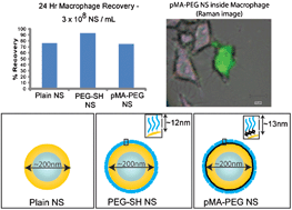
In this study, the relative toxicity of native gold-silica nanoshells (NS) has been compared to nanoshells modified with poly(ethylene glycol)-thiol (PEG-SH) and a Raman-active PEG, p-mercaptoaniline-poly(ethylene glycol) (pMA-PEG), in mouse alveolar macrophage cell cultures (RAW 264.7). The results from toxicity profiling using an MTT assay demonstrate that cell viability post-particle exposure is a function of three factors: nanoshell concentration, surface functionalization, and incubation time. By minimizing particle concentrations and incubation times, cell cultures are able to recover within 24 h of nanoshell removal, indicative of nanoshells having more of a cytostatic versus cytotoxic effect on macrophage cells. The mechanism of the cytostatic effect has been investigated by imaging the presence of reactive oxygen species (ROS) using a fluorescence assay kit (Image-iT™ LIVE) after the introduction of NS to the cell cultures. Elevated ROS signals are seen in the cells containing higher concentration of NS, and indicate that the major reason of toxicity may due to the oxidative stress caused by excess NS particles. Raman imaging experiments with pMA-PEG coated nanoshells showed that cells exposed for even short exposure times (∼2 h) retained those particles up to 24 h after exposure, while migration experiments suggest that surviving cells retain their nanoshells and may reallocate them to progeny cells upon cell division.
You have access to this article
 Please wait while we load your content...
Something went wrong. Try again?
Please wait while we load your content...
Something went wrong. Try again?
