Sonochemical selective synthesis of ZnO/CdS core/shell nanostructures and their optical properties
Jun
Geng
ab,
Xiang-Dong
Jia
ac and
Jun-Jie
Zhu
*a
aKey Laboratory of Analytical Chemistry for Life Science, School of Chemistry and Chemical Engineering, Nanjing University, Nanjing, 210093, P. R. China. E-mail: jjzhu@nju.edu.cn; Fax: +86-25-83594976; Tel: +86-25-83594976
bDepartment of Chemistry, Jiangsu Institute of Education, Nanjing, 210013, P. R. China
cCollege of Science, Nanjing Forestry University, Nanjing, 210037, P. R. China
First published on 27th August 2010
Abstract
The core/shell-type ZnO nanosphere/CdS nanorod and ZnO nanosphere/CdS nanoparticle composites have been selectively prepared through a simple ultrasound-assisted solution phase conversion process using monodispersed ZnO nanospheres as a starting reactant and in situ template. The formation mechanism of the products is closely connected with the sonochemical effect of ultrasonic irradiation. The photoluminescence and electrogenerated chemiluminescence properties of the as-prepared core/shell structures were investigated.
1. Introduction
The shape, phase, and size of inorganic nanocrystals are important elements in varying their electrical, optical, and other properties.1 Consequently, the design and controlled fabrication of nanomaterials with functional properties are required to meet the ever-increasing demands.Surface coating or modification has been recognized as one of the most advanced and intriguing methods to build tailored nanomaterials.1,2 Materials are coated for a number of reasons: Coatings can alter the charge, functionality, and reactivity of the surface, and enhance their thermal, mechanical, or chemical stability.1,2 Much effort has recently been invested to create new class of nanomaterials through surface coating, and different kinds of core/shell nanostructures have been fabricated by various synthetic methods, including the CdSe/ZnS nanocrystals obtained by a biomimetic method using a bi-functional peptide,3 FePt/ZnO core/shell nanoparticles synthesized by a seed-mediated growth,4 SnO2/TiO2 hollow nanostructures prepared via a colloid seeded deposition process,5 Pd@CeO2 core-shell nanostructures by self-assembly,6 ZnO@Cd(OH)2 core-shell nanoparticles by a sol–gel method,7 fluorescent core/shell CdTe@SiO2 particles via a water-in-oil reverse microemulsion method,8 CdTe/CdS/ZnS core/shell/shell quantum dots synthesized in the aqueous phase assisted by microwave irradiation,9 branched core/shell bismuth telluride/bismuth sulfide nanorods prepared by using a biomolecular surfactant,10 and so on.
Among the various non-linear optical (NLO) materials investigated, the direct band gap semiconductors, especially ZnO and CdS, have attractive non-linear properties that make them as the ideal candidates for NLO based devices. ZnO is a transparent oxide semiconductor that possesses piezoelectric properties, which has been widely used for solar cells,11 nanolasers,12 optoelectronic devices,13 and electromechanical coupled sensors and transducers.14 Moreover, ZnO is biocompatible and can be directly used for biomedical applications.14 CdS nanocrystals are one of the most studied systems among all the semiconducting nanocrystals.15 The bulk CdS has a direct band gap of 2.4 eV at 300 K, and the typical Bohr exciton diameter of CdS is around 5.8 nm; consequently, CdS nanocrystals in the size range of 1–6 nm show sizable quantum confinement effects with remarkably different optical properties.
The surface coating of ZnO nanomaterials with a preferred CdS can promote the construction of ZnO-based core/shell-type composites with novel optical and electrical properties. A few approaches have been reported for the surface coating of ZnO nanomaterials with CdS. For example, ZnO nanowire/CdS nanoparticle heterostructure arrays were fabricated by a two-step chemical solution deposition method,16 core/shell-type ZnO nanorod/CdS nanoparticle composites were prepared via a sonochemical route,17 ZnO nanowire arrays coated with CdS quantum dots were successfully fabricated with a chemical bath deposition process,18 CdS quantum dots were deposited on the vertically aligned ZnO nanorods electrode by chemical bath deposition,19 10 nm sized hexagonal CdS nanoparticles were decorated on the surface of well-aligned ZnO nanowall through a facile hydrothermal approach,20 ZnO/CdS nanocomposites were obtained by colloidal chemical synthesis,21 and so on. However, all these reports involved coating core structure with nanoparticles, no other coating status with different morphology was reported.
In this paper, core/shell-type ZnO nanosphere/CdS nanorod and ZnO nanosphere/CdS nanoparticle composites are selectively achieved by a simple ultrasonic irradiation of a mixture of monodispersed ZnO nanospheres, cadmium chloride, and thiourea or TAA in an aqueous medium. The use of different sulfur sources resulted in different surface coating statuses. The ultrasonic irradiation played an important role in the controllable synthesis of the core/shell nanostructures. The photoluminescence and electrogenerated chemiluminescence of the obtained ZnO/CdS composites have been investigated to show the interaction between the ZnO cores and the external CdS nanorod or nanoparticle shells.
2. Experimental
2.1 Materials
All the reagents used were of analytical purity and were used without further purification. Zn(NO3)2·6H2O, PEG-20000, triethanolamine (TEA), thioacetamide (TAA), and thiourea were purchased from Beijing Chemical Reagents Ltd. Co. of China.2.2 Synthesis
ZnO nanospheres were synthesized following ref. 22. Typically, Zn(NO3)2·6H2O (0.0015 mol) was dissolved in H2O (100 mL), and then PEG-20000 (1 g) and TEA (2 mL) were introduced sequentially with stirring to form a clear solution. The transparent mixture solution was exposed to high-intensity ultrasound irradiation under ambient air for 30 min. Ultrasound irradiation was generated with a high-intensity ultrasonic probe (Xinzhi Co., China, JY92-2D, 0.6 cm diameter; Ti-horn, 20 kHz, 60 W cm−2) immersed directly in the reaction solution. A white precipitate was centrifuged, washed with distilled water and absolute ethanol in sequence, and finally dried in air. The final products were collected for characterizations and further preparations.ZnO/CdS core/shell structures. ZnO nanospheres (0.001 mol) and CdCl2 (0.001 mol) were well dispersed in EtOH (50 mL), and then thiourea (0.001 mol) or TAA (0.001 mol) was introduced. The mixture was exposed to high-intensity ultrasound irradiation for 30 min to prepare ZnO/CdS core/shell structures.
2.3 Characterization
The X-ray powder diffraction (XRD) analysis was performed on a Philips X'-pert X-ray diffractometer at a scanning rate of 4° min−1 in the 2θ range from 10–80°, with graphite-monochromatized Cu-Kα radiation (λ = 0.15418 nm). The scanning electron micrographs (SEM) were taken on a LEO-1530VP field-emission scanning electron microscope. Transmission electron microscopy (TEM) was carried out on a JEOLJEM 200CX transmission electron microscope, using an accelerating voltage of 200 kV. High-resolution transmission electron micrographs (HRTEM) were obtained by employing a JEOL-2010 high-resolution transmission electron microscope with an accelerating voltage of 200 kV. Photoluminescence spectra (PL) were measured on a SLM48000DSCF/AB2 fluorescence spectrometer made by American SLM Incorporation at room temperature.Cyclic voltammetry (CV) and electrogenerated chemiluminescence (ECL) curves were obtained simultaneously using a model MPI-A electrochemiluminescence analyzer systems (Xi'An Remax Electronic Science & Technology Co. Ltd., Xi'An, China). The photomultiplier tube (PMT) used in the above analyzer was held at 800 V during the whole process of detection. Carbon-paste electrodes with a diameter of 4 mm were used as the working electrode.23 A conventional three-electrode electrochemical cell that was used for all measurements consisted of a carbon-paste working electrode (0.5 cm2), a Pt wire counter electrode, and an Ag/AgCl reference electrode.
3. Results and discussion
3.1 Characterization of the final products
Fig. 1 shows the XRD patterns of the as-prepared products. The obtained ZnO sample is of wurtzite structure (hexagonal phase, space group P63mc). All of the diffraction peaks in the XRD pattern (Fig. 1a) are well assigned to hexagonal-phase ZnO as reported in JCPDS card (no. 36-1451). The XRD patterns of the core/shell nanostructures using thiourea and TAA as sulfur sources are shown in Fig. 1b and c, respectively. Hexagonal phase CdS structure (JCPDS card no. 41-1049) is found coexistent with hexagonal phase ZnO in the XRD pattern of the core/shell structures. The apparently broadening of CdS diffraction peaks indicates that the samples are composed of nanosized particles. | ||
| Fig. 1 The XRD patterns of (a) ZnO spheres, (b) ZnO nanosphere/CdS nanorod core/shell structure using thiourea as a sulfur source, (c) ZnO nanosphere/CdS nanoparticle core/shell structure using TAA as a sulfur source. | ||
The as-prepared ZnO sample appeared as uniform solid nanospheres with diameter of ca. 400 nm, as shown in Fig. 2a, b and c. With this nanoscale ZnO spheres as template, ZnO/CdS core/shell structures were obtained. When Cd2+ reacted with thiourea under ultrasonic irradiation in the existence of ZnO template, CdS nanorods with diameter of ca. 40–50 nm and length of about 300 nm were found on the shell of ZnO nanospheres (Fig. 2d, e, f) to form a special core/shell structure with size of about 600 nm. While TAA substituted thiourea as sulfur source, an ordinary core/shell structure with CdS nanoparticles as shell coating on the ZnO nanospheres (Fig. 2g, h, i) could be obtained. The size of this core/shell structure is about 500 nm.
 | ||
| Fig. 2 The low-magnification (left column), high-magnification (middle column) SEM images and TEM images (right column) of (a, b, c) ZnO spheres, (d, e, f) ZnO nanosphere/CdS nanorod core/shell structure using thiourea as sulfur source, and (g, h, i) ZnO nanosphere/CdS nanoparticle core/shell structure using TAA as sulfur source. | ||
The HRTEM image recorded on the tip of CdS nanorods coated on the ZnO nanospheres (Fig. 3a) shows lattice fringes with interplanar spacing of 0.33 nm for the (002) faces of hexagonal CdS, indicating the rod grew preferentially along the c axis. The HRTEM image recorded on the shell of ZnO nanosphere/CdS nanoparticle structure (Fig. 3b) shows that the shells are polycrystalline nature. The interplanar spacings of about 0.33 nm and 0.36 nm, which correspond to the lattice spacing for the (002) and (100) faces of hexagonal CdS, respectively, could be detected on the shell of ZnO nanosphere/CdS nanoparticle structure.
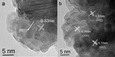 | ||
| Fig. 3 The HRTEM images recorded (a) on the tip of CdS nanorods coated on the ZnO nanospheres, (b) on the shell of ZnO nanosphere/CdS nanoparticle structure. | ||
3.2 Sonochemical formation mechanisms
In recent years, ultrasonic irradiation has been extensively used in the synthesis of nanomaterials.24 The effects of high intensity ultrasound result primarily from acoustic cavitation:25 the formation, growth, and implosive collapse of bubbles in liquids. During the acoustic cavitation process, very high temperatures (>5000 K), pressures (>20 MPa), and cooling rates (>1010 K s−1) can be achieved upon the collapse of the bubble, which provides a unique platform for the growth of nanostructures including 0D nanoparticles,26 1D nanorods,27 to 2D nanoplates,27 and even mesoporous28 or hollow structures.29On account of the enhanced surface effects of the shock waves generated from ultrasound, a two-steps sonochemical route was developed to assemble ZnO/CdS core/shell nanostructures via a simple template method: Firstly, the ZnO nanospheres were prepared through a sonochemical route as templates; then, 1D CdS nanorods or 0D CdS nanoparticles were generated and attached on the ZnO templates after the surface nucleation and crystal growth processes.
During the sonochemical process in aqueous solution, the elevated temperatures and pressures inside the collapsing bubbles caused water to vaporize and further pyrolyze into H˙ and OH˙ radicals.30 In the present case, the sonochemical formation processes of the as-prepared nanostructures are probably related to the radical species generated from water molecules by the absorption of ultrasonic energy. The ZnO nanospheres are formed through the sonohydrolysis mechanism which have been formulated.31,22 The likely reaction step is shown as follow:
| Zn2+(aq) + H2O(l) → ZnO(s) + 2H+(aq) | (1) |
The formation of CdS nanorods or nanoparticles using different sulfur sources may be related with the different formation process and different release speed of S2−. In the system using thiourea as a sulfur source, thiourea may act as not only the sulfur source but also a bidentate ligand to form relatively stable Cd-thiourea complexes. The complex ions lead to a high remaining monomer concentration after the nucleation stage. Thus, a non-equilibrium growth for the elongated crystals is facilitated.32
So the possible growth mechanism of ZnO nanosphere/CdS nanorod may be described as follows. Firstly, the complex action between Cd2+ and thiourea leads to the formation of Cd–thiourea complexes, which prevent the production of a large number of free S2− in the solution, and will be favorable for the formation of the nanorods.33 Secondly, the Cd–thiourea complexes undergo a decomposition process under ultrasonic irradiation for a certain period of time to produce CdS nuclei. Owing to the slow release of reaction ions, the elongated growth along the [001] direction of rodlike crystals is favored.32 Thirdly, when there exists a supporter such as ZnO nanospheres, the sonochemically generated CdS clusters would be attached on its surface to form a composite nanomaterial with core/shell-type geometry.34 The formation process can be expressed as the following:
| Cd2+ + thiourea → [Cd(thiourea)2] 2+ → CdS(nanorod) | (2) |
| n(CdS) + ZnO → ZnO nanosphere/CdS nanorod | (3) |
While using TAA as a sulfur source, the reaction process follows a different mechanism. The whole process can be described as below:
| H2O ))) H˙ + OH˙ | (4) |
| H˙ + OH˙ + CH3CSNH2 → CH3C(NH2)(OH)-SH | (5) |
| H˙ + OH˙ + CH3C(NH2)(OH)-SH → CH3C(NH2)(OH)2 + H2S | (6) |
| CH3C(NH2)(OH)2 → CH3CO(NH2) + H2O | (7) |
| Cd2+ + H2S → CdS (nanoparticle) + H2O | (8) |
| n(CdS) + ZnO → ZnO nanosphere/CdS nanoparticle | (9) |
The quicker release of the intermediate gas leads to nucleation occurring at an outburst speed right after the reaction solution was irradiated, resulting in a large quantity of CdS nuclei and extremely low monomer concentration in the solution, so small dot-shaped product was obtained under this situation.35
3.3 PL study
Fig. 4a illustrates the PL spectrum of the spherical ZnO nanostructure under photon excitation of 325 nm. A strong emission at 581 nm in the yellow region, which can be attributed to intrinsic defects in ZnO as oxygen interstitials,36 dominates the PL spectrum. The PL spectrum of ZnO nanosphere/CdS nanorod composite (Fig. 4b) shows a strong and broad green emission peaked around 525 nm and a shoulder peak centered at 473 nm, which can be attributed to the defect states related emission band and the near band emission of CdS, respectively.37,38 The PL spectrum of ZnO nanosphere/CdS nanoparticle structure (Fig. 4c) shows a broad emission band centered at 540 nm, which can be attributed to the emission from the defect states, such as cadmium interstitials or sulfur vacancies in CdS nanoparticles.38 Compared with the above ZnO nanosphere/CdS nanorod composite, the emission intensity is reduced. We suggest that the reduced PL in the green region of CdS could be attributed to the quenching by interfacial charge transfer.20 We also assume that rodlike crystals on the surface would possess more defects due to the faster 1D crystal growth. These results could indicate that luminescence properties of the semiconductors are very sensitive to their structures and strongly dependent on the surface state and structural defects. According to the structural characteristics of the ZnO/CdS composites, the emission band shift in the PL spectrum could be attributed to the interaction between the two semiconductors of ZnO and CdS. | ||
| Fig. 4 PL spectra of (a) ZnO nanospheres, (b) ZnO nanosphere/CdS nanorod composite and (c) ZnO nanosphere/CdS nanoparticle composite. | ||
3.4 Electrogenerated chemiluminescence study
ECL is generated by relaxation of excited-state molecules that are produced through electron-transfer annihilation of electrogenerated anion and cation radicals.39 As the electrode potential is made more negative, electrons are injected into the surface CdS nanocrystals and electrogenerated anion radicals (CdS˙−) are formed. The addition of co-reactants can help overcome either the limited potential window of a solvent or the poor stability of electrogenerated oxidized or reduced species.39 In our experiments, persulfate was used as the co-reactant. In this case, the reduction of persulfate produces a strong oxidant SO4−˙. This intermediate can react with the negatively charged CdS nanocrystals to produce excited states and then generate high-intensity light emission.| S2O82− + e− → SO42− + SO4−˙ | (10) |
| CdS−˙ + SO4−˙ → CdS* + SO42− | (11) |
| CdS* → CdS + hv | (12) |
Fig. 5 shows the ECL emission of ZnO nanospheres, ZnO nanosphere/CdS nanorod and ZnO nanosphere/CdS nanoparticle in 0.1 M KOH and 0.1 M K2S2O8 aqueous solution containing 0.1 M KCl as the supporting electrolyte. The electrode potential was cycled between 0.1 and −1.5 V at a scan rate of 100 mV s−1. The ECL light emissions of the crystals all show quite good stability and enhanced intensity. As shown in Fig. 5, the ECL intensities of the two ZnO/CdS composites are both higher than that of pure ZnO nanospheres, which imply that surface coating could enhance the ECL intensity of ZnO nanospheres. It is interesting that the ECL intensity of ZnO nanosphere/CdS nanorod composite is higher than that of ZnO nanosphere/CdS nanoparticle composite. As proved previously, the ECL emission is much more sensitive to their surface electronic structure than the PL, and is dependent on the surface properties and the presence of surface defects.39,40 We assume that the ZnO nanosphere/CdS nanorod structure would possess more defects due to the faster crystal formation which might result in higher ECL intensity. These results could indicate that ECL properties are closely related to the structure of these nanocrystals. Therefore, more detailed investigation on ECL of these prepared core/shell semiconductor nanocrystals to find the relationship between structure and ECL emission is urgently needed and still in progress. The optical measurement conditions need to be optimized in further work to improve the stability of ECL signals.
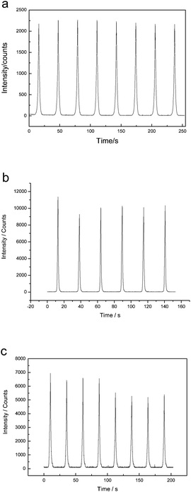 | ||
| Fig. 5 ECL emission from (a) pure ZnO nanospheres, (b) ZnO nanosphere/CdS nanorod and (c) ZnO nanosphere/CdS nanoparticle in 0.1 M K2S2O8 + 0.1 M KOH + 0.1 M KCl aqueous solution with potential cycles between 0.1 V and −1.5 V (scan rate: 100 mV s−1). | ||
4. Conclusions
In summary, the core/shell-type ZnO nanosphere/CdS nanorod and ZnO nanosphere/CdS nanoparticle composites have been successfully synthesized via a facile sonochemical route. By using ZnO nanospheres as template, core/shell nanostructures have been constituted. Different sulfur sources used in reaction systems resulted in different surface coating statuses. The formation mechanism of these nanocrystals is connected with the sonochemical effect of ultrasonic irradiation. This method might also be of potential to synthesize other spherical nanostructured functional materials by reacting with appropriate compounds. The PL spectra of the core/shell nanostructures displayed enhanced green emission. The ECL properties of these semiconductor core/shell nanostructures were researched, for the first time, and the experimental results showed that these composites had enhanced ECL intensity compared with pure ZnO nanospheres which shows surface coating may result in interesting photonic properties.Acknowledgements
This work is supported by the National Natural Science Foundation of China (grant no. 20635020 and 20805022). We thank Bo Liu and Guifen Jie for their kind help.References
- (a) A. P. Alivisatos, Science, 1996, 271, 933 CrossRef CAS; (b) F. Caruso, Adv. Mater., 2001, 13, 11 CrossRef CAS.
- R. A. Caruso and M. Antonietti, Chem. Mater., 2001, 13, 3272 CrossRef CAS.
- S. Singh, K. Bozhilov and A. Mulchandani, Chem. Commun., 2010, 46, 1473 RSC.
- T. J. Zhou, M. H. Lu and Z. H. Zhang, Adv. Mater., 2010, 22, 403 CrossRef CAS.
- S. Q. Liu, Y. X. Li, M. J. Xie, X. F. Guo, W. J. Ji and W. P. Ding, Mater. Lett., 2010, 64, 402 CrossRef CAS.
- M. Cargnello, N. L. Wieder and T. Montini, J. Am. Chem. Soc., 2010, 132, 1402 CrossRef CAS.
- R. Mishra, R. S. Yadav, A. C. Pandey, S. S. Sanjay and C. Dar, J. Lumin., 2010, 130, 365 CrossRef CAS.
- L. H. Jing, C. H. Yang, R. R. Qiao, M. Niu, M. H. Du, D. Y. Wang and M. Y. Gao, Chem. Mater., 2010, 22, 420 CrossRef CAS.
- Y. He, H. T. Lu, L. M. Sai, Y. Y. Su, M. Hu, C. H. Fan, W. Huang and L. H. Wang, Adv. Mater., 2008, 20, 3416 CrossRef CAS.
- A. Purkayastha, Q. Y. Yan and M. S. Raghuveer, Adv. Mater., 2008, 20, 2679 CrossRef CAS.
- Z. W. Pan, Z. R. Dai and Z. L. Wang, Science, 2001, 291, 1947 CrossRef CAS.
- J. C. Johnson, H. Q. Yan, R. D. Schaller, L. H. Haber, R. J. Saykally and P. D. Yang, J. Phys. Chem. B, 2001, 105, 11387 CrossRef CAS.
- R. F. Service, Science, 1997, 276, 895 CrossRef CAS.
- W. L. Wang, Mater. Today, 2004, 7, 26 CrossRef.
- L. E. Brus, J. Chem. Phys., 1983, 79, 5566 CrossRef CAS.
- Y. Tak, S. J. Hong, J. S. Lee and K. Yong, Cryst. Growth Des., 2009, 9, 2627 CrossRef CAS.
- T. Gao, Q. H. Li and T. H. Wang, Chem. Mater., 2005, 17, 887 CrossRef CAS.
- Y. Zhang, T. F Xie, T. F Jiang, X. Wei, S. Pang, X. Wang and D. J. Wang, Nanotechnology, 2009, 20, 155707 CrossRef.
- W. Lee, S. K. Min, V. Dhas, S. B. Ogale and S. H. Han, Electrochem. Commun., 2009, 11, 103 CrossRef CAS.
- F. Fang, D. X. Zhao, B. H. Li, Z. Z. Zhang, J. Y. Zhang and D. Z. Shen, Appl. Phys. Lett., 2008, 93, 233115 CrossRef.
- L. Irimpan, V. P. N. Nampoori and P. Radhakrishnan, J. Appl. Phys., 2008, 103, 094914 CrossRef.
- J. Geng, B. Liu, L. Xu, F. N. Hu and J. J. Zhu, Langmuir, 2007, 23, 10286 CrossRef CAS.
- T. Ren, J. Xu, Y. Tu, S. Xu and J. J. Zhu, Electrochem. Commun., 2005, 7, 5 CrossRef CAS.
- (a) M. M. Mdleleni, T. Hyeon and K. S. Suslick, J. Am. Chem. Soc., 1998, 120, 6189 CrossRef CAS; (b) A. Gedanken, Ultrason. Sonochem., 2004, 11, 47 CrossRef CAS.
- K. S. Suslick, S. B. Choe, A. A. Cichowlas and M. W. Grinstaff, Nature, 1991, 353, 414 CrossRef CAS.
- A. Gedanken, Ultrason. Sonochem., 2004, 11, 47 CrossRef CAS.
- J. Geng, W. H. Hou, Y. N. Lv, J. J. Zhu and H. Y. Chen, Inorg. Chem., 2005, 44, 8503 CrossRef CAS.
- R. K. Rana, Y. Mastai and A. Gedanken, Adv. Mater., 2002, 14, 1414 CrossRef CAS.
- J. Geng, J. J. Zhu, D. J. Lu and H. Y. Chen, Inorg. Chem., 2006, 45, 8403 CrossRef CAS.
- K. S. Suslick, Ultrasound: Its Chemical, Physical and Biological Effects; VCH: Weinhein, Germany, 1988 Search PubMed.
- R. V. Kumar, Y. Diamant and A. Gedanken, Chem. Mater., 2000, 12, 2301 CrossRef CAS.
- (a) Z. L. Xiao, C. Y. Han, W. K. Kwok, H. H. Wang, U. Welp, J. Wang and G. W. Crabtree, J. Am. Chem. Soc., 2004, 126, 2316 CrossRef CAS; (b) X. J. Zhang, Q. R. Zhao, Y. P. Tian and Y. Xie, Cryst. Growth Des., 2004, 4, 355 CrossRef CAS.
- X. H. Liao, H. Wang, J. J. Zhu and H. Y. Chen, Mater. Res. Bull., 2001, 36, 2339 CrossRef CAS.
- (a) T. Gao and T. H. Wang, Chem. Commun., 2004, 2558 RSC; (b) N. A. Dhas and A. Gedanken, Appl. Phys. Lett., 1998, 72, 2514 CrossRef.
- (a) X. G. Peng, Adv. Mater., 2003, 15, 459 CrossRef CAS; (b) J. Geng, J. J. Zhu and H. Y. Chen, Cryst. Growth Des., 2006, 6, 321 CrossRef CAS.
- (a) P. Yang, H. Yan, S. Mao, R. Russo, J. Johnson, R. Saykally, N. Morris, J. Pham, R. He and H. J. Choi, Adv. Funct. Mater., 2002, 12, 319 CrossRef; (b) J. J. Wu and S. C. Liu, Adv. Mater., 2002, 14, 215 CrossRef CAS; (c) Y. B. Li, Y. Bando and D. Golberg, Appl. Phys. Lett., 2004, 84, 3603 CrossRef CAS.
- M. Agata, H. Kurase and S. Hayashi, Solid State Commun., 1990, 76, 1061 CrossRef CAS.
- N. A. Dhas and A. Gedanken, Appl. Phys. Lett., 1998, 72, 2514 CrossRef.
- Z. Ding, B. M. Quinn, S. K. Haram, L. E. Pell, B. A. Korgel and A. J. Bard, Science, 2002, 296, 1293 CrossRef CAS.
- (a) N. Myung, Z. Ding and A. J. Bard, Nano Lett., 2002, 2, 1315 CrossRef CAS; (b) N. Myung, Y. Bae and A. J. Bard, Nano Lett., 2003, 3, 1053 CrossRef CAS.
| This journal is © The Royal Society of Chemistry 2011 |