In situ observations of fullerene fusion and ejection in carbon nanotubes†
Sandeep
Gorantla
*a,
Felix
Börrnert
a,
Alicja
Bachmatiuk
a,
Maria
Dimitrakopoulou
a,
Ronny
Schönfelder
a,
Franziska
Schäffel
b,
Jürgen
Thomas
a,
Thomas
Gemming
a,
Ewa
Borowiak-Palen
c,
Jamie H.
Warner
b,
Boris I.
Yakobson
d,
Jürgen
Eckert
ae,
Bernd
Büchner
a and
Mark H.
Rümmeli
*ae
aIFW Dresden, P.O. Box 270116, Dresden, D-01171, Germany. E-mail: s.gorantla@ifw-dresden.de; m.rümmeli@ifw-dresden.de
bDepartment of Materials, University of Oxford, Parks Rd, Oxford, OX1 3PH, UK
cInstistute of Chemical Engineering and Chemical Technology, West Pomeranian University of Technology, Pulaskiego St. 10, 70 322, Szczecin, Poland
dRichard E. Smalley Inst. for Nanoscale Science and Technology and Dept. of Mechanical Engineering and Materials Science, Rice University, Houston, Texas 77005-1892, USA
eTechnische Universität Dresden, Dresden, D-01062, Germany
First published on 16th August 2010
Abstract
We present insitu experimental observations of fullerenes seamlessly fusing to single-walled carbon nanotubes. The morphing-entry of a fullerene to the interior of a nanotube is also captured. The confined (1D) motion of the newly-encapsulated fullerene within its host attests to the actual change from the exterior to interior.
Synthesizing new hybrid nanostructures at the molecular level by combining single-walled carbon nanotubes (SWNTs) and C60 fullerenes is not only fascinating but is also essential to advancing our understanding for the formation of multicomponent hybrid nanostructures with unusual morphologies. These nanostructures may play a promising role in the development of future molecular nanoelectronic devices.1,2 The pioneering work on the encapsulation of C60 molecules inside SWNTs forming so called peapods, by Smith et al. marked the beginning of carbon nanotube based hybrid molecular structures.3 More recently, fullerenes have been covalently attached to the outer sidewalls of SWNT via oxygen bridges (nanobuds).4 Despite renewed theoretical interest in studying the properties of these hybrid carbon nanostructures,5 there has been limited experimental progress in fusing fullerenes to SWNT. While, there have been numerous works on electron beam irradiation driven manipulation and engineering of carbon nanostructures,6 as far as we are aware, there are no reports on the direct seamless fusing of a fullerene to a SWNT outer wall. This is mainly due to knock-on damage being incurred when using electron accelerating voltages above 86 kV.7 This effect is worsened in the presence of impurities.8 Theoretically, Zhao et al. have shown a fullerene can fuse to a SWNT wall (without bridging atoms) through Stone-Wales bond rotation paths. Moreover they showed that this process can go further with the fullerene re-emerging on the other side of the wall, viz. a fullerene can penetrate a SWNT.9
In this manuscript we present in situ low voltage high resolution transmission electron microscopy (HREM) observations, experimentally confirming the formation of fused fullerenes with a nanotube sidewall, forming a hump. Moreover, we also observe the ejection of a fullerene within a SWNT from a defect hump-like structure. In this study, the TEM was operated using an 80 kV accelerating voltage. TEM investigations of our annealed peapod samples revealed a variety of structures. Typically the sample contained many bundles of tubes. Some of the nanotubes have inner tubes (e.g. Fig. S1a of the ESI†) formed from coalesced fullerenes, similar to previous reports.10 In addition, we noticed a relatively large number of tubes had hump-like structures on their surface (Fig. S1a and b). We refer to these structures as humped carbon nanotubes (hSWNT). Remarkably, we also observed fullerenes interact with the outer wall of some of the SWNT and hSWNT. One such example is presented in Fig. 1 (and movie S1†). In this in situ study we observed a fullerene dock to the surface of a double-walled carbon nanotube (DWNT) with a short inner tube. The chirality of the host SWNT is determined to be [19,3] and the diameter of the fullerene is 0.69 ± 0.03 nm which corresponds to a C60 structure. The inner tube diameter varies over its length. Once the fullerene attaches to the surface of the DWNT (Fig. 1a) it apparently sticks to the outer tube surface. It is not clear what the basis for the initial docking mechanism is, since the outer wall appears unbroken at this stage. Perhaps a defect site aids this process, since previous studies of ours suggest possible attractive forces such as van der Waals and Casimir effects from defect free tubes are insufficient to trap fullerenes.11 It is worth noting that the knock-on damage threshold for fullerenes is 40 kV.12 Hence, electron-beam irradiation induced defects on fullerenes, may also aid in docking to SWNT. The inner tube is then observed to oscillate to and fro within the outer tube. This oscillation process allows it to dissipate energy gained from the electron beam irradiation.6 As the inner tube moves past the region in which the fullerene is trapped, a point in time is reached in which the inner tube distorts at this location. The tube wall apparently opens and closes several times and the fullerene structure also distorts during this process. Fig. 1b presents an image of this interactive progression. Greater detail can be obtained by watching the movie S1.† By frame c, the inner tube has moved fully away from the region of interest and the fullerene has now seamlessly fused with the tube. The excess carbon from the wall opening to the hump may have gone to the hump structure, the inner tube or both. However, the size of the hump is enlarged relative to the initial fullerene making it likely that at least some of the excess carbon transferred to the hump. The observed welding of a spherical fullerene to a carbon nanotube to form a hump has been examined in various theoretical works.9,13 Our observations confirm the fusing process experimentally. We also observed fullerenes interact around humps. An example is provided in Fig. S2 of the ESI. Here a fullerene moves around the hump (see movie S2†) and so differs from the previous example in which the entrapped fullerene remained stationary on the surface. A previous study of ours showed enhanced π–π interactions between a bent tube and fullerene is able to trap the fullerene while allowing some motion to occur under the electron beam irradiation.11 This same argument can be applied to explain the dancing motion of the fullerene around the hump. With continued irradiation (12 min) the fullerene is seen to fuse into the hump resulting in an elongated hump (cf. Fig. 4 in ref. 13). With yet further electron irradiation (16 min) the elongated hump restructures into a broader and flatter hump. Zhao et al., have shown that the flattening of the hump reduces the total energy. The total energy can be further reduced through deformation or defects in the wall.9 Their study not only showed that a fullerene could fuse to the exterior of a tube wall and form a hump structure, but that the topological versatility of the Stone–Wales bond rotations involved could ultimately allow the fullerene to be released in the interior of the tube. Fig. 2 (and movie S3) show remarkable in situ observations of a defect structure in the tube wall re-arranging and releasing a fullerene into the interior of the tube in the process. Fig. 2a1 shows a SWNT with a ring-like feature at the centre of the tube. Its chirality is determined to be [19,1]. The ring probably corresponds to a large hump as viewed from the top and is supported by an image simulation (Fig. 2a2). The hump is larger than that formed by a single fullerene and this is easily observed by comparing Fig. 2a1–a3, which is an image simulation of a hump formed from a single C60 fullerene.9Fig. 2a4 shows an end view of a hump on a SWNT to aid visualization. After 8 mins of irradiation the ring like structure starts to alter forming a lump on the right side of the SWNT and two small crest-like shapes can be observed in-between the tube walls. These shapes are very similar to image simulations made from the theoretical predictions of Zhao et al. (Fig. 2b3), and can be attributed to concavity formation (see Fig. 2b4), which occurs to minimize energy. With continued electron beam irradiation the concavity formation deepens forming an inverted hump (viz. the hump now resides within the tube – Fig. 3 c1–c4). The inverted hump has a large strain, which can be released by necking-off, ultimately leading to a nested fullerene inside the tube (Figs. d1–d4). In our in situ study, after the ejection of a fullerene, there remains a smaller hump on the left wall of the host tube forming a bulbous region. The measured diameter of the newly formed nested-fullerene is 0.78 ± 0.03 nm, this corresponds closely to a C80 fullerene with Ih symmetry.14 The fullerene then oscillates within the confines of the bulbous region before suddenly shooting further up the SWNT (Fig. 3 and movie S3). The oscillating behavior of the fullerene within the hump region and its confined one dimensional motion up the core of the tube verify the fullerene is indeed nested within the tube. The bulbous region in which the fullerene initially oscillates is in essence a defect area that can trap a fullerene.11 None the less the fullerene is able to escape. The process of escaping and shooting up the tube allows the fullerene to release energy supplied by the electron beam. As it shoots up the tube it encounters another defect area which again traps it.
![(a)–(c) In situ HREM observations of covalent fusing of a C60 molecule with the sidewall of a DWNT (a′)–(c′) Simulated HREM images of hybrid nanostructure comprising [19,3] SWNT and C60 fullerene, corresponding to experimentally observed interaction dynamics. The scale bar is 2 nm.](/image/article/2010/NR/c0nr00426j/c0nr00426j-f1.gif) | ||
| Fig. 1 (a)–(c) In situ HREM observations of covalent fusing of a C60 molecule with the sidewall of a DWNT (a′)–(c′) Simulated HREM images of hybrid nanostructure comprising [19,3] SWNT and C60 fullerene, corresponding to experimentally observed interaction dynamics. The scale bar is 2 nm. | ||
![(a1)–(d1) In situ HREM observations of a fullerene ejection in the host SWNT, through penetration and necking-off of a hump. (a2)–(d2) Simulated HREM images of supercell structures comprising [19,1] SWNT and hump. (a3)–(d3) Simulated HREM images of theoretically calculated penetration structures of [10,10] SWNT and C60 fullerene show close resemblance with the contrast observed in our experimental images, (a4)–(d4) stick-and-ball structure models along the end view of corresponding theoretically calculated structures. The scale bar is 2 nm.](/image/article/2010/NR/c0nr00426j/c0nr00426j-f2.gif) | ||
| Fig. 2 (a1)–(d1) In situ HREM observations of a fullerene ejection in the host SWNT, through penetration and necking-off of a hump. (a2)–(d2) Simulated HREM images of supercell structures comprising [19,1] SWNT and hump. (a3)–(d3) Simulated HREM images of theoretically calculated penetration structures of [10,10] SWNT and C60 fullerene show close resemblance with the contrast observed in our experimental images, (a4)–(d4) stick-and-ball structure models along the end view of corresponding theoretically calculated structures. The scale bar is 2 nm. | ||
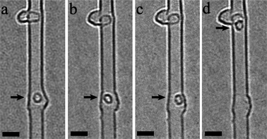 | ||
| Fig. 3 In situ HREM observations of confined motion of a fullerene after ejection. The scale bar is 2 nm. | ||
Our observations bear strong similarities to the theoretically predicted endocytosis process described by Zhao et al.9 In this process, they argue that despite the overall barrier being relatively high (12 eV), the endocytotic entry of a fullerene can be reduced by bending deformations. This can be facilitated by defects and/or, as in our case, electron irradiation. Moreover, estimates show that the final state energy (fullerene within a tube) to be lower than the initial (hump) energy because of the stronger van der Waals attraction within the tube.15 This, they argue, is the overall driving force for the concavity formation and ultimately the release of a fullerene within the tube. Since the 80 kV beam used in this study is above the knock-on threshold for fullerenes, as mentioned above, it is reasonable to anticipate the humps may also be susceptable to knock-on effects which may aid their restructuring. Another potential mechanism which can help fusion may be localized heating. Studies by Warner et al. suggest localized temperatures can reach 1100–1300 K.6
In summary, experimental in situ low voltage HREM observations showing the seamless fusion of a fullerene to carbon nanotubes forming a hump are shown. In addition, our studies show a defect-hump structure maintained under continued electron irradiation to re-structure. The restructuring process leads to the release of a fullerene leaving a small hump remaining in the tube wall. The released buckyball then shuttles up the tube with confined 1D motion, confirming it is nested within the tube. Our in situ morphing-entry observation of a fullerene into a nanotube, strongly resembles previous theoretical predictions.
Acknowledgements
SG acknowledges the “Pakt für Forschung und Innovation”, FB thanks the DFG RU (1540/8-1), AB and FS thank the Alexander von Humboldt Foundation, MD the DAAD and MHR the EU (ECEMP) and the Freistaat Sachsen.References
- C. Joachim, J. K. Gimzewski and A. Aviram, Nature, 2000, 408, 541 CrossRef CAS.
- K. Moth-Poulsen and T. Bjornholm, Nat. Nanotechnol., 2009, 4, 551 CrossRef CAS.
- B. W. Smith, M. Monthioux and D. E. Luzzi, Nature, 1998, 396, 323 CrossRef CAS.
- A. G. Nasibulin, P. V. Pikhitsa, H. Jiang, D. P. Brown, A. V. Krasheninnikov, A. S. Anisomov, P. Queipo, A. Moisala, D. Gonzalez, G. Lientschnig, A. Hassanien, S. D. Shandakov, G. Lolli, D. E. Resasco, M. Choi, D. Tomanek and E. I. Kauppinen, Nat. Nanotechnol., 2007, 2, 156 CrossRef CAS.
- T. Meng, C. Y. Wang and S. Y. Wang, Phys. Rev. B: Condens. Matter Mater. Phys., 2008, 77, 033415 CrossRef; X. Wu and X. C. Zeng, Nano Lett., 2009, 9, 250 CrossRef CAS; X. Zhu and H. Su, Phys. Rev. B: Condens. Matter Mater. Phys., 2009, 79, 165401 CrossRef.
- F. Banhart, J. Li and M. Terrones, Small, 2005, 1, 953 CrossRef CAS; J. H. Warner, Y. Ito, M. H. Rümmeli, T. Gemming, B. Büchner, H. Shinohara and G. A. D. Briggs, Phys. Rev. Lett., 2009, 102, 195504 CrossRef; M. S. Wang, Y. Bando, J. A. R. Manzo, F. Banhart and D. Golberg, ACS Nano, 2009, 3, 2632 CrossRef CAS; J. H. Warner, Y. Ito, M. H. Rümmeli, B. Büchner, H. Shinohara and G. A. D. Briggs, ACS Nano, 2009, 3, 3037 CrossRef CAS.
- B. W. Smith and D. E. Luzzi, J. Appl. Phys., 2001, 90, 3509 CrossRef CAS.
- J. H. Warner, F. Schäffel, G. Zhong, M. H. Rümmeli, B. Büchner, J. Robertson and G. A. D. Briggs, ACS Nano, 2009, 3, 1557 CrossRef CAS.
- Y. Zhao, Y. Lin and B. I. Yakobson, Phys. Rev. B: Condens. Matter Mater. Phys., 2003, 68, 233403 CrossRef.
- S. Bandow, M. Takizawa, K. Hirahara, M. Yudasaka and S. Iijima, Chem. Phys. Lett., 2001, 337, 48 CrossRef CAS; E. Hernández, V. Meunier, B. W. Smith, R. Rurali, H. Terrones, M. Buongiorno Nardelli, M. Terrones, D. E. Luzzi and J.-C. Charlier, Nano Lett., 2003, 3, 1037 CrossRef CAS.
- S. Gorantla, S. Avdoshenko, F. Börrnert, A. Bachmatiuk, M. Dimitrakopoulou, F. Schäffel, R. Schönfelder, J. Thomas, T. Gemming, J. H. Warner, G. Cuniberti, J. Eckert, B. Büchner and M. H. Rümmeli, Nano Res., 2010, 3, 92 Search PubMed.
- T. Füller and F. Banhart, Chem. Phys. Lett., 1996, 254, 372 CrossRef.
- Y. Zhao, B. I. Yakobson and R. E. Smalley, Phys. Rev. Lett., 2002, 88, 185501 CrossRef.
- M. S. Dresselhaus, G. Dresselhaus and P. C. Eklund, in Science of Fullerenes and Carbon Nanotubes, Academic Press, New York, 1996, vol. 1, ch. 3, pp. 70 Search PubMed.
- L. A. Girifalco, M. Hodak and R. S. Lee, Phys. Rev. B: Condens. Matter Mater. Phys., 2000, 62, 13104 CrossRef CAS.
Footnote |
| † Electronic supplementary information (ESI) available: Experimental details and videos. See DOI: 10.1039/c0nr00426j |
| This journal is © The Royal Society of Chemistry 2010 |