Fabrication of micropatterns of nanoarrays on a polymeric gel surface
Peng
Liu
,
Jianguo
Sun
,
Jinghuan
Huang
,
Rong
Peng
,
Jian
Tang
and
Jiandong
Ding
*
Key Laboratory of Molecular Engineering of Polymers of Ministry of Education, Department of Macromolecular Science, Laboratory of Advanced Materials, Fudan University, Shanghai 200433, China. E-mail: jdding1@fudan.edu.cn; Fax: +86-21-65640293; Tel: +86-21-65643506
First published on 30th September 2009
Abstract
Micro–nano patterns of gold on the surface of poly(ethylene glycol) (PEG) hydrogels were prepared. The approach combines the technique of conventional photolithography (a top-down method for micropatterns), block copolymer micelle nanolithography (a bottom-up method for gold nanopatterns), and a linker-assistant technique to transfer a pattern on a hard surface to a polymeric surface. Hybrid micro–nano patterns on hydrogels were characterized using scanning electron microscopy, atomic force microscopy and X-ray photoelectron spectroscopy. The patterned Au nanoparticles were further modified by a peptide containing arginine-glycine-aspatate (RGD). The cell-adhesion contrast of the patterned hydrogel surface was confirmed by preliminary cell experiments.
Introduction
Cell–material interaction is an important issue in tissue engineering and biomaterials.1–3 Surface patterning offers an attractive way to precisely manipulate chemical composition and topographical properties of surface microenvironments to mimic the extracellular matrix (ECM) for investigation of cell–material interactions.4–22 Topographic micropatterns such as ridges and grooves have, for a long time, been used to alter cell alignment.4–7 Chemical micropatterns with a cell-adhesion contrast have experienced a rapid growth in recent years to control the position, shape and orientation of cells.8–13Despite the encouraging advances made with micropatterning techniques, patterns merely on a microscale are insufficient to mimic the ECM. For example, integrins are receptors that link ligands in the ECM with some specific peptide sequences such as arginine-glycine-aspatate (RGD). These transmembrane receptors mediate cellular interactions with the underlying ECM and form adhesion sites that play an important role in governing many aspects of cellular behavior such as focal adhesion. An integrin is about 10–12 nm in size and faces the extracellular side.23,24 So, a surface pattern with ligands at such a nanoscale is very helpful for understanding cell–ECM interactions. Some recent research has shifted to surface nanopatterning for further insights into cell behavior.14–22
Although nanopatterning is more powerful for manipulating extracellular signaling molecules to investigate cell responses at a molecular or supermolecular level, micropatterning is still meaningful for controlling the spatial distribution of a single cell or a group of cells. Both micropatterning and nanopatterning have played a critical role in discovering interactions between cells and their supporting surface. To date, various patterning techniques on substrates have been set up, not limited to the field of biomaterials.25 Fabrication of micropatterns mostly relies on conventional photolithography,26 which serves as a typical top-down approach. As far as surface nanopatterning is concerned, both top-down and bottom-up techniques have been developed such as advanced ray lithography,26–28nanoimprint lithography,29dip-pen nanolithography,30 molecular self-assembly,31–35colloidal crystal templating36 and so on.37,38 For preparation of a pattern with nanodots less than 20 nm in diameter, block copolymer micelle nanolithography presents a unique way of patterning metal nanoparticles.39–41
In contrast to the many studies of preparation techniques for either metal nanopatterns or micropatterns, few reports concern fabrication of hybrid micro–nano patterns of metals,42–44 and the so-far micropatterning of metal nanostructures is limited to inorganic substrates, such as silicon. The present paper will report a micro–nano pattern on a polymeric hydrogel . As a demonstration, a gold micro–nano pattern on the surface of crosslinked poly(ethylene glycol) (PEG) hydrogel will be prepared.
Why hydrogels ? As a biomimetic soft matter and wet material, hydrogels have been a rich topic in material science during the latest decade.45–52 It is facile to adjust the mechanical properties of hydrogels to several orders of magnitude,53 and the substrate rigidity has recently been found to even serve as a strong biological signal to cells.54,55 Meanwhile PEG molecules are well known for their bio-fouling resistance and are often used to passivate background to resist cell adhesion.8–11PEGhydrogels are more stable than self-assembly of PEG molecules and offer long-term cell resistance.
To the best of our knowledge, a micropattern of a regular metal nanoarray on a polymer surface has never been reported due to some “inherent” difficulties. For instance, plasma treatment, which is used in the fabrication of a metal nanoarrays viablock copolymer micelle nanolithograthy, may destroy the corresponding polymeric substrate. As the first paper in our series of fundamental research, this paper is focused upon the fabrication technique of a micro–nano pattern of a metal fixed on the surface of a polymeric hydrogel . Combination of bottom-up, top-down, and transfer techniques will be put forward to solve this material problem. Our fabrication approach is schematically presented in Fig. 1. The nanostructured micropatterns of gold on PEGhydrogels is prepared in a three-stage process: first, a conventional photolithography technique is used to generate a frame of a micropattern; second, the block copolymer micelle nanolithography technique and the lift-off technique are combined to further generate micropatterns of gold nanostructures on a hard substrate; third, a linker-assistant technique is employed to transfer the micro–nano pattern of gold on the hard substrate to a polymeric gel surface. The micro–nano pattern of gold will then be modified with an RGDpeptidevia the thiol group before cell culture.
 | ||
| Fig. 1 Schematic presentation of the fabrication of a micro–nano pattern of gold on surface of a PEGhydrogel . (a) With standard photolithography, micropatterns of photoresist are first formed on a piece of glass. (b) Subsequently, the monolayer of block copolymers (PS-b-P2VP) micelles loaded with gold precursor is dip-coated onto glass surfaces. After lift-off, an oxygen plasma treatment is performed, and consequently, gold nanoparticles are reduced and deposited in desired micro-islands. (c) In the following transfer nanolithography step, gold nanoparticles are modified by the linker, propene thiol, which has a thiol end-group and a double-bond end-group. Then, poly(ethylene glycol) diacrylate (PEGDA) macromers are pre-coated and photopolymerized. The linkers are reacted with the gold via Au–S bonds and with the PEG network via joining polymerization. After peel-off, Au nanostructures on PEGhydrogels with the designed micropatterns are obtained; gold nanoparticles were further functionalized with a RGD-thiol reagent. | ||
Results and discussion
Fabrication of micro–nano patterns of gold on PEGhydrogels
When polystyrene-block-poly(2-vinylpyridine) (PS-b-P2VP) was dissolved in toluene, spherical reverse micelles were formed with the hydrophilic P2VP blocks forming cores and the lipophilic PS blocks forming coronae. Next, a metal salt (HAuCl4) was added to the suspension. The gold precursors migrated into the cores of the micelles due to their lipophobic character. In order to group the micelle arrays into a designed micropattern, the standard photolithography was first used to produce micropatterns of photoresist on a solid substrate. The thickness of photoresist was controlled in the range 30–100 nm to alleviate aggregation of micelles in the boundary of micro-islands. PS-b-P2VP micelles loaded with gold precursors were deposited on glass with a pre-designed micropattern of photoresist by dip-coating. After lift-off of the photoresist by acetone, an oxygen plasma was then used to remove the remaining block copolymers and meanwhile reduce the gold precursors in the micellar cores to gold nanoparticles. Thus micropatterns of gold nanostructures on glass were obtained.A transfer technique was further introduced to convey micropatterns of gold nanostructures from a solid substrate to a hydrogel . Propene thiol was used as the linker. Its thiol end-group was covalently bonded to the gold nanoparticles. Poly(ethylene glycol) diacrylate (PEGDA) macromers mixed with photoinitiators were coated, and then irradiated with UV light. After the PEGDAs were cross-linked and reacted with propene thiol, the gel was separated from the solid substrate. As a result, micropatterns of gold nanostructures on a hydrogel surface were obtained.
The resultant micro–nano patterns of gold are shown in Fig. 2. The diameter of the micro-islands was about 80 μm and the interval between two neighboring micro-islands was about 100 μm, which was determined mainly by the mask used in the photolithography. Gold nanoparticles were arranged hexagonally as preservation of the pseudo-hexagonal order of the original micelles. Fig. 2c shows a field-emission scanning electron microscopy (FE-SEM) image of a micropattern of gold nanostructures on a hydrogel surface. The size and distribution of the micropattern in Fig. 2c were reminiscent of those on the solid substrate in Fig. 2a. Fig. 2d illustrates that gold nanoparticles still remain on the gel surface after transfer from glass.
![FE-SEM images of micro–nano patterns of gold prepared on a piece of glass [(a) and (b)] and transferred to a PEGhydrogel [(c) and (d)]. (a) and (c) are low-magnification images showing micropatterns, and (b) and (d), are high-magnification images of one micro-island showing nanopatterns.](/image/article/2010/NR/b9nr00124g/b9nr00124g-f2.gif) | ||
| Fig. 2 FE-SEM images of micro–nano patterns of gold prepared on a piece of glass [(a) and (b)] and transferred to a PEGhydrogel [(c) and (d)]. (a) and (c) are low-magnification images showing micropatterns, and (b) and (d), are high-magnification images of one micro-island showing nanopatterns. | ||
The hexagonal arrangement of gold nanodots is further demonstrated by atomic force microscopy (AFM) imaging, as shown in Fig. 3. Although not perfectly ordered, the nanoparticles exhibited a high degree of short-range hexagonal order as evident from the corresponding autocorrelation function (inset in Fig. 3a). The height of the gold nanodots in AFM measurements gave the sizes of the gold nanoparticles, which were around 9 nm. The distance between gold nanoparticles was controlled by the diblock copolymer micellar template, and the resultant period in this study was found to be about 55 nm. Such an interval is much larger than the nanoparticles themselves, and cannot be achieved simply by close-packing of gold nanoparticles. The linker molecule was necessary for the transfer technique, for only nanopits were produced on hydrogel surfaces without the employment of the linker (Fig. 3c). The AFM image of a gold nanopattern on a PEGhydrogel (Fig. 3d) indicates the success of the transfer technique.
 | ||
| Fig. 3 AFM images showing the nanostructures: (a) The gold nanopattern on glass. The inset in the top right corner shows the corresponding auto-correlation image; (b) a height profile along a line in (a); (c) nanopits on a hydrogel without the linker assistance in transferring from glass to hydrogel ; (d) gold nanodots on a hydrogel with the linker used in transfer. | ||
X-Ray photoelectron spectroscopy (XPS) analysis of the sample indicates the presence of gold in metallic Au0 state (Fig. 4), located at 84.3 eV and 88.0 eV for the 4f7/2 and 4f5/2 levels, respectively,39,40 despite the low amount of gold on the PEGhydrogel surface.
 | ||
| Fig. 4 XPS spectra of a PEGhydrogel (a) with and (b) without decoration of micro–nano patterns of gold. | ||
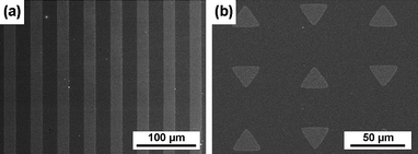
With changing the pre-designed photolithography mask, various micropatterns of gold nanoarrays on PEGhydrogel surface can be easily obtained. In Fig. 5a, bright microstripes correspond to the location of gold nanoparticles. The width of microstripes was about 20 μm and the space between two stripes was 25 μm. Fig. 5b shows a regular triangular micropattern (20 × 20 × 20 μm) of gold nanostructures on a PEGhydrogel surface.
 | ||
| Fig. 5 FE-SEM of (a) striped and (b) triangled micropatterns of nanostructured gold on PEGhydrogels . | ||
Cell adhesion on micro–nano patterns on hydrogels
A reagent containing a cyclic peptide and a thiol group is available to assemble on Au nanodots. The RGD sequence is recognized by integrin and can thus promote cell adhesion. An Au nanodot of about 9 nm diameter after functionalization with RGD provides a single anchor point to bind one integrin molecule, since the size of an integrin molecule in the cell membrane is about 10–12 nm.23,24Previous report showed that cells can not adhere well when RGD spacing is larger than a given value, say, 70 nm.20,22 In this experiment, the spacing of the Au nanodots is about 55 nm. Since RGD and PEG are available for, and resistant to, cell adhesion respectively, micro–nano hybrid patterns on a PEGhydrogel fabricated in our experiment present a surface with a significant cell adhesion contrast. Cell experiments confirmed that 3T3 fibroblasts adhered well on micro–nano patterns (Fig. 6). The viability of 3T3 fibroblasts on the micro-islands was also investigated using a commercially available fluorescence live/dead assay that stained live cells green and dead cells red. The result as shown in Fig. 6b indicates that most of cells were alive on the islands of the patterned substrate. 4,6-diamidino-2-phenylindole (DAPI) was employed to stain cell nuclei, which were clearly visualized in Fig. 6c. Phallaoidin conjugated by tetramethyl rhodamine isothiocyanate (TRITC) was used to stain F-actin in the cytoskeleton, and Fig. 6d outlines the organization of actin filaments. All of these observations during the cell experiments confirm that cells can adhere and spread on such a micro–nano pattern of gold functionalized with RGD when the spacing between the nanodots is 55 nm. Meanwhile with assistance from the micropattern, we can control cell location in a micro-island with a diameter of 80 μm.
 | ||
| Fig. 6 Optical micrographs of cells on PEGhydrogels with micropatterns of gold nanoparticles functionalized with RGD. The micro-islands were, according to the mask in photolithography, of 80 μm diameter with 100 μm intervals. (a) A phase-contrast micrograph of 3T3 fibroblasts after 12 h in culture; (b) the corresponding fluorescence micrograph after viability/cytotoxicity staining, with the green and red colors indicating alive and dead cells respectively; (c) a fluorescence micrograph upon DAPI staining showing cell nuclei; (d) another fluorescence micrograph showing F-actins in the cytoskeleton. | ||
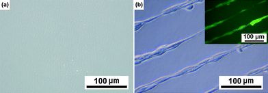
It should be indicated that the micro–nano patterns are themselves invisible in an optical microscope, as indicated in Fig. 7a without cell culture, but indirectly visualized after cell culture, as demonstrated in Fig. 6a–c and 7b. Taking advantage of our micro–nano hybrid patterns, both cell adhesion and orientation can be controlled. Hence, our present technique affords a possibility to extensively investigate cell–biomaterial interactions with multi-scale material features. The interval of nanodots could be tuned by block copolymer composition and dipping condition etc.,22 and it is well known to adjust the microscale surface feathers via selection of photolithography masks and adjust the macroscopic hydrogel rigidity via macromonomer concentration etc. It is also worth noting that due to chemical linking of gold nanoparticles on the polymeric surface, our micro–nano patterns of gold on PEGhydrogels is very stable in water and cell culture media for a long time (at least one month according to our observations).
 | ||
| Fig. 7 Optical micrographs of PEGhydrogels with micropatterns of gold nanoparticles functionalized with RGD. The microstripes were, according to the mask in photolithography, of 20 μm width and 50 μm intervals. (a) and (b) refer to a micro–nano patterned hydrogel without and with 3T3 fibroblasts cultured, respectively. The inset in the top right corner of (b) shows the corresponding fluorescence micrograph after viability/cytotoxicity staining. | ||
Conclusions
In conclusion, we have presented an approach to generate micro–nano patterns of gold on PEGhydrogels . The method combines photolithography, block copolymer micelle nanolithography and a transfer technique to fabricate hierarchical patterns on hydrogels . Those metallic nano- and microscale structures on hydrogels cannot be well prepared merely by the present nanofabrication or microfabrication methods. Cell experiments showed that this approach was biocompatible. Micro–nano hybrid patterns functionalized with RGD were used to control the density and distance of ligands. Such a hybrid pattern also manipulated cell location and orientation. The dependence of cell behavior upon the surface geometry on the micro- and nanoscales and upon substrate rigidity on the macroscopic scale will be investigated in the future. We reasonably expect that hybrid micro–nano patterns of metals on the surfaces of polymeric hydrogels might be very powerful for extensive researches in biomaterial and related fields, and then, fruitful results might be obtained via adjustment of the parameters of micropatterns, nanopatterns and substrate rigidity.Experimental
Materials used in experiments
Block copolymers of polystyrene and poly(2-vinylpyridine), PS-b-P2VP were from Polymer Source Inc. Canada. The molecular weights of PS and P2VP blocks were 52![[hair space]](https://www.rsc.org/images/entities/char_200a.gif) 400 and 28
400 and 28![[hair space]](https://www.rsc.org/images/entities/char_200a.gif) 100, respectively. The polydispersity index (Mw/Mn) was 1.07. The photoresist was purchased from Suzhou Ruihong Electronic Chemicals Co. Ltd. China (RZJ-304). PEGDA (Mw 700) and the photoinitiator 2-hydroxy-1-[4-(hydroxyethoxy)phenyl]-2-methl-1-propanone (D2959) were products of Aldrich Chemical Co. USA. Cyclo(Arg-Gly-Asp-(D)Phe-Lys)-(PEG)-COCH2CH2SH (c(RGDfK)-PEG-Mpa) (Mw 836.97) was obtained from Peptides International USA. Microscope glass cover slips were used after extensive and fresh cleaning.
100, respectively. The polydispersity index (Mw/Mn) was 1.07. The photoresist was purchased from Suzhou Ruihong Electronic Chemicals Co. Ltd. China (RZJ-304). PEGDA (Mw 700) and the photoinitiator 2-hydroxy-1-[4-(hydroxyethoxy)phenyl]-2-methl-1-propanone (D2959) were products of Aldrich Chemical Co. USA. Cyclo(Arg-Gly-Asp-(D)Phe-Lys)-(PEG)-COCH2CH2SH (c(RGDfK)-PEG-Mpa) (Mw 836.97) was obtained from Peptides International USA. Microscope glass cover slips were used after extensive and fresh cleaning.
Fabrication of micropatterns of photoresist on a solid substrate
Glass substrates were cut from larger substrates and immersed into H2SO4/30% H2O2 (3 : 1 vol.) for 30 min, washed for 10 min with Milli-Q water and for 5 min more with acetone, both in the ultrasonic bath. Micropatterns of photoresist (30–100 nm thick) on the substrates were fabricated by standard photolithography.Fabrication of micropatterns of gold nanostructures on glass
A solution (5 mg mL−1) of PS-b-P2VP was made by dissolving the polymer in 3 mL of anhydrous toluene. Micelles were thus formed with P2VP blocks as cores and PS blocks as coronae. After stirring for 5 h, the metal precursor (HAuCl4·4H2O) with a given molar ratio over pyridine unit in PS-b-P2VP (L = 0.5) was added and stirred for at least 24 h until the colloid became transparent, and the gold acid was accumulated in the polar P2VP cores. Substrates with micropatterns of photoresist were dip-coated with micelles loaded with gold precursors using a self-built pulling machine and dried by exposure to air. The photoresist was lifted off by acetone. The substrates were then treated by an oxygen plasma to reduce the metal precursors to gold nanoparticles and to remove polymers. The size of a Au nanoparticle was determined mainly by the loading amount of gold precursors in the corresponding micelle core. Finally, gold nanostructures were positioned in the micropattern on glass.Transfer of gold nanostructured micropatterns to the hydrogel surface
The micro–nano patterns of gold on glass were treated with the linker (propene thiol) for 1 h. PEGDA macromers mixed with photoinitiator were then coated and cross-linked by UV irradiation for 1 h. Separation of polymer from glass resulted in a hydrogel with its surface modified by micropatterns of gold nanostructures.Characterization of surface patterns
FE-SEM observations were carried out in a Hitachi S-4800 microscope at an acceleration voltage of 2 kV. AFM (Dimension 3100, Digital Instruments) with a silicon cantilever (40 N m−1) was used to image sample surfaces under ambient conditions in tapping mode. XPS measurements were carried out on a RBD upgraded PHI-5000C ESCA system (Perkin Elmer) using monochromatic Mg Kα X-rays at 1253.6 eV was operated at 250 W, and spectrum calibration was performed by taking the C 1s electron peak (BE = 284.6 eV) as the internal reference. The data were analyzed using the RBD AugerScan 3.21 software provided by RBD Enterprises.Cell culture on hydrogels decorated with micropatterns of nanostructured Au
Before cell culture, we first functionalized gold via a reagent of a cyclic peptide containing the RGD sequence and a thiol group with a procedure similar to the literature.22 For immobilization of RGD on Au dots, the PEG-functionalized substrates were immersed in a 25 μmol c(RGDfK)-PEG-Mpa aqueous solution for 24 h to link the peptide to Au nanodots via the thiol group. The samples were then rinsed extensively with Milli-Q water and shaken for at least 18 h with several water exchanges to remove noncovalently-bound RGD reagents.Prior to cell seeding, the PEGhydrogels with micropatterns of gold nanoparticles were sterilized by immersion into 70% ethanol for 30 min and then rinsed with PBS solution for 30 min three times. 3T3 fibroblasts at a population density 17![[hair space]](https://www.rsc.org/images/entities/char_200a.gif) 000 cells cm−2 on samples were cultured in Dulbecco's Modified Eagle medium supplemented with 10% calf bovine serum at 37 °C in a humidified atmosphere of 95% air and 5% CO2. The pH of the medium was adjusted to 7.4. An inverted optical microscope (Zeiss Axiovert 200) equipped with an integrated digital camera was used to capture images.
000 cells cm−2 on samples were cultured in Dulbecco's Modified Eagle medium supplemented with 10% calf bovine serum at 37 °C in a humidified atmosphere of 95% air and 5% CO2. The pH of the medium was adjusted to 7.4. An inverted optical microscope (Zeiss Axiovert 200) equipped with an integrated digital camera was used to capture images.
Cell viability assays
A live/dead viability/cytotoxicity fluorescence assay was used to investigate cell viability on patterned hydrogels . This assay contains two fluorophores: one is green and membrane-permeable (calcein AM), and the other is red and only stains cells with compromised membranes (ethidium homodimer-1). After 12 h of incubation, the medium was removed and cells were rinsed with phosphate-buffered saline (PBS) solution. The fluorophore mixture was dropped into the sample, incubated in darkness for 7 min at 37 °C. The samples were imaged using fluorescence microscopy.Other immunofluorescence staining
After 12 h of incubation, the culture medium was removed and the surface was washed with PBS (pH 7.4). Samples were then fixed in a 1.5% formaldehyde solution in PBS for 10 min and permeabilized for 5 min with 0.1% Triton X-100. Nuclei were stained by DAPI for 8 min. In another experiments, the filamentous actins were labeled with TRITC-conjugated phalloidin for 10 min to evidence the organization of microfilaments. Cells were rinsed in PBS and observed in a fluorescence microscope.Acknowledgements
The authors are grateful for financial support from the Chinese Ministry of Science and Technology (973 Program No. 2009CB930000), NSF of China (Grants No. 50533010 and No. 20774020), the Science and Technology Developing Foundation of Shanghai (Grant No. 07JC14005), and the Shanghai Education Committee (Project No. B112).References
- B. D. Boyan, T. W. Hummert, D. D. Dean and Z. Schwartz, Biomaterials, 1996, 17, 137–146 CrossRef CAS.
- D. C. Miller, A. Thapa, K. M. Haberstroh and T. J. Webster, Biomaterials, 2004, 25, 53–61 CrossRef CAS.
- C. J. Bettinger, Z. T. Zhang, S. Gerecht, J. T. Borenstein and R. Langer, Adv. Mater., 2008, 20, 99–103 CrossRef CAS.
- T. A. E. Gehrke, X. F. Walboomers and J. A. Jansen, Tissue Eng., 2000, 6, 505–517 CrossRef CAS.
- A. Curtis and C. Wilkinson, Biomaterials, 1997, 18, 1573–1583 CrossRef CAS.
- C. Oakley and D. M. Brunette, J. Cell Sci., 1993, 106, 343–354.
- E. T. den Braber, J. E. de Ruijter, L. A. Ginsel, A. F. von Recum and J. A. Jansen, J. Biomed. Mater. Res., 1998, 40, 291–300 CrossRef CAS.
- C. S. Chen, M. Mrksich, S. Huang, G. M. Whitesides and D. E. Ingber, Science, 1997, 276, 1425–1428 CrossRef CAS.
- D. Falconnet, A. Koenig, F. Assi and M. Textor, Adv. Funct. Mater., 2004, 14, 749–756 CrossRef CAS.
- M. Veiseh, B. T. Wickes, D. G. Castner and M. Q. Zhang, Biomaterials, 2004, 25, 3315–3324 CrossRef CAS.
- Y. Li, B. Yuan, H. Ji, D. Han, S. Q. Chen, F. Tian and X. Y. Jiang, Angew. Chem., Int. Ed., 2007, 46, 1094–1096 CrossRef CAS.
- J. G. Sun, S. V. Graeter, L. Yu, S. F. Duan, J. P. Spatz and J. D. Ding, Biomacromolecules, 2008, 9, 2569–2572 CrossRef CAS.
- K. Jang, K. Sato, K. Mawatari, T. Konno, K. Ishihara and T. Kitamori, Biomaterials, 2009, 30, 1413–1420 CrossRef CAS.
- N. Idota, T. Tsukahara, K. Sato, T. Okano and T. Kitamori, Biomaterials, 2009, 30, 2095–2101 CrossRef CAS.
- L. Richert, F. Vetrone, J. H. Yi, S. F. Zalzal, J. D. Wuest, F. Rosei and A. Nanci, Adv. Mater., 2008, 20, 1488–1492 CrossRef CAS.
- D. K. Hoover, E. W. L. Chan and M. N. Yousaf, J. Am. Chem. Soc., 2008, 130, 3280–3281 CrossRef CAS.
- R. T. Petty, H. W. Li, J. H. Maduram, R. Ismagilov and M. Mrksich, J. Am. Chem. Soc., 2007, 129, 8966–8967 CrossRef CAS.
- S. V. Graeter, J. H. Huang, N. Perschmann, M. Lopez-Garcia, H. Kessler, J. D. Ding and J. P. Spatz, Nano Lett., 2007, 7, 1413–1418 CrossRef CAS.
- M. Arnold, E. A. Cavalcanti-Adam, R. Glass, J. Blummel, W. Eck, M. Kantlehner, H. Kessler and J. P. Spatz, ChemPhysChem, 2004, 5, 383–388 CrossRef CAS.
- K. Y. Lee, E. Alsberg, S. Hsiong, W. Comisar, J. Linderman, R. Ziff and D. Mooney, Nano Lett., 2004, 4, 1501–1506 CrossRef CAS.
- G. Maheshwari, G. Brown, D. A. Lauffenburger, A. Wells and L. G. Griffith, J. Cell Sci., 2000, 113, 1677–1686 CAS.
- J. H. Huang, S. V. Graeter, F. Corbellinl, S. Rinck, E. Bock, R. Kemkemer, H. Kessler, J. D. Ding and J. P. Spatz, Nano Lett., 2009, 9, 1111–1116 CrossRef CAS.
- J. P. Xiong, T. Stehle, B. Diefenbach, R. G. Zhang, R. Dunker, D. L. Scott, A. Joachimiak, S. L. Goodman and M. A. Arnaout, Science, 2001, 294, 339–345 CrossRef CAS.
- J. P. Xiong, T. Stehle, R. G. Zhang, A. Joachimiak, M. Frech, S. L. Goodman and M. A. Aranout, Science, 2002, 296, 151–155 CrossRef CAS.
- M. Geissler and Y. N. Xia, Adv. Mater., 2004, 16, 1249–1269 CrossRef CAS.
- T. Ito and S. Okazaki, Nature, 2000, 406, 1027–1031 CrossRef CAS.
- R. F. W. Pease, J. Vac. Sci. Technol., B, 1992, 10, 278–285 CrossRef CAS.
- L. Geppert, IEEE Spectrum, 1996, 33, 33–38 CrossRef.
- S. Y. Chou, P. R. Krauss and P. J. Renstrom, Science, 1996, 272, 85–87 CrossRef CAS.
- R. D. Piner, J. Zhu, F. Xu, S. H. Hong and C. A. Mirkin, Science, 1999, 283, 661–663 CrossRef CAS.
- M. Park, C. Harrison, P. M. Chaikin, R. A. Register and D. H. Adamson, Science, 1997, 276, 1401–1404 CrossRef CAS.
- B. H. Sohn, S. I. Yoo, B. W. Seo, S. H. Yun and S. M. Park, J. Am. Chem. Soc., 2001, 123, 12734–12735 CrossRef CAS.
- J. W. Park and E. L. Thomas, J. Am. Chem. Soc., 2002, 124, 514–515 CrossRef CAS.
- S. Park, B. Kim, J. Y. Wang and T. P. Russell, Adv. Mater., 2008, 20, 681–685 CrossRef CAS.
- Y. Zhang, E. A. Matsumoto, A. Peter, P. C. Lin, R. D. Kamien and S. Yang, Nano Lett., 2008, 8, 1192–1196 CrossRef CAS.
- G. Zhang, D. Y. Wang and H. Mohwald, Nano Lett., 2005, 5, 143–146 CrossRef CAS.
- Y. N. Xia and G. M. Whitesides, Angew. Chem., Int. Ed., 1998, 37, 550–575 CrossRef CAS.
- Y. L. Loo, R. L. Willett, K. W. Baldwin and J. A. Rogers, J. Am. Chem. Soc., 2002, 124, 7654–7655 CrossRef CAS.
- J. P. Spatz, S. Mossmer, C. Hartmann, M. Moller, T. Herzog, M. Krieger, H. G. Boyen, P. Ziemann and B. Kabius, Langmuir, 2000, 16, 407–415 CrossRef CAS.
- H. G. Boyen, G. Kastle, F. Weigl, B. Koslowski, C. Dietrich, P. Ziemann, J. P. Spatz, S. Riethmuller, C. Hartmann, M. Moller, G. Schmid, M. G. Garnier and P. Oelhafen, Science, 2002, 297, 1533–1536 CrossRef CAS.
- J. Peng, W. Knoll, C. Park and D. H. Kim, Chem. Mater., 2008, 20, 1200–1202 CrossRef CAS.
- J. P. Spatz, V. Z. H. Chan, S. Mossmer, F. M. Kamm, A. Plettl, P. Ziemann and M. Moller, Adv. Mater., 2002, 14, 1827–1832 CrossRef CAS.
- S. H. Yun, B. H. Sohn, J. C. Jung, W. C. Zin, M. Ree and J. W. Park, Nanotechnology, 2006, 17, 450–454 CrossRef CAS.
- B. Gorzolnik, P. Mela and M. Moeller, Nanotechnology, 2006, 17, 5027–5032 CrossRef CAS.
- B. Jeong, Y. H. Bae, D. S. Lee and S. W. Kim, Nature, 1997, 388, 860–862 CrossRef CAS.
- N. A. Peppas, Y. Huang, M. Torres-Lugo, J. H. Ward and J. Zhang, Annu. Rev. Biomed. Eng., 2000, 2, 9–29 CrossRef CAS.
- A. S. Hoffman, Adv. Drug Delivery Rev., 2002, 54, 3–12 CrossRef CAS.
- M. P. Lutolf, G. P. Raeber, A. H. Zisch, N. Tirelli and J. A. Hubbell, Adv. Mater., 2003, 15, 888–892 CrossRef CAS.
- X. Q. Jia, G. Colombo, R. Padera, R. Langer and D. S. Kohane, Biomaterials, 2004, 25, 4797–4804 CrossRef CAS.
- L. Yu, H. Zhang and J. D. Ding, Angew. Chem., Int. Ed., 2006, 45, 2232–2235 CrossRef CAS.
- L. Yu and J. D. Ding, Chem. Soc. Rev., 2008, 37, 1473–1481 RSC.
- R. X. Yuan and X. T. Shuai, J. Polym. Sci., Part B: Polym. Phys., 2008, 46, 782–790 CrossRef CAS.
- J. P. Gong, Y. Katsuyama, T. Kurokawa and Y. Osada, Adv. Mater., 2003, 15, 1155–1158 CrossRef CAS.
- R. J. Pelham and Y. L. Wang, Proc. Natl. Acad. Sci. U. S. A., 1997, 94, 13661–13665 CrossRef CAS.
- D. E. Discher, P. Janmey and Y. L. Wang, Science, 2005, 310, 1139–1143 CrossRef CAS.
| This journal is © The Royal Society of Chemistry 2010 |
