Direct rapid prototyping of PDMS from a photomask film for micropatterning of biomolecules and cells†
Hyundoo
Hwang‡
a,
Gyumin
Kang‡
a,
Ju Hun
Yeon
a,
Yoonkey
Nam
*a and
Je-Kyun
Park
*ab
aDepartment of Bio and Brain Engineering, KAIST, 335 Gwahangno, Yuseong-gu, Daejeon 305-701, Korea. E-mail: jekyun@kaist.ac.kr; ynam@kaist.ac.kr; Fax: +82-42-350-4310; Tel: +82-42-350-4315 Tel: +82-42-350-4322
bDepartment of Biological Sciences, KAIST, 335 Gwahangno, Yuseong-gu, Daejeon 305-701, Korea
First published on 20th October 2008
Abstract
The soft lithographic technique is a collection of simple and cost-effective patterning techniques which applies an elastomeric stamp to transfer a nano/micro-scale pattern. Patterning biological materials using soft lithography provides procedurally simple control of the surface chemistry and the cell environments. However, conventional methods for generating microstructures on a substrate require expensive clean room facilities and skillful training. Here we report a simple and inexpensive clean-room free process using a conventional photomask film as a master to fabricate elastomeric stamps or microfluidic channels. This ultra rapid prototyping technique was applied to print FITC labeled poly-L-lysine with a 10 µm feature size on a glass substrate using soft lithographic processes, such as micro-contact printing and micromolding in capillaries, for patterning human hepatocellular carcinoma cells, human skin fibroblasts and hippocampal neurons from E-18 Sprague-Dawley rat. This novel technique using a photomask film as a master would be very useful ‘hands-on’ tool for the generation of micro-patterned chemical or biological assays using cells and proteins.
Introduction
Micropatterning of biological materials such as proteins and cells is a powerful technique for several applications in biology and chemistry. The ability to pattern proteins at desired locations on a substrate is essential in a number of emerging technologies, including biosensors and biochips for biochemical analysis and diagnosis.1 In addition, the patterning of cells is also necessary, especially in cell biology, not only for studying intercellular interactions,2cell shape changes,3apoptosis4 and differentiation,5 but also for creating cell-based biosensors6 and microarrays.7Although several patterning techniques such as photochemical methods,1,8 electrokinetic methods9 and direct spraying10 or spotting11,12 have been reported, soft lithographic methods, including micro-contact printing (µCP) and micromolding in capillaries (MIMIC), have attracted much attentions as one of the most simple, cost-effective, and convenient methods for patterning biological materials on a substrate.13 Soft lithographic techniques use an elastomeric stamp or mold to transfer a nano-/micro-scale pattern.14Patterning proteins and cells using soft lithography provides procedurally simple control of the surface chemistry and the cell environments.13,15 However, conventional methods such as photolithography and electron beam lithography for generating microstructures on a silicon wafer require expensive clean-room facilities and skillful technicians, which are usually not available for the real potential user group in biology and chemistry community.
For the simple rapid fabrication of microstructures, direct printing methods using an office laser printer have been reported.16–19 Bao et al. have directly utilized the laser-printed film as a master for a poly(dimethylsiloxane) (PDMS) microchannel which has applied for capillary electrophoresis.17 However, the laser-printed master, which was constructed with laser-printed toners on a polyester film, had too rough surface and low resolution (the minimum line width ≥ 100 µm) to apply for other biological and chemical applications. Very recently, a toner was directly printed onto the copper sheet as a mask for the simple fabrication of master by selective wet-etching.19 However, the minimum feature size by this technique was still in the order of hundreds of micrometers as well as the etching processes are certainly required.
In this report, we demonstrate a simple, inexpensive, and clean-room free process to fabricate elastomeric stamps and molds, which have both smooth surface and high resolution, for soft lithographic micropatterning of biomaterials. A commercially available high-resolution photomask film was directly used as a master to produce PDMS stamps or microfluidic channels, and micrometer-scale cell patterns, including human hepatocellular carcinoma cells, human skin fibroblasts and hippocampal neurons from E-18 Sprague-Dawley rat, were created by patterning a cell-adhesive biomolecule, FITC-labeled poly-L-lysine (PLL-FITC) on a glass substrate with µCP and MIMIC.
Experimental
The desired micropatterns–spots, lines, and grids–were generated using a conventional computer-aided design (CAD) program and printed on a photomask film (0.25 mm-thickness; Kodak Co., Ltd., NY) using a conventional laser film printer (Laser Plotter RG 8500; Dainippon Screen Mfg. Co., Ltd., Japan). According to the data sheet, the minimum laser beam size was 4 µm and the minimum pitch was 1 µm at the highest resolution (25400 dpi). The printing processes were ordered by a company for a conventional photomask film printing service (Han & All Technology, Korea). There is no problem to use an expensive laser film printer because many customer services for manufacturing and delivering photomask films are available elsewhere. A 5:1 mixture of PDMS (Sylgard 184; Dow Corning Co., MI) and curing agent was poured on the printed photomask film and cured in a convection oven. After about 1 ∼ 2 h at 65 °C, the PDMS stamps and molds for the soft lithographic patterning of PLL-FITC (Sigma-Aldrich, St. Louis, MO) were obtained by peeling off the cured PDMS from the printed photomask film. The surface profiles of printed photomask film master and molded PDMS structures were obtained by using a scanning electron microscope (SEM 535M; Philips, the Netherlands). The topographic images were obtained and the thickness was measured using a white-light scanning interferometry (PWM-T300; Pemtron Co. Ltd., Korea).On the basis of the molded PDMS structures, µCP and MIMIC was used to obtain PLL-FITC micropatterns on a glass substrate (22 mm × 22 mm, Corning, NY). Glass substrates and the PDMS stamps (or molds) were rinsed by ultrasonicator with acetone, isopropyl alcohol, and de-ionized (DI) water sequentially each for 5 min and then blow-dried by air-stream. To coat PLL-FITC on the surface of the PDMS stamps using µCP, they were soaked in 0.1 mg/ml PLL-FITC solution for 30 min and again dried. The PDMS stamp was gently placed on the glass substrate to transfer PLL-FITC. After 20 s, that is an optimal time to prevent sagging of the PDMS stamp, the PLL-FITC patterned substrate was sterilized with 70% ethanol solution.
For MIMIC, we first attached the PDMS mold to a dummy glass slide and treated with the oxygen plasma for 10 min to selectively switch PDMS surfaces inside the channel to hydrophilic while retaining the hydrophobicity of the PDMS surfaces that make direct contact with the sample surface. Then, the PDMS mold was transferred to the actual glass surface and performed the MIMIC process. In this way, we were able to avoid any irreversible bonding between the PDMS mold and glass surfaces. The larger capillary force by the hydrophilic channel surfaces makes the spontaneous injection of 0.1 mg/ml PLL-FITC solution into the PDMS-glass channel easy. After overnight incubating in room temperature, the PDMS mold was detached from the glass substrate and the residual PLL-FITC was washed out using phosphate-buffered saline (PBS). After the patterning processes, the substrate was sterilized with 70% ethanol solution before plating cells.
Fig. 1 shows the overall process for patterning PLL-FITC with the soft lithographic patterning process such as µCP and MIMIC using the ultra-rapidly prototyped elastomers. The whole processes could be performed in office and chemical wet room without the expensive and huge clean-room facilities. The fluorescence images of PLL-FITC patterns were obtained by using a fluorescence microscope (Axiovert 200MAT; Carl Zeiss Inc., Switzerland) and the intensity profiles were analyzed by using a conventional image analysis program (i-solution; IMT i-Solution Inc., Korea). To investigate viability of neurons on the patterned substrate, LIVE/DEAD Viability/Cytotoxicity Kit (L-3224; Invitrogen, CA) was used and the fluorescence image of calcein-AM and ethidium homodimer-1 stained neurons was obtained from a confocal microscope (LSM510; Carl Zeiss Inc.).
 | ||
| Fig. 1 Schematic diagram of the overall processes for patterning proteins using the rapid prototyped PDMS stamp and mold. | ||
Results and discussion
Fig. 2a and 2b show the SEM images of the photomask film as a master and the PDMS mold which has 10 µm-wide grid or spot array pattern. The topographic images and cross-section profiles could also be obtained using a white-light scanning interferometry (Fig. 3a and 3b). The photomask film is composed of a transparent polyester film and a dark silver halide emulsion layer which typically functions as a masking layer in photolithography but functions as a microstructure in our method. The measured height of emulsion structures from the polyester surface of the photomask film was 1.03 ± 0.11 µm (mean ± S.D., n = 15). Although the measured thickness was relatively low, it was thick enough to make a stamp or a microchannel for patterning biomolecules and cells using soft lithographic methods. In addition, the wide applicable size of the film, the maximum is 28 × 32 in, allowed us to obtain much larger patterning area as well as much more stamping or molding units at a time than the conventional photolithographic method which uses generally 4 in silicon wafer as a master substrate. After submitting the mask design, it was delivered in a day. Once the photomask film arrived, the PDMS mold could be casted immediately. By eliminating the clean-room access for the master fabrication, the overall prototyping process from the mask design to PDMS molding could be done in 24 hours. Despite repeated molding processes, the photomask film, which acts as a master in this method, was reusable without any degradation. In addition, the manufacturing costs would also be reduced since the expensive conventional lithographic equipments are not required. This simple rapid prototyping method costs less than $40, which includes manufacture and a fast delivery service from companies for photomask film printing. | ||
| Fig. 2 SEM images of the printed photomask film as a master (left) and the molded PDMS microstructures (right). (a) 10 µm-wide grid and (b) spot array patterns. | ||
 | ||
| Fig. 3 Topographic images and cross-section profiles of the printed photomask film as a master (left) and the molded PDMS microstructures (right). (a) 10 µm-wide grid and (b) spot array patterns. | ||
To determine the minimum feature size of the photomask film, we designed a test pattern with lines of various sizes of width and spacing (gap)–1 µm to 10 µm. The minimum line width and the gap obtained were 7 µm and 2 µm, respectively. The obtained resolution of the line width was 1 µm. The minimum line width limited the minimum channel width of the PDMS mold for MIMC and the minimum gap size limited the minimum contact printing sizes for µCP. The height of the structures was also affected by the gap among the master patterns–about 0.45, 0.65, 0.75, and 1 µm height for 2, 3, 4, and ≥ 5 µm gap spacings. These height changes affect the minimum feature size of stamped molecule patterns in consequence.
The grid (Fig. 2a and 3a) and spot array (Fig. 2b and 3b) patterns were used to demonstrate MIMIC and µCP, respectively. The line pattern was used for both MIMIC and µCP of the target biomolecule, PLL-FITC. The relatively low height of microstructure, which is about 1 µm, often resulted in sagging of the elastomeric structures. To avoid the problem, we adjusted the ratio of base agent to curing agent to 5:1 for increasing the elastic modulus of PDMS.20 Especially in the case of µCP process, we needed to shorten the stamping time below 20 s. After patterning PLL-FITC on a glass substrate with MIMIC and µCP, the fluorescence intensity profile was investigated. The measurements indicate that the fluorescence biomolecule was patterned well on the substrate with a feature size of 10 µm (Fig. 4a and 4b).
To investigate the minimum feature size of molecule patterns, we used the test pattern mentioned above and printed the PLL-FITC by µCP. The minimum printable pattern using the PDMS mold, which is casted from the test pattern mask, was 4 µm-wide lines with 7 µm spacings. Patterns with smaller line width and wider spacing did not produce well-defined PLL-FITC patterns. At the available ranges of pattern size, we could successfully control the pattern dimensions with a micrometer scale. However, at the pattern smaller than 4 µm, the edge of the patterns was rugged and uncontrollable. For this research, however, wherein the biological cells were patterned, the order of tens of micrometer was reasonable resolution. Under these results, we can conclude this rapid prototyping technique based on the photomask film can be applicable for micropatterning of proteins.
Three different cell types–hepatocytes, fibroblasts and primary hippocampal neurons–were cultured on the PLL-FITC patterned substrate as shown in Fig.5. Poly-L-lysine has been widely used for promoting cell attachment.21 When the target cells were plated on the PLL-FITC patterned substrates, they showed preferential adhesion on the patterned areas, which resulted in patterned cell cultures on 10 ∼ 30 µm wide patterns. Consequently, after the washing process which was performed after 1 h since we seeded the HepG2/C3A cells and CCD-986sk fibroblasts, only the cells attached onto the PLL-coated region remained. After the cultivation for 1 to 3 days, they grew along the PLL-FITC patterns which are square and line shapes (Fig. 5a-5c). The orientation of the cells on 20 µm-wide lines was more uniformly aligned along the line pattern than them on 30 µm-side squares which are relatively wide and undirectional pattern. In the case of the cells on the bare glass substrate, the attachment and the differentiation were not well appeared (data not shown). The HepG2/C3A cells and CCD-986sk fibroblasts on the PLL-FITC coated–but not patterned–area grew with random distribution, orientation and shapes. These results provided us possibilities to control the shape, size, proliferation and differentiation of biological cells using various biomolecule patterns created by the proposed technique.
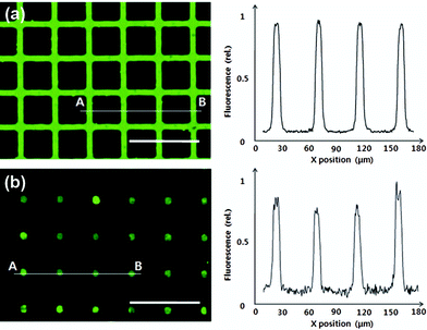 | ||
| Fig. 4 Fluorescence images (left) and intensity profiles (right) of (a) 10 µm-wide grid and (b) spot array patterns of PLL-FITC, which was patterned with micromolding in capillaries (MIMIC) and micro-contact printing (µCP) processes, respectively (Scale bar = 100 µm). | ||
 | ||
| Fig. 5 Microscopic pictures of the cells cultured on the PLL-FITC patterned substrates. (a) HepG2/C3A cells on the 30 µm-side square array after 1 day and (b) on the 20 µm-wide line pattern after 3 days. (c) CCD-986sk fibroblasts on the 30 µm-wide lines after 2 days. (d) Live cell staining (calcein-AM) of neurons patterned on the 10 µm-wide grid pattern at 3 days in vitro. All scale bars are 150 µm. | ||
Fig. 5d shows dissociated rat hippocampal neurons cultured on the PLL-FITC grid pattern in Fig. 4a, which was created by utilizing MIMIC with the PDMS mold in Fig. 2a. Neurite outgrowth as well as somata adhesion was well confined by the 10 µm patterns and resulted in the ordered neuronal networks in vitro. Calcein-AM staining indicated that most of the neurons were live on the patterns at 3 days after the culture. The selective cell adhesion and the effect of guided neurite growth were comparable to previously reported similar reports using conventional lithographic techniques.22,23 Judging from the obtained minimum feature size of biomolecule patterns and neuronal growth, we believe that the presented rapid prototyping method can be applied to design various neurobiological assays such as axon guidance and structured neuronal networks.24
The high degree of freedom of photomask film design is very valuable to make the complicate micro-patterns. Also the high resolution printing technology using a laser film printer is in still development to reduce the minimum pattern width. Now anyone can easily access to a photomask film with one's own design from widely distributed photomask printing service companies without buying an expensive laser film printer. We expect this direct rapid prototyping of PDMS from a photomask film can be widely applicable in soft lithography.
Conclusions
In conclusion, we have demonstrated a simple and inexpensive clean-room free process to fabricate elastomeric stamps for soft lithography. A conventional high-resolution photomask film was directly used as a master to produce PDMS stamps or molds, and micrometer-scale cell patterns were created by µCP and MIMIC. By utilizing this technique, we could print a cell adhesive biomolecule, PLL-FITC, with a minimum feature size of 10 µm on glass substrates reliably, and pattern several types of cells such as human hepatocytes, human fibroblasts and hippocampal neurons. This ultra rapid prototyping method using a photomask film as a master would be very useful technique for patterning cells and proteins with simple, rapid and inexpensive processes as well as with high resolution using soft lithography.Acknowledgements
This work was supported by the Korea Science and Engineering Foundation (KOSEF) NRL Program grant funded by the Korea government (MEST) (R0A-2008-000-20109-0), by the Nano/BioScience and Technology Program (2005-01291) of the MEST, and by the System IC 2010 program (10030554) of the Ministry of Knowledge and Economy (MKE). The authors also thank the Chung Moon Soul Center for BioInformation and BioElectronics, KAIST.References
- A. S. Blawas and W. M. Reichert, Biomaterials, 1998, 19, 595–609 CrossRef CAS.
- J. Fukuda, A. Khademhosseini, J. Yeh, G. Eng, J. Cheng, O. C. Farokhzad and R. Langer, Biomaterials, 2006, 27, 1479–1486 CrossRef CAS.
- K. K. Parker, A. L. Brock, C. Brangwynne, R. J. Mannix, N. Wang, E. Ostuni, N. A. Geisse, J. C. Adams, G. M. Whitesides and D. E. Ingber, FASEB J, 2002, 16, 1195–1204 CrossRef CAS.
- C. S. Chen, M. Mrksich, S. Huang, G. M. Whitesides and D. E. Ingber, Science, 1997, 276, 1425–1428 CrossRef CAS.
- A. Tourovskaia, X. Figueroa-Masot and A. Folch, Lab Chip, 2005, 5, 14–19 RSC.
- C. Ziegler, Anal Bioanal Chem, 2000, 366, 552–559 CAS.
- C. J. Flaim, S. Chien and S. N. Bhatia, Nat Methods, 2005, 2, 119–125 CrossRef CAS.
- I. S. Carrico, S. A. Maskarinec, S. C. Heilshorn, M. L. Mock, J. C. Liu, P. J. Nowatzki, C. Franck, G. Ravichandran and D. A. Tirrell, J Am Chem Soc, 2007, 129, 4874–4875 CrossRef CAS.
- N. Mittal, A. Rosenthal and J. Voldman, Lab Chip, 2007, 7, 1146–1153 RSC.
- V. N. Morozov and T. Y. Morozova, Anal Chem, 1999, 71, 3110–3117 CrossRef CAS.
- E. Zubritsky, Anal Chem, 2000, 72, 761A–767A CAS.
- D. A. Chang-Yen, D. G. Myszka and B. K. Gale, J Microelectromech Syst, 2006, 15, 1145–1151 CrossRef CAS.
- R. S. Kane, S. Takayama, E. Ostuni, D. E. Ingber and G. M. Whitesides, Biomaterials, 1999, 20, 2363–2376 CrossRef.
- Y. Xia and G. M. Whitesides, Angew Chem Int Ed, 1998, 37, 550–575 CrossRef CAS.
- S. Kim, K. W. Kwon, M. C. Park, S. H. Lee, S. M. Kim and K. Y. Suh, Biochip J, 2008, 2, 1–11 Search PubMed.
- C. L. d. Lago, H. Dominguez, T. d. Silva, C. A. Neves and J. G. A. Brito-Neto, Anal Chem, 2003, 75, 3853–3858 CrossRef.
- N. Bao, Q. Zhang, J.-J. Xu and H.-Y. Chen, J Chromatography A, 2005, 1089, 270–275 CrossRef CAS.
- A. Liu, F. He, K. Wang, T. Zhou, Y. Lu and X. Xia, Lab Chip, 2005, 5, 974–978 RSC.
- M. Abdelgawad, M. W. L. Watson, E. W. K. Young, J. M. Mudrik, M. D. Ungin and A. R. Wheeler, Lab Chip, 2008, 8, 1379–1385 RSC.
- F. M. Sasoglu, A. J. Bohl and B. E. Layton, J Micromech Microeng, 2007, 17, 623–632 CrossRef CAS.
- E. Yavin and Z. Yavin, J Cell Biol, 1974, 62, 540–546 CrossRef CAS.
- C. D. James, R. Davis, M. Meyer, A. A. T. A. Turner, S. A. T. S. Turner, G. A. W. G. Withers, L. A. K. L. Kam, G. A. B. G. Banker, H. A. C. H. Craighead, M. A. I. M. Issacson, J. A. T. J. Turner and W. A. S. W. Shain, IEEE Trans Biomed Eng, 2000, 47, 17–21 CrossRef CAS.
- Y. Nam, D. W. Branch and B. C. Wheeler, Biosens Bioelectron, 2006, 22, 589–597 CrossRef CAS.
- T. Pearce and J. C. Williams, Lab Chip, 2006, 7, 30–40 Search PubMed.
Footnotes |
| † Electronic supplementary information (ESI) available: Cell culture method. See DOI: 10.1039/b810341k |
| ‡ These authors contributed equally to this work. |
| This journal is © The Royal Society of Chemistry 2009 |