Nucleic acids in two dimensions: layers of base pairs linked by carboxylate
Eva
Corral
a,
Huub
Kooijman
b,
Anthony L.
Spek†
b and
Jan
Reedijk
*a
aLeiden Institute of Chemistry, Gorlaeus Laboratories, Leiden University, PO Box 9502, 2300 RA Leiden, The Netherlands. E-mail: reedijk@chem.leidenuniv.nl; Fax: +31 71 527 4671
bBijvoet Center for Biomolecular Research, Crystal and Structural Chemistry, Utrecht University, Padualaan 8, NL-3584 CH Utrecht, The Netherlands
First published on 27th November 2006
Abstract
The formation of a planar 2D hydrogen-bonded network between DNA bases and formate residues is reported, leading to unprecedented parallel sheets of DNA analogues.
Nucleic acids, such as DNA, RNA and their fragments, occur naturally in 3D chain-based structures derived from the double chain structure first described by Watson and Crick in 1953.1 The principal forces holding this spatial organization together are the Watson–Crick base pairing and the stacking between the bases; the chains being built of sugar–phosphate links. Several deviations from these structures are known to naturally occur. These abnormalities are the subject of intense studies, and in some cases they have been provoked in a search for therapeutic applications.2–5 Bends and kinks usually arise because of the presence of special sequences or mismatching, for instance in some RNAs.6–8 Triple helix chains are also known,4,5 as well as some quadruplex structures,3,9–12 knots and features such as hammerhead and other junctions.2 In all cases, the 1D organisation is one of the factors that determine the structure.
Much work has been done to go beyond these derivations and create new artificial base associations. Different approaches, such as metal-assisted hydrogen bonding13,14 and incorporation of artificial bases into DNA,15,16 have been used to develop new DNA base pairs or duplexes, many of which can be enzymatically replicated in the search for possible new biological applications.17
More recently, research has been reported on the synthesis of ion channels that consist of self-assembled supramolecular rosettes. These rosettes contain nucleic acids and other DNA-based artificial nucleosides that associate with each other in unusual ways. The rosettes pile up due to π-stacking.18–20
Following these lines of investigation, some supramolecular helical,21 linear,22 and macrocyclic structures23–25 have also been obtained.
To date, a complete, 2D-organized flat structure of nucleic acid bases has never been achieved by the self-assembly of nucleobases in solution, and it has been questioned whether such a structure, with hydrogen bonding within the plane only, would be possible. In fact, when looking closely at the common nucleic acid bases, it is not difficult to imagine that such structures should be possible, either with neutral bases, or with cationic or anionic bases in combination with small cations or flat anions, respectively.
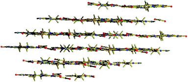
To explore this possibility in detail, a simple DNA model base that resembles a nucleotide and which has been used in many model systems, namely 9-ethylguanine,26–28 was selected, in combination with the smallest bifunctional flat anion, i.e.formate. Simple model building shows that, in this case, all strong hydrogen bond donors and hydrogen bond acceptors would match. Indeed when 9-ethylguanine (eg) was crystallised from a formic acid solution at rt, crystals of (Heg)(HCOO) could be isolated (see Fig. 1).

The asymmetric unit contains two (Heg)(HCOO) ion pairs. The packing environment of these pairs is virtually identical. The formate anion plays an indispensable role in the formation of a hydrogen bond network (see Table 1 and Fig. 2), in which the 9-ethylguaninium residues are associated to each other by the unusual so-called 12-trans sugar edge/sugar edge interactions, as described in the common Leontis/Westhof classification.29,30 These base pairs belong to the so-called class IV of the Saenger classification,31 which a more recent designation classifies as GG N3-amino, symmetric.32 To the best of our knowledge, only one example is known of an organism containing this kind of base pairing in a cellular organelle, the Haloarcula marismortuiribosome in its pairs G315:G336 and G2428:G2466.33 This base pair association has never before been achieved artificially without a simultaneous inclusion of metal atoms in the structure, such as gold13 or cadmium,34 or the blockage of the N7 of the purine ring with a metal atom or a methyl group.14,35
| Interatomic distances | Angles/° | ||
|---|---|---|---|
| Donor-H⋯Acceptor | D⋯A/Å | ||
| a Subscripts: I = (1 – x, 1 – y, 1 – z), II = (1 – x, –y, 1 – z), III = (1 – x, 1 – y, –z). | |||
| N(24)–H(24)⋯O(31II) | 2.545(3) | N(21)–C(21)–N(22) | 117.0(2) |
| N(21)–H(21)⋯O(41I) | 2.776(3) | N(22)–C(21)–N(23) | 119.4(2) |
| N(22)–H(22A)⋯O(42I) | 2.883(3) | O(41I)–N(21)–C(21) | 116.09(16) |
| N(22)–H(22B)⋯N(23III) | 3.026(3) | N(22)–O(42I)–C(41I) | 112.04(17) |
| O(42I)–N(22)–H(22B) | 115.5 (2) | ||
 | ||
| Fig. 2 Detail from Fig. 1, with numbering of the major atoms. The subscripts in atom labels are the same as in Table 1. | ||
The nucleoside–formate sheets described herein were found to allow a very close base pair stacking, with a distance between parallel layers of only 3.288(1) Å (see Fig. 3) (the distance between base pair planes in B-DNA is 3.46 Å).
The structure described in this paper is not the only possible example of 2D nucleobase packing that could be imagined. Current work is focusing on such systems by changing both the nucleic acid bases and the counterions. Formate has proved to be a valid example of a counterion which, due to its simplicity as much as to its planar geometry, could help to build these systems. Although, in principle, nitrate could also be considered suitable to yield a planar crystal structure, it does not have a hydrophobic part in proximity to the ethyl group and cannot form such a lattice. However, the formate hydrogen fits perfectly into the gap, while the corresponding nitrate oxygen atom would provoke repulsion forces that would distort the 2D structure.
The self-organisation of organic molecules into non-covalently bonded nanostructures, such as flat solid surfaces, gives structures with a high degree of order. This opens up a wide range of applications, for example in electronic and optical devices,36 in corrosion inhibition37 and in supramolecular chemistry.38 In molecular electronics, gold nanoparticles are embedded in ultrathin organic films, which could be used to interconnect gold nanoelectrodes in a molecular scale electronic device, as suggested by Samori and co-workers.39
The possible uses of these nucleoside layers in nanotechnology are only just starting to emerge,40 and much research is currently being done in fields such as DNA computation. DNA biosensors could be made by taking advantage of the specificity in the binding of the base pairs.41,42
Ribbon-like architectures have been described, which are formed by the self-assembly of guanosines in both solution and the solid state.43–45 Different applications of these ribbon structures in fields such as surface chemistry and photochemistry are being studied.46–49 The exploitation of DNA fragments and their mutual hydrogen bonding interactions for material purposes was extensively reviewed by Seeman.50
From a theoretical point of view, this kind of structure is of interest in studies on the emergence of life.51 It has been suggested that purine and pyrimidine monolayers could be candidates for a stationary phase in organic molecule separation systems and as templates for the assembly of higher ordered polymers at the prebiotic solid–liquid interface.52,53
In conclusion, a new type of arrangement of DNA bases in layers by hydrogen bonding is reported; this provides insights of novel templates for nanotechnology based on the 2D structures of nucleosides linked by a very simple carboxylate-containing molecule.
Experimental
Materials and reagents
9-Ethylguanine was purchased from Sigma and used as supplied. All other chemicals and solvents were reagent grade commercial materials and used as received without further purification.Physical measurements
C, H and N determinations were performed on a Perkin-Elmer 2400 Series II analyzer. NMR spectra were recorded on a Bruker DPX-300 spectrometer operating at a frequency of 300 MHz. Chemical shifts were calibrated against tetramethylsilane (TMS).Experimental procedure
9-Ethylguanine was crystallized as a white solid from a 0.014 M solution in formic acid–benzyl alcohol (1 : 1). The crystals obtained were found to be suitable for X-ray diffraction measurements. The product was collected by filtration, washed with a little (about 3 mL) ice cold water and dried in vacuo over silica. Anal. found: C, 42.4; H, 5.0; N, 30.8. C7H10N5O·CHO2 requires: C, 42.7; H, 4.9; N, 31.1%. δH (300 MHz, DMSO-d6,Me4Si) 10.46 (1 H, s, NH), 8.12 (1 H, s, HCOO), 7.67 (1 H, s, C(8)H), 6.37 (2 H, s, NH2), 3.94 (2 H, dd, J1 = 7.3, J2 = 14.5, CH2), 1.31 (3 H, t, J = 7.3, CH3).X-ray structural determination
![[1 with combining macron]](https://www.rsc.org/images/entities/char_0031_0304.gif) (no. 2), a = 7.4575(12), b = 11.6882(12), c = 12.8664(15) Å, α = 114.651(10), β = 94.767(11), γ = 101.729(10)°, V = 980.1(2) Å3, Z = 4, Dc = 1.5263(3) g cm–3, µ(Mo-Kα) = 0.120 mm–1, T = 150 K, 23598 reflections measured, 3550 independent, Rint = 0.1231 (before de-twinning), Rσ = 0.0559. The measured crystal was a twin, with a two-fold rotation around the b–c plane as the twin operation. Data were de-twinned using PLATON.54 Refinement of 356 parameters converged at a final wR2 value of 0.1540 (all data), R1 = 0.0515 (for 2847 reflections with I > 2σ(I)), S = 1.085, –0.29 < Δρ < 0.27 e Å–3. CCDC 612070. For crystallographic data in CIF or other electronic format see DOI: 10.1039/b613845d
(no. 2), a = 7.4575(12), b = 11.6882(12), c = 12.8664(15) Å, α = 114.651(10), β = 94.767(11), γ = 101.729(10)°, V = 980.1(2) Å3, Z = 4, Dc = 1.5263(3) g cm–3, µ(Mo-Kα) = 0.120 mm–1, T = 150 K, 23598 reflections measured, 3550 independent, Rint = 0.1231 (before de-twinning), Rσ = 0.0559. The measured crystal was a twin, with a two-fold rotation around the b–c plane as the twin operation. Data were de-twinned using PLATON.54 Refinement of 356 parameters converged at a final wR2 value of 0.1540 (all data), R1 = 0.0515 (for 2847 reflections with I > 2σ(I)), S = 1.085, –0.29 < Δρ < 0.27 e Å–3. CCDC 612070. For crystallographic data in CIF or other electronic format see DOI: 10.1039/b613845d
Acknowledgements
The authors would like to thank the Graduate Research School HRSMC, a joint activity of Leiden University and the two Universities in Amsterdam. This research has been financially supported by the Council for Chemical Sciences of the Netherlands Organisation for Scientific Research (CW-NWO). The support and sponsorship concerted by COST Actions D20/0001/00, D20/0002/00 and D20/003/01 is kindly acknowledged.References
- J. D. Watson and F. H. C. Crick, Nature, 1953, 171, 737 CAS.
- L. Citti and G. Rainaldi, Curr. Gene Ther., 2005, 5, 11 Search PubMed.
- M. A. Keniry, Biopolymers, 2001, 56, 123 CrossRef CAS.
- R. V. Guntaka, B. R. Varma and K. T. Weber, Int. J. Biochem. Cell Biol., 2003, 35, 22 Search PubMed.
- J. S. Sun, T. Garestier and C. Helene, Curr. Opin. Struct. Biol., 1996, 6, 327 CrossRef CAS.
- C. M. Duarte, L. M. Wadley and A. M. Pyle, Nucleic Acids Res., 2003, 31, 4755 CrossRef CAS.
- D. J. Klein, T. M. Schmeing, P. B. Moore and T. A. Steitz, EMBO J., 2001, 20, 4214 CrossRef CAS.
- L. M. Wadley and A. M. Pyle, Nucleic Acids Res., 2004, 32, 6650 CrossRef CAS.
- D. Rhodes and R. Giraldo, Curr. Opin. Struct. Biol., 1995, 5, 311 CrossRef CAS.
- E. H. Blackburn, Cell, 1994, 77, 621 CrossRef CAS.
- D. Sen and W. Gilbert, Nature, 1988, 334, 364 CrossRef CAS.
- W. I. Sundquist and A. Klug, Nature, 1989, 342, 825 CrossRef CAS.
- A. Schimanski, E. Freisinger, A. Erxleben and B. Lippert, Inorg. Chim. Acta, 1998, 283, 223 CrossRef CAS.
- R. K. O. Sigel, E. Freisinger, S. Metzger and B. Lippert, J. Am. Chem. Soc., 1998, 120, 12000 CrossRef CAS.
- J. A. Piccirilli, T. Krauch, S. E. Moroney and S. A. Benner, Nature, 1990, 343, 33 CrossRef CAS.
- E. T. Kool, Acc. Chem. Res., 2002, 35, 936 CrossRef CAS.
- C. Y. Switzer, S. E. Moroney and S. A. Benner, Biochemistry, 1993, 32, 10489 CrossRef CAS.
- F. Rakotondradany, A. Palmer, V. Toader, B. Z. Chen, M. A. Whitehead and H. F. Sleiman, Chem. Commun., 2005, 5441 RSC.
- N. Sakai, Y. Kamikawa, M. Nishii, T. Matsuoka, T. Kato and S. Matile, J. Am. Chem. Soc., 2006, 128, 2218 CrossRef CAS.
- M. S. Kaucher, W. A. Harrell and J. T. Davis, J. Am. Chem. Soc., 2006, 128, 38 CrossRef CAS.
- T. Giorgi, S. Lena, P. Mariani, M. A. Cremonini, S. Masiero, S. Pieraccini, J. P. Rabe, P. Samori, G. P. Spada and G. Gottarelli, J. Am. Chem. Soc., 2003, 125, 14741 CrossRef CAS.
- E. Mezzina, P. Mariani, R. Itri, S. Masiero, S. Pieraccini, G. P. Spada, F. Spinozzi, J. T. Davis and G. Gottarelli, Chem.–Eur. J., 2001, 7, 388 CrossRef CAS.
- J. T. Davis, Angew. Chem., Int. Ed., 2004, 43, 668 CrossRef CAS.
- J. L. Sessler, J. Jayawickramarajah, M. Sathiosatham, C. L. Sherman and J. S. Brodbelt, Org. Lett., 2003, 5, 2627 CrossRef CAS.
- J. L. Sessler, M. Sathiosatham, K. Doerr, V. Lynch and K. A. Abboud, Angew. Chem., Int. Ed., 2000, 39, 1300 CrossRef CAS.
- N. Grover, T. W. Welch, T. A. Fairley, M. Cory and H. H. Thorp, Inorg. Chem., 1994, 33, 3544 CrossRef CAS.
- P. M. Vanvliet, J. G. Haasnoot and J. Reedijk, Inorg. Chem., 1994, 33, 1934 CrossRef CAS.
- K. van der Schilden, F. Garcia, H. Kooijman, A. L. Spek, J. G. Haasnoot and J. Reedijk, Angew. Chem., Int. Ed., 2004, 43, 5668 CrossRef CAS.
- N. B. Leontis and E. Westhof, RNA, 2001, 7, 499 CrossRef CAS.
- N. B. Leontis, J. Stombaugh and E. Westhof, Nucleic Acids Res., 2002, 30, 3497 CrossRef CAS.
- W. Saenger, Principles of Nucleic Acid Structure, Springer-Verlag, New York, 1984 Search PubMed.
- U. Nagaswamy, N. Voss, Z. D. Zhang and G. E. Fox, Nucleic Acids Res., 2000, 28, 375 CrossRef CAS.
- N. Ban, P. Nissen, J. Hansen, P. B. Moore and T. A. Steitz, Science, 2000, 289, 905 CrossRef CAS.
- P. A. Ochoa, M. I. Rodriguez-Tapiador, S. S. Alexandre, C. Pastor and F. Zamora, J. Inorg. Biochem., 2005, 99, 1540 CrossRef CAS.
- R. K. O. Sigel, E. Freisinger, M. Abbate and B. Lippert, Inorg. Chim. Acta, 2002, 339, 355 CrossRef CAS.
- J. D. Swalen, D. L. Allara, J. D. Andrade, E. A. Chandross, S. Garoff, J. Israelachvili, T. J. McCarthy, R. Murray, R. F. Pease, J. F. Rabolt, K. J. Wynne and H. Yu, Langmuir, 1987, 3, 932 CrossRef CAS.
- T. Kowalik, H. J. P. Adler, A. Plagge and M. Stratmann, Macromol. Chem. Phys., 2000, 201, 2064 CrossRef CAS.
- V. A. Russell and M. D. Ward, Chem. Mater., 1996, 8, 1654 CrossRef CAS.
- P. Samori, V. Francke, K. Mullen and J. P. Rabe, Chem.–Eur. J., 1999, 5, 2312 CrossRef CAS.
- H. Yan, Science, 2004, 306, 2048 CrossRef CAS.
- A. Ferancova, R. Ovadekova, M. Vanickova, A. Satka, R. Viglasky, J. Zima, J. Barek and J. Labuda, Electroanalysis, 2006, 18, 163 CrossRef CAS.
- S. G. Wang, R. Wang, P. J. Sellin and Q. Zhang, Biochem. Biophys. Res. Commun., 2004, 325, 1433 CrossRef CAS.
- T. Giorgi, F. Grepioni, I. Manet, P. Mariani, S. Masiero, E. Mezzina, S. Pieraccini, L. Saturni, G. P. Spada and G. Gottarelli, Chem.–Eur. J., 2002, 8, 2143 CrossRef CAS.
- K. Araki, R. Takasawa and I. Yoshikawa, Chem. Commun., 2001, 1826 RSC.
- G. Gottarelli, S. Masiero, E. Mezzina, S. Pieraccini, J. P. Rabe, P. Samori and G. P. Spada, Chem.–Eur. J., 2000, 6, 3242 CrossRef CAS.
- G. Maruccio, P. Visconti, V. Arima, S. D’Amico, A. Blasco, E. D’Amone, R. Cingolani, R. Rinaldi, S. Masiero, T. Giorgi and G. Gottarelli, Nano Lett., 2003, 3, 479 CrossRef CAS.
- T. Kato, Science, 2002, 295, 2414 CrossRef CAS.
- R. Rinaldi, E. Branca, R. Cingolani, S. Masiero, G. P. Spada and G. Gottarelli, Appl. Phys. Lett., 2001, 78, 3541 CrossRef CAS.
- G. Gottarelli, S. Masiero, E. Mezzina, S. Pieraccini, G. P. Spada and P. Mariani, Liq. Cryst., 1999, 26, 965 CrossRef CAS.
- N. C. Seeman, Nature, 2003, 421, 427 CrossRef.
- S. J. Sowerby, M. Edelwirth and W. M. Heckl, J. Phys. Chem. B, 1998, 102, 5914 CrossRef CAS.
- S. J. Sowerby and W. M. Heckl, Origins Life Evol. Biosphere, 1998, 28, 283 CrossRef CAS.
- S. J. Sowerby, W. M. Heckl and G. B. Petersen, J. Mol. Evol., 1996, 43, 419 CrossRef CAS.
- A. L. Spek, J. Appl. Crystallogr., 2003, 36, 7 CrossRef CAS.
Footnote |
| † Address correspondence concerning the crystallography to this author, a.l.spek@chem.uu.nl. |
| This journal is © The Royal Society of Chemistry and the Centre National de la Recherche Scientifique 2007 |