Reduction of polyspermic penetration using biomimetic microfluidic technology during in vitro fertilization
Sherrie G.
Clark
ab,
Kathyrn
Haubert
d,
David J.
Beebe
c,
C. Edward
Ferguson
a and
Matthew B.
Wheeler
*abc
aDepartments of Animal Sciences and Bioengineering, University of Illinois at Urbana-Champaign, Urbana, IL 71801, USA. E-mail: mbwheele@uiuc.edu; Fax: +1 217-333-8286; Tel: +1 217-333-2239
bDepartment of Veterinary Clinical Medicine, University of Illinois at Urbana-Champaign, Urbana, IL 61802, USA. E-mail: sgclark@uiuc.edu; Fax: +1 217-333-7126; Tel: +1 217-244-5704
cDepartment of Biomedical Engineering, University of Wisconsin, Madison, WI 53706, USA. E-mail: djbeebe@wisc.edu; Fax: +1 608-265-9239; Tel: +1 608-262-2260
dVitae LLC, Madison, WI 53703, USA. E-mail: haubert@vitaellc.com; Tel: +1 608-256-9979
First published on 19th August 2005
Abstract
Efforts to improve the in vitro embryo production process in pigs have included modifying culture medium and number of spermatozoa inseminated in order to reduce the incidence of polyspermy. Polyspermy is a pathological condition which results in aberrant embryonic development. The microchannels are designed to more closely mimic the function of the oviduct and create a flow pattern of spermatozoa past the oocytes similar to the pattern in the oviduct. In vitro fertilization of porcine oocytes in the microchannels has produced a higher incidence of monospermic penetration (p < 0.05) as compared to the oocytes fertilized in the traditional microdrop system with comparable penetration and male pronucleus formation rates. Additionally, cleavage rates of the embryos as well as development to the blastocyst stage are similar. Here we demonstrate that the biomimetic microchannel in vitro fertilization system can reduce polyspermy and, therefore, increase the number of potentially viable embryos without reducing the overall in vitro production efficiency.
Introduction
Polyspermic penetration of oocytes fertilized in vitro remains one of the major reasons for the failure to produce viable porcine embryos. This phenomenon has continually haunted the porcine IVF system.1 In the in vitro system, there are a large number of capacitated sperm cells present near the matured oocytes. In contrast, with in vivo-produced oocytes there are very few spermatozoa in the immediate vicinity of the oocytes in the oviduct. The oviduct acts as a reservoir for sperm and then serves as a “gate” which allows sperm cells to enter the ampullary-isthmus junction near the time that the oocytes are arrested at metaphase II and ready to be fertilized.2–4 The mechanisms by which sperm cells are maintained in the reservoir may be binding of the spermatozoa to the oviductal epithelium, physical obstruction to forward progression by thick, viscous secretions, and decreased motility of the sperm tails.5,6The geometrically constrained microenvironment present in microchannels provides another tool to study cell biology.7 The application of microchannel technology to assisted reproduction is one illustration of how microchannels can improve upon existing methods. Microchannels have been used to perform assisted reproductive techniques involving in vitro maturation (IVM),8,9in vitro fertilization (IVF),10,11 embryo culture12–14 and manipulation.15–17
The microchannels (Fig. 1a) are manufactured using polydimethylsiloxane (PDMS) micromolding techniques18,19 and are utilized in the laminar flow regime to transport sperm towards the oocytes. The design of the microchannel14 is such that oocytes and embryos can be “parked” at a particular location, called the constriction region, to allow for continual visualization (Fig. 1b) as they develop.
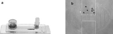 | ||
| Fig. 1 The microfluidic culture environment (microchannel). (a) A photograph of the PDMS-borosilicate device containing a straight channel 1000 µm wide and 250 µm high (photo courtesy of Vitae, LLC). (b) Microscopic view of porcine oocytes at the constriction region. | ||
The channel design is similar in important ways to the in vivo structure (see Fig. 2 a,b). Not only has the microchannel provided a means by which oocytes matured similarly to the traditional microdrop system,8 but also could allow sperm cells to flow past the oocytes in a manner more similar to that of the oviduct. This study was conducted to evaluate in vitro fertilization of in vitro-matured porcine oocytes in a microfluidic device compared to the microdrop system, the effect of each fertilization system on male pronucleus formation and polyspermic penetration of the oocytes and the development of the embryos to the blastocyst stage after fertilization in each system.
 | ||
| Fig. 2 The microchannel device mimics the in vivo functionality. (a) In vivo the sperm is guided to the oocyte via a narrowing fluid path in the oviduct. (b) The microchannel design recapitulates the in vivo function. The microchannel routes the sperm to the “parked” oocyte. | ||
Experimental
A. Oocyte collection and IVM
The in vitro-matured oocytes were obtained by culturing follicular oocytes obtained from the ovaries of prepuberal gilts. Follicles (3–6 mm in diameter) were aspirated using an 18 gauge needle attached to a 10 mL syringe. Cumulus-oocyte complexes (COCs) were selected based on being surrounded by at least 3 layers of cumulus cells with a homogenous cytoplasm. The COCs were selected in TCM-199 HEPES medium supplemented with 0.1% PVA. Then they were washed three times in TCM-199 maturation medium supplemented with 0.25 µg LH, 0.25 µg FSH, 5 ng EGF, 0.57 mM cysteine, 0.1% polyvinyl alcohol, 25 µg streptomycin and 37.5 µg potassium penicillin G. Fifty oocytes were placed in a well of a Nunc 4-well multidish (Nunc, Roskilde, Denmark) filled with 500 µL of the pre-equilibrated supplemented TCM-199 maturation medium covered with warm paraffin oil. The COCs were cultured in an atmosphere of 100% humidified 5% CO2 in air at 39 °C for 44 h.B. Semen preparation for IVF
Semen was collected from crossbred boars using the gloved hand technique and extended in commercial extender (Androhep; Minitube of America, Verona, WI) in the on-farm processing laboratory and stored at 16–17 °C until used within 1–2 days. Extended semen was centrifuged at 200 × g for 3 min to remove dead sperm cells, washed and centrifuged at 1200 × g for 3 min three consecutive times with mTBM [113.1 mM NaCl, 3 mM KCl, 7.5 mM CaCl2·2H2O, 5 mM sodium pyruvate, 11 mM glucose, 20 mM Tris (crystallized free base; Fisher Scientific, Fair Lawn, NJ), 1 mM caffeine, and 0.4% BSA (Fraction V, A2138; Sigma)]. The sperm cells were re-suspended to a concentration of 6 × 10⁵ cells mL−1 and the sperm cell suspension was placed in a 100% humidified 5% CO2 in air atmosphere at 39 °C for 90 min prior to addition to cumulus-free oocytes to allow for capacitation to occur.C. Microchannel fabrication and design
The microchannel devices were fabricated using standard photolithography and micromolding techniques.18,19 Briefly, EPON SU-8 photoresist was patterned onto a three inch silcon wafer to create a master. Polydimethylsiloxane (PDMS) from a Dow-Corning Sylgard 184 elastomer kit was poured onto the master and cured, creating device tops with the channel structure molded into them. A funnel was also molded into each device top to facilitate oocyte loading. The PDMS tops were then plasma bonded to glass microscope slides (1 × 3 in), which formed the base of the device and the bottom of the channels. Finally, a 10 mm tall by 10 mm id borosilicate reservoir was glued over the funnel inlet, and a female luer connector was glued over the outlet hole. Fig. 1a and 2b provide illustrations of the key device components. The luer allows connection of a syringe for loading medium in addition to serving as an open reservoir during passive, gravity driven flow. Each device contains a single straight microchannel. The channel is 1000 µm wide × 250 µm tall × 38 mm long. A 20 µm high constriction holds oocytes in place near the center of the channel, while still allowing media and sperm to flow through to the outlet.D. Oocyte and sperm handling techniques involving the microchannels
After 44 h of culture, cumulus cells were removed from the oocytes by vortexing them in 0.1 mg mL−1 hyaluronidase in mTBM. Denuded oocytes were washed in mTBM and fifteen oocytes were placed into a microchannel that has been filled with 200 µL of mTBM and equilibrated at 39 °C under 100% humidified 5% CO2 in air. Oocytes were transferred using traditional micropipette techniques and carefully placed near the opening of the channel within the funnel. The oocytes were allowed to roll down the inside of the funnel towards the opening to the channel and enter the channel. Assistance was provided in the manner of gentle tapping and placing the microchannel device on a slight incline to advance the oocytes toward the constriction site.After oocytes were successfully loaded into the microchannels, 200 µL of capacitated sperm cells (6 × 10⁵ sperm cells mL−1) were added to the funnel. This addition of fluid to the inlet reservoir created a slight pressure head, resulting in gravity driven flow that carried sperm past the oocytes at the constriction. In the microdrops, the addition of the same volume (50 µL) of sperm cells at a concentration of 6 × 10⁵ cells mL−1 would be added to the 50 µL fertilization drop. The sperm cells would be relatively evenly distributed throughout the microdrop and produce a final concentration of 3 × 10⁵ cells mL−1. In the microchannel IVF system, the same volume (200 µL) of 6 × 10⁵ sperm cells mL−1 would be added to the 200 µL of fertilization medium in the microchannel.
The oocytes and sperm cells were co-cultured for 6 h. At this time, potential zygotes were removed from the channels by reversing the flow towards the funnel and placed into culture drops of 100 µL of North Carolina State University 23 (NCSU-23) under paraffin oil. The embryos were cultured in NCSU-23 containing 0.4% BSA (bovine serum albumin) until day 4 and then the medium was changed to NCSU-23 containing FBS (fetal bovine serum) and cultured to the blastocyst stage.
E. Fixing and staining
Approximately 18 h after the beginning of the co-incubation period of oocytes with sperm, presumptive zygotes from each treatment (microdrop and microchannel) were removed from culture in NCSU-23 by conventional micropipetting techniques and vortexed to remove any remaining cumulus cells and attached spermatozoa. Denuded zygotes were mounted on slides and fixed with 25% (v/v) glacial acetic acid in absolute ethanol for 48–72 h. After 48–72 h of fixation, the slides containing the zygotes were stained with 1% aceto-orcein and examined under phase contrast microscopy (40×–400×). Once stained, slides were examined and parameters including maturation, penetration, monospermic penetration, male pronucleus formation, and number of sperm penetrating per oocyte were recorded. Penetration was determined by the presence of one or more swollen sperm heads and/or male pronuclei and their corresponding sperm tails. The presence of three or more pronuclei was designated as polyspermic. Aceto-orcein staining has been shown to be a repeatable and reliable methodology in our laboratory. We have therefore chosen to use this method over immunofluoresence of pronuclei and/or DIC imaging of the sperm tail to evaluate polyspermy.F. Statistics and data analysis
For all experiments, data are reported as mean ± SEM. For all other experiments, each replicate had an analysis of variance performed using the mixed model in a randomized complete block design in the SAS program (SAS Institute, Inc., Cary, NC). Data analyzed for Experiment 1 was from 5 replicates with 10 or 15 oocytes per treatment group per replicate (the number of oocytes per subclass (n) is shown in Table 1 and values are mean ± SEM. Data analyzed for Experiment 2 was from 6 replicates with 15 oocytes per treatment group per replicate (the number of oocytes per subclass (n) is shown in Fig. 3 and values are mean ± SEM. Differences were considered significant with an alpha level of p ≤ 0.05. | ||
| Fig. 3 Experiment 2: Comparison of fertilization parameters in each IVF system. The different superscripts a and b within columns represent significant differences (p < 0.05). Values are mean ± SEM (n = 138 oocytes for the control microdrop and 128 oocytes for the microchannel, respectively). Mat: maturation (% of total), pene: penetration (% of matured), mono: monospermic penetration (% of penetrated), mpf: male pronuclear formation (% of penetrated). | ||
| Treatment | Oocyte number | Maturation (% of total) | Penetration (% of matured) | Monospermic (% of penetrated) | MPFc (% of penetrated) | # Sperm/oocyte |
|---|---|---|---|---|---|---|
| a Values with different superscripts within columns represent significant statistical differences (p < 0.05). b Values with different superscripts within columns represent significant statistical differences (p < 0.05). c MPF = male pronucleus formation. d Data shown from 5 replicates and values are mean ± SEM. Control 10 or 15: microdrop IVF system containing 10 or 15 oocytes, respectively; MC 10 or 15: microchannel IVF system containing 10 or 15 oocytes, respectively. | ||||||
| Control 10 | 49 | 83.14 ± 4.8 | 96.67 ± 4.9 | 22.33 ± 12.4a | 78.13 ± 10.5 | 6.47 ± 0.4a |
| MC 10 | 48 | 72.67 ± 5.0 | 88.06 ± 5.1 | 55.42 ± 7.6b | 82.50 ± 10.5 | 2.03 ± 0.1b |
| Control 15 | 50 | 91.71 ± 4.8 | 97.50 ± 4.9 | 22.32 ± 10.7a | 80.24 ± 9.4 | 7.39 ± 0.5a |
| MC 15 | 42 | 87.17 ± 5.0 | 87.17 ± 5.1 | 63.89 ± 7.1b | 73.15 ± 9.9 | 1.66 ± 0.1b |
Results
The results of a preliminary study (Experiment 1) to examine the effect of oocyte number (10 vs. 15) within each system on fertilization parameters is shown in Table 1. There was no significant difference observed between maturation, penetration and male pronucleus formation percentages in either the microdrop and microchannel systems irrespective of oocyte number. However, the monospermic penetration percentage was higher (p < 0.05) in the microchannel system when compared to the microdrop system. Correspondingly, there were significantly less sperm penetrating each oocyte (p < .0.05) in the embryos produced in the microchannels.Based on the results above, Experiment 2 was designed to examine the effect of treatment on fertilization parameters with a larger number of oocytes. Several important differences were also observed in this portion of the study between microchannel and microdrop fertilization. Aceto-orcein staining data again revealed a higher incidence of monospermic penetration and a lower number of spermatozoa per oocyte in the embryos fertilized in the microchannels as compared to the controls (Fig. 3). Additionally, the number of sperm penetrating each oocyte was counted. The mean number of sperm in control oocytes was 6.65 ± 0.4 in contrast to the 1.77 ± 0.1 sperm in the microchannel oocytes.
These data together demonstrate that the microchannel environment reduces the incidence of polyspermy during IVF of porcine oocytes (p < 0.05) while maintaining comparable penetration and male pronuclear formation rates.
Discussion
The differences in the physical environments (microchannel vs. control) are important and may be responsible for the observed differences in fertilization. In the microdrop, a dome-shaped volume of liquid, that can be approximated by a hemisphere of radius of 6.27 mm for a 50 µL drop, covers the oocyte. In the microchannel, the oocyte is surrounded on four sides (top, bottom, and both sides) by a physical wall. In a microdrop, the sperm move at random through the large hemispherical volume of medium. In microdrop IVF, one strives to achieve (via changing the sperm concentration) a balance between having enough sperm present to fertilize the egg while avoiding sperm concentrations that are too high leading to polyspermy. In practice that has been difficult to achieve and polyspermy is a persistent problem in porcine IVF.1 There are several factors contributing to the reduced polyspermy in the microchannel. The physical differences between a microdrop and a microchannel play role. Due to the geometry and methods, it is likely that the concentration and dynamics of sperm near the oocyte are different in the microchannel than in the microdrop. However, previous results with different sperm concentrations in microdrops suggest that concentration alone would not explain the results (i.e. reduced polyspermy while maintaining appropriate oocyte penetration rates). The presence of flow may also influence the fertilization process. In fact, the microchannel device mimics the in vivo functionality. In vivo, the oocyte is held (“parked”) at the ampullary–isthmic junction (the site of fertilization) between the ampulla and the isthmus of the oviduct. The sperm flow to the junction guided by a narrowing of the isthmus of the oviduct (see Fig. 2a). The microchannel with constriction region serves a similar function guiding the sperm to the “parked” oocytes. The combination of the different physical conditions present in the microchannel create a fertilization system that, based on our knowledge of in vivo fertilization, mimics some of the in vivo fertilization conditions contributing to the observed reduction in polyspermy. Additional studies are needed to fully elucidate the interaction between the physical factors that influence fertilization. In fact, the biomimetic microchannel system may provide insights into natural behavior that is difficult to study in vivo.Conclusion
The results are important from both commercial and scientific perspectives. The significant reduction in polyspermy has direct and immediate implications for porcine production. Just as importantly, the results combined with previous culture studies continue to support the idea that microchannel culture systems provide a useful tool for understanding the basic mechanisms of reproduction by mimicking the physical conditions present in vivo.Acknowledgements
The authors would like to thank Jim Baltz and Synthia Lane from the Department of Animal Sciences at UIUC for help with the illustrations and technical assistance respectively. This material is based upon work supported by the Cooperative State Research, Education, and Extension Service, U.S. Department of Agriculture, under USDA Multi-state Project W-1171 through UIUC and Vitae, LLC (Madison, WI).References
- L. R. Abeydeera, Theriogenology, 2002, 57, 257 CrossRef CAS.
- R. H. F. Hunter, J. Reprod. Fertil., 1981, 63, 109 Search PubMed.
- R. H. F. Hunter, J. Reprod. Fertil., 1984, 72, 203 Search PubMed.
- E. Topfer-Peterson, M. Petrounkina and M. Ekhlasi-Hundrieser, Anim. Reprod. Sci., 2000, 60–61, 653 CrossRef.
- S. S. Raychoudhury and S. S. Suarez, Theriogenology, 1991, 36, 1059 CrossRef.
- J. N. Mburu, S. Einarsson, N. Lundeheim and H. Rodriguez-Martinez, Anim. Reprod. Sci., 1996, 45, 109 CrossRef CAS.
- G. M. Walker, H. C. Zeringue and D. J. Beebe, Lab Chip, 2004, 4, 91 RSC.
- P. N. Hester, H. M. Roseman, S. G. Clark, E. M. Walters, D. J. Beebe and M. B. Wheeler, Theriogenology, 2002, 57, 723.
- E. M. Walters, D. J. Beebe and M. B. Wheeler, Theriogenology, 2001, 55, 497.
- S. G. Clark, E. M. Walters, D. J. Beebe and M. B. Wheeler, Biol. Reprod., 2002, 66(Suppl. 1), 528 Search PubMed.
- S. G. Clark, E. M. Walters, D. J. Beebe and M. B. Wheeler, Theriogenology, 2003, 59, 441.
- D. Beebe, W. Wheeler, H. Zeringue, E. Walters and S. Raty, Theriogenology, 2002, 57, 125 CrossRef CAS.
- M. B. Wheeler, D. J. Beebe, E. M. Walters and S. Raty, in Second International IEEE EMBS Special Topic Conference on Microtechnology in Medicine and Biology, 2002, p. 104 Search PubMed.
- S. Raty, E. M. Walters, J. A. Davis, H. C. Zeringue, D. J. Beebe, S. L. Rodriguez-Zas and M. B. Wheeler, Lab Chip, 2004, 4, 186 RSC.
- H. C. Zeringue, M. B. Wheeler and D. J. Beebe, Lab Chip, 2005, 5, 108 RSC.
- H. C. Zeringue, J. J. Rutledge and D. J. Beebe, Lab Chip, 2005, 5, 108 RSC.
- R. S. Suh, N. Phadke, D. A. Ohl, S. Takayama and G. D. Smith, Hum. Reprod. Update, 2003, 9, 451 Search PubMed.
- B. H. Jo, L. M. Van Lerberghe, K. M. Motsegood and D. J. Beebe, J. Microelectromech. Syst., 2000, 9, 76 CrossRef CAS.
- J. C. McDonald and G. M. Whitesides, Acc. Chem. Res., 2002, 35, 491 CrossRef CAS.
| This journal is © The Royal Society of Chemistry 2005 |