Electrostatically arranged cytochrome c–fullerene photoelectrodes†
Israel
Zilbermann
a,
Andrew
Lin
a,
Maria
Hatzimarinaki
b,
Andreas
Hirsch
*b and
Dirk M.
Guldi
*a
aRadiation Laboratory, University of Notre Dame, Notre Dame, IN 46556, USA. E-mail: guldi.1@nd.edu; Fax: 1 574 631 8068; Tel: 1 574 631 7441
bInstitut für Organische Chemie, Universität Erlangen-Nürnberg, Henkestr. 42, 91054 Erlangen, Germany. E-mail: hirsch@organik.uni-erlangen.de; Fax: 0049 9131 85 26864; Tel: 0049 9131 85 22537
First published on 28th November 2003
Abstract
We have developed a molecular-level switch—a C60/cytochrome c modified ITO electrode—that reversibly transmits and processes solar energy.
Bioengineering of photovoltaic entities is a rapidly progressing field of interdisciplinary interest, providing important information about the integration of biomolecules into functional systems.1 The layer-by-layer technique emerged as a powerful technique towards functional systems and photoactive electrodes due to a fine-tuned interplay of interactions at the molecular level.2 In this work we wish to describe a simple molecular-level switch—a C60/cytochrome c modified ITO electrode—that reversibly transmits/processes solar energy.
The strategy involves the use of mitochondrial cytochrome c (FeCytc), which is a polycationic redox protein. It plays a dominating role as a charge carrier and/or charge mediator complex in numerous biological3 and synthetic systems.4 FeCytc is then electrostatically linked to a negatively-charged dendritic fullerene (C60DF)—see Scheme 1.5 The function of the construct is given by the redox activity of the protein's heme group, that is, the FeIIICytc/FeIICytc transformation.
 | ||
| Scheme 1 Electron transfer processes occurring in photoexcited ITO/C60DF/FeIICytc and structure of C60DF. | ||
Once FeIICytc/C60DF is deposited onto an ITO electrode, the fullerene constitutes the primary electron acceptor (A1; C60DF/C60DF˙−: −0.70 ± 0.05 V vs. SCE; 1*(C60DF)/C60DF˙−: +1.07 ± 0.05 V vs. SCE); it accepts the electron from the electron donating FeIICytc redox protein (D; FeIICytc/FeIIICytc: +0.25 ± 0.05 V vs. SCE) and relays it to the electrode. In the current context the ITO electrode serves as a secondary electron acceptor (A2: −0.25 V vs. SCE)6 that ultimately accumulates and stores the electrons. Scheme 1 underlines the aspect of a fine-tuned redox gradient in the integrated donor–acceptor–acceptor (i.e., D–A1–A2) construct.
The stepwise assembly of substrate/C60DF/FeIICytc includes the following sequence. First, deposition of the negatively-charged dendritic fullerene necessitates a base layer of poly(diallyl-dimethylammonium) chloride (PDDA), adsorbed onto the hydrophilic substrate.‡ Mainly Coulombic forces, which are pH and ionic strength sensitive, govern the physisorption process between the opposite charged ions of PDDA and C60DF.5 Similar principles apply then for implanting a layer of FeIIICytc on top of the C60DF surface. The distinct absorption features of C60DF (i.e., ∼260 and 330 nm) and FeIIICytc (i.e., Soret- and Q-bands around 410 nm and 525 nm, respectively) facilitate their convenient detection—see Supporting Information Fig. S1.
After completing each deposition step—PDDA, C60DF, FeIIICytc—the surface structure was analyzed by AFM. While the image of the PDDA base layer can be best described by a fairly flat surface with occasional pores—see Fig. 1,7 the visualization of the dendritic C60DF layer and subsequently the FeIIICytc layer led to different representations—see Fig. 1. Now, fine-grained structures of C60DF and FeIIICytc layers are clearly discernible. Fig. 1 shows that in the case of the dendritic C60DF, the surface coverage, that is, characteristic 20–50 nm 2D nanoaggregates, amalgamates on a macroscopic picture into a continuous film. Layers of FeIIICytc reveal, despite the amorphous nature of the protein matrix, a similar texture.
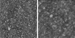 | ||
| Fig. 1 AFM images of (left) silicon/PDDA/C60DF and (right) silicon/PDDA/C60DF/FeIIICytc (1 µm × 1 µm). | ||
Repetition of the C60DF/FeIIICytc sequencing leads to substrate/(C60DF/FeIIICytc)n where n could be as large as 10. To confirm the regularities of the sequential sandwich packing we followed the layer-by-layer build-up via absorption spectrophotometric and ellipsometric means on quartz and silicon wafers, respectively. Figures S2 and S3 illustrate that both variables, namely, C60DF/FeIIICytc visible transitions and film thickness, gave rise to linear dependences.8 We conclude we have achieved success in fabricating layered, closely-packed ITO-surfaces with tailorable absorption-cross sections throughout the visible region and, thus, enhancing an efficient use of the solar spectrum. For example, fairly high absorbances of up to 0.2 (i.e., Soret-band region) were accomplished for the sandwich structures.
Linear sweep wave voltammetry performed with ITO/C60DF/FeIIICytc electrodes revealed, on the cathodic side, two distinct and well-separated reduction steps.§ Linear scan rate dependences substantiated surface-based, thin-layer electrochemical processes rather than diffusion controlled events. The FeIIICytc/FeIICytc redox process appeared first, at −0.25 ± 0.05 V—Fig. S4—followed by the one-electron reduction of C60DF/C60DF˙− at −0.70 ± 0.05 V—both potentials are given versus SCE. These values are in excellent agreement with measurements conducted with the individual constituents and confirm that both chromophores sustain their redox characteristics in the layered films. From these redox potentials we estimate the energy of the C60DF˙−/FeIIICytc charge-separated state as ∼ 0.7 eV, sufficiently less energetic than the photoexcited chromophores; > 1.80 eV.
Owing to the good conductivity that layered films of C60DF exhibit, we probed the electrochemical FeIIICytc/FeIICytc9 processes as a function of ITO/(C60DF/FeIIICytc)n (n = 0–6). What is most interesting is that we found a satisfying linear dependence of the currents, which relate to the FeIIICytc reduction, in films of up to 4 sandwich layers. Beyond this point, however, the currents level off and any further current increase is suppressed. The latter observation is consistent with a strongly impacted connection between the ITO electrode, on one end, and (FeIIICytc)5 or (FeIIICytc)6, on the opposite end, in ITO/(C60DF/FeIIICytc)n.
The photocurrent generation of an ITO/C60DF/FeIIICytc cell is quite small—about 0.1 µA (Fig. 2)—a deficit that finds many causes. Most importantly, the electron donating ability of FeIIICytc, that is, its one-electron oxidation to form presumably FeIVCytc, renders it an inappropriate choice for reducing C60DF upon illumination. This picture should change dramatically, once the electrochemical reduction of the trivalent iron center is carried out. In fact, after electrolyzing the ITO/C60DF/FeIIICytc cell at −0.25 V, a potential that leaves C60DF as a primary electron acceptor intact, a nearly 10-fold amplification of the photocurrent was registered—see Fig. 2. The photocurrent was measured after the completion of preparative electrolysis—5 minutes—without any active bias potential on during the photocurrent measurements. The designed gradient in the integrated ITO/C60DF/FeIICytc construct ensures the efficient formation of C60DF˙−/FeIIICytc in the excited device (i.e., singlet excited state) plus a thermodynamically supported electron injection from C60DF˙− into the secondary electron acceptor, ITO.7a The photoaction spectrum—Fig. S5—reveals broadening relative to the ITO/C60DF/FeIICytc ground state features, indicating electronic interactions between the moieties. To confirm the photocurrent results 1*C60DF fluorescence experiments were carried out, which indicate the presence and absence of electron transfer quenching in 1*C60DF/FeIICytc and 1*C60DF/FeIIICytc, respectively were conducted. Reversing the electrode's bias and to reoxidize the redox protein at +0.2 V led again to the termination of the photocurrent, that is, reverting back to about 0.1 µA. Again, the photocurrents were recorded without any bias, but after 5 minutes of preparative electrolysis. We would like to emphasise that these switching features are completely reproducible: the photoelectrochemical cell performance depends solely on either a negative (i.e., switching ON via reducing FeIIICytc) or a positive (i.e., switching OFF via oxidizing FeIICytc) bias, applied to the electrode.
 | ||
| Fig. 2 Photocurrent of (C60DF/FeIICytc)—filled circle—and (C60DF/FeIIICytc)—open circle—assembled on ITO electrode. Experimental conditions: deoxygenated aqueous solutions containing 0.1 M NaCl as supporting electrolyte; either −0.25 V or +0.2 V bias voltage is applied. | ||
The above-mentioned electrochemical results point to fairly good contacts between the ITO electrode, the ultimate electron acceptor, and FeIICytc, the electron donor, situated in one of the outermost layers for n up to 4. Interestingly, the photocurrents in the corresponding ITO/(C60DF/FeIIICytc)n (n = 0–4) give rise to a similar trend, that is, they are linearly dependent on the number of layers (n) and, hereby, confirm the electrochemistry. Also, intermolecular interactions within the individual layers, as evidenced by the broadening of the spectra, ensure good charge migration/delocalization. The reversible ON/OFF switching remains reproducible even in ITO/(C60DF/FeIIICytc)4.
In conclusion, specific integration of ITO, C60DF and FeIICytc building blocks yields an environmentally very stable molecular switch. The function of the redox protein-based system, that is, reversible switching between two defined states, is attained by altering the redox state of the iron center: applying different electrochemical potentials switches the photoperformance ON (FeIICytc) or OFF (FeIIICytc). A promising asset of our work is that, for the first time, stacking donor–acceptor sandwich layers (i.e., C60DF/FeIIICytc) helps to enhance the photosensitivity of the switch nearly linearly with the absorption cross section of the modified ITO electrodes.
This work was supported by the Office of Basic Energy Sciences of the U.S. Department of Energy (NDRL 4494) and DFG (SFB 583 Redoxaktive Metallkomplexe—Reaktivitätssteuerung durch molekulare Architekturen). I. Z. acknowledges his sabbatical leave from the Nuclear Research Centre Negev, Beer Sheva, Israel.
Notes and references
- (a) E. Braun, Y. Eichen, U. Sivan and G. Ben-Yoseph, Nature, 1998, 391, 775 CrossRef CAS; (b) I. Willner and E. Katz, Angew. Chem., Int. Ed., 2000, 39, 1180 CrossRef; (c) J. Fritz, M. K. Baller, H. P. Lang, H. Rothuizen, P. Vettiger, E. Meyer, H. J. Guntherodt, C. Gerber and J. K. Gimzewski, Science, 2000, 288, 316 CrossRef CAS; (d) A. N. Shipway and I. Willner, Acc. Chem. Res., 2001, 34, 421 CrossRef CAS; (e) T. Chen, S. C. Barton, G. Binyamin, Z. Q. Gao, Y. C. Zhang, H. H. Kim and A. Heller, J. Am. Chem. Soc., 2001, 123, 8630 CrossRef CAS; (f) J. J. Wei, H. Y. Liu, D. E. Khoshtariya, H. Yamamoto, A. Dick and D. H. Waldeck, Angew. Chem., Int. Ed., 2002, 41, 4700 CrossRef CAS; (g) I. Willner, Science, 2002, 298, 2407 CrossRef CAS; (h) N. Mano, F. Mao and A. Heller, J. Am. Chem. Soc., 2002, 124, 12962 CrossRef CAS; (i) S.-J. Park, T. A. Taton and C. A. Mirkin, Science, 2002, 295, 1503 CrossRef CAS.
- (a) M. Ferreira, J. H. Cheung and M. F. Rubner, Thin Solid Films, 1994, 244, 806 CrossRef CAS; (b) J. H. Fendler and F. C. Meldrum, Adv. Mater., 1995, 7, 607 CrossRef CAS; (c) S. W. Keller, S. A. Johnson, E. S. Brigham, E. H. Yonemoto and T. E. Mallouk, J. Am. Chem. Soc., 1995, 117, 12879 CrossRef CAS; (d) G. Decher, Science, 1997, 277, 1232 CrossRef CAS; (e) N. A. Kotov, Nanostruct. Mater., 1999, 12, 789 CrossRef.
- (a) G. W. Pettigrew, G. R. Moore, Cytochromes c: Biological Aspects, Springer, Berlin, 1987 Search PubMed; (b) N. M. Kostic, Met. Ions Biol. Syst., 1991, 27, 129 Search PubMed; (c) G. McLendon and R. Hake, Chem. Rev., 1992, 92, 481 CrossRef CAS; (d) J. R. Winkler and H. B. Gray, Chem. Rev., 1992, 92, 369 CrossRef; (e) J. M. Nocek, J. S. Zhou, S. D. Forest, S. Priyadarshy, D. N. Beratan, J. N. Onuchic and B. M. Hoffman, Chem. Rev., 1996, 96, 2459 CrossRef CAS; (f) R. H. Holm and E. I. Solomon, Chem. Rev., 1996, 96, 2237 CrossRef.
- (a) S. S. Isied, G. Worosila and S. J. Artherton, J. Am. Chem. Soc., 1982, 104, 7659 CrossRef CAS; (b) S. S. Isied, Adv. Chem. Ser., 1997, 253, 331 Search PubMed; (c) J. V. McArdle, H. B. Gray, C. Creutz and N. Sutin, J. Am. Chem. Soc., 1974, 96, 5737 CrossRef CAS; (d) J. V. McArdle, K. Yocon and H. B. Gray, J. Am. Chem. Soc., 1977, 12, 4141 CrossRef; (e) M. Meier and R. van Eldik, Inorg. Chim. Acta, 1994, 225, 95 CrossRef CAS; (f) M. Meier, R. van Eldik, I. J. Chang, G. A. Mines, D. S. Wuttke, J. R. Winkler and H. B. Gray, J. Am. Chem. Soc., 1994, 116, 1577 CrossRef CAS; (g) M. Meier, J. Sun, R. van Eldik and J. F. Wishart, Inorg. Chem., 1996, 35, 1564 CrossRef CAS; (h) M. Meier and R. van Eldik, Inorg. Chim. Acta, 1996, 242, 185 CrossRef CAS; (i) M. Meier and R. van Eldik, Chem. Eur. J., 1997, 3, 39 CrossRef CAS.
- (a) A. Hirsch and O. Vostrowsky, Eur. J. Org. Chem., 2001, 829 CrossRef CAS; (b) M. Braun, S. Atalick, D. M. Guldi, H. Lanig, M. Brettreich, S. Burghardt, M. Hatzimarinaki, E. Ravanelli, M. Prato, R. van Eldik and A. Hirsch, Chem. Eur. J., 2003, 9, 3867 CrossRef CAS.
- F. Fromherz and W. Arden, J. Am. Chem. Soc., 1980, 102, 6211 CrossRef.
- (a) C. Luo, D. M. Guldi, M. Maggini, E. Menna, S. Mondini, N. A. Kotov and M. Prato, Angew. Chem., Int. Ed., 2000, 39, 3905 CrossRef CAS; (b) D. M. Guldi, C. Luo, D. Koktysh, N. A. Kotov, T. Da Ros, S. Bosi and M. Prato, Nano Lett., 2002, 2, 775 CrossRef CAS.
- Reproducible images were recorded for all the following C60DF and FeIIICytc layers.
- (a) Z. Zhang, A. E. F. Nassar, Z. Lu, J. B. Schenkman and J. F. Rusling, J. Chem. Soc., Faraday Trans., 1997, 93, 1769 RSC; (b) Y. M. Lvov, Z. Lu, J. B. Schenkmann, X. Zu and J. F. Rusling, J. Am. Chem. Soc., 1998, 120, 4073 CrossRef CAS.
Footnotes |
| † Electronic supplementary information (ESI) available: experimental details. See http://www.rsc.org/suppdata/cc/b3/b310289k/ |
| ‡ The sequence of operations resulting in production of layered films was the following: (i) dipping of the substrate into a solution of 0.5% PDDA at pH 3 for 10 minutes; (ii) rinsing with water for 1 minute; (iii) dipping of the substrate into a solution of C60DF at pH 7.6 for 40 minutes; (iv) rinsing with water again for 1 minute; (v) dipping of the substrate with the C60DF layer into a solution of FeIIICytc for 120 minutes; (vi) final washing for 1 minute with water. |
| § Platinum auxiliary electrode; ITO working electrode; 1 cm × 1 cm film area; Xe-lamp – 500 mW output; 350 nm cut off filter. |
| This journal is © The Royal Society of Chemistry 2004 |