Issue 4, 2014

Two-color SERS microscopy for protein co-localization in prostate tissue with primary antibody–protein A/G–gold nanocluster conjugates

Abstract

SERS microscopy is a novel staining technique in immunohistochemistry, which is based on antibodies labeled with functionalized noble metal colloids called SERS labels or nanotags for optical detection. Conventional covalent bioconjugation of these SERS labels cannot prevent blocking of the antigen recognition sites of the antibody. We present a rational chemical design for SERS label–antibody conjugates which addresses this issue. Highly sensitive, silica-coated gold nanoparticle clusters as SERS labels are non-covalently conjugated to primary antibodies by using the chimeric protein A/G, which selectively recognizes the Fc part of antibodies and therefore prevents blocking of the antigen recognition sites. In proof-of-concept two-color imaging experiments for the co-localization of p63 and PSA on non-neoplastic prostate tissue FFPE specimens, we demonstrate the specificity and signal brightness of these rationally designed primary antibody–protein A/G–gold nanocluster conjugates.
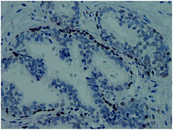
Graphical abstract: Two-color SERS microscopy for protein co-localization in prostate tissue with primary antibody–protein A/G–gold nanocluster conjugates

Supplementary files

Article information

Article type
Paper
Submitted
05 Nov 2013
Accepted
14 Dec 2013
First published
02 Jan 2014

Nanoscale, 2014,6, 2361-2367

Author version available

Two-color SERS microscopy for protein co-localization in prostate tissue with primary antibody–protein A/G–gold nanocluster conjugates

M. Salehi, L. Schneider, P. Ströbel, A. Marx, J. Packeisen and S. Schlücker, Nanoscale, 2014, 6, 2361 DOI: 10.1039/C3NR05890E

To request permission to reproduce material from this article, please go to the Copyright Clearance Center request page.

If you are an author contributing to an RSC publication, you do not need to request permission provided correct acknowledgement is given.

If you are the author of this article, you do not need to request permission to reproduce figures and diagrams provided correct acknowledgement is given. If you want to reproduce the whole article in a third-party publication (excluding your thesis/dissertation for which permission is not required) please go to the Copyright Clearance Center request page.

Read more about how to correctly acknowledge RSC content.

Social activity

Spotlight

Advertisements