
Stimulated Raman scattering microscopy with spectral phasor analysis: applications in assessing drug–cell interactions†
 a
Liam T.
Wilson,
b
Connie
An,
‡a
Aristea A.
Leventi,
a
Alastair W.
Wark,
a
Nicholas C. O.
Tomkinson,
*b
Karen
Faulds
*a
and
Duncan
Graham
*a
a
Liam T.
Wilson,
b
Connie
An,
‡a
Aristea A.
Leventi,
a
Alastair W.
Wark,
a
Nicholas C. O.
Tomkinson,
*b
Karen
Faulds
*a
and
Duncan
Graham
*a
Abstract
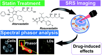
Statins have displayed significant, although heterogeneous, anti-tumour activity in breast cancer disease progression and recurrence. They offer promise as a class of drugs, normally used for cardiovascular disease control, that could have a significant impact on the treatment of cancer. Understanding their mode of action and accurately assessing their efficacy on live cancer cells is an important and significant challenge. Stimulated Raman scattering (SRS) microscopy is a powerful, label-free imaging technique that can rapidly characterise the biochemical responses of live cell populations following drug treatment. Here, we demonstrate multi-wavelength SRS imaging together with spectral phasor analysis to characterise a panel of breast cancer cell lines (MCF-7, SK-BR-3 and MDA-MB-231 cells) treated with two clinically relevant statins, atorvastatin and rosuvastatin. Label-free SRS imaging within the high wavenumber region of the Raman spectrum (2800–3050 cm−1) revealed the lipid droplet distribution throughout populations of live breast cancer cells using biocompatible imaging conditions. A spectral phasor analysis of the hyperspectral dataset enables rapid differentiation of discrete cellular compartments based on their intrinsic SRS characteristics. Applying the spectral phasor method to studying statin treated cells identified a lipid accumulating phenotype in cell populations which displayed the lowest sensitivity to statin treatment, whilst a weaker lipid accumulating phenotype was associated with a potent reduction in cell viability. This study provides an insight into potential resistance mechanisms of specific cancer cells towards treatment with statins. Label-free SRS imaging provides a novel and innovative technique for phenotypic assessment of drug-induced effects across different cellular populations and enables effective analysis of drug–cell interactions at the subcellular scale.
- This article is part of the themed collections: Imaging, biosensing and diagnostics: 2025 Chemical Science symposium collection and Most popular 2022 analytical chemistry articles
 Please wait while we load your content...

