
On-chip perivascular niche supporting stemness of patient-derived glioma cells in a serum-free, flowable culture†

Abstract
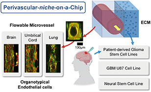
Glioblastoma multiforme (GBM) is the most common and the most aggressive type of primary brain malignancy. Glioblastoma stem-like cells (GSCs) can migrate in vascular niches within or away from the tumour mass, increasing tumour resistance to treatments and contributing to relapses. To study individual GSC migration and their interactions with the perivasculature of the tumour microenvironment, there is a need to develop a human organotypic in vitro model. Herein, we demonstrated a perivascular niche-on-a-chip, in a serum-free condition with gravity-driven flow, that supported the stemness of patient-derived GSCs and foetal neural stem cells grown in a three-dimensional environment (3D). Endothelial cells from three organ origins, (i) human brain microvascular endothelial cells (hCMEC/D3), (ii) human umbilical vein endothelial cells (HUVECs) and, (iii) human lung microvascular endothelial cells (HMVEC-L) formed rounded microvessels within the extracellular-matrix integrated microfluidic chip. By optimising cell extraction protocols, systematic studies were performed to evaluate the effects of serum-free media, 3D cell cultures, and the application of gravity-driven flow on the characteristics of endothelial cells and their co-culture with GSCs. Our results showed the maintenance of adherent and tight junction markers of hCMEC/D3 in the serum-free culture and that gravity-driven flow was essential to support adequate viability of both the microvessel and the GSCs in co-culture (>80% viability at day 3). Endpoint biological assays showed upregulation of neovascularization-related genes (e.g., angiopoietins, vascular endothelial growth factor receptors) in endothelial cells co-cultured with GSCs in contrast to the neural stem cell reference that showed insignificant changes. The on-chip platform further permitted live-cell imaging of GSC – microvessel interaction, enabling quantitative analysis of GSC polarization and migration. Overall, our comparative genotypic (i.e. qPCR) and phenotypic (i.e. vessel permeability and GSC migration) studies showed that organotypic (brain cancer cells–brain endothelial microvessel) interactions differed from those within non-tissue specific vascular niches of human origin. The development and optimization of this on-chip perivascular niche, in a serum-free flowable culture, could provide the next level of complexity of an in vitro system to study the influence of glioma stem cells on brain endothelium.
 Please wait while we load your content...

