Diabetic wound regeneration using heparin-mimetic peptide amphiphile gel in db/db mice†
Abstract
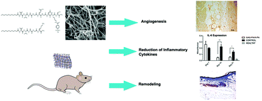
There is an urgent need for more efficient treatment of chronic wounds in diabetic patients especially with a high risk of leg amputation. Biomaterials capable of presenting extracellular matrix-mimetic signals may assist in the recovery of diabetic wounds by creating a more conducive environment for blood vessel formation and modulating the immune system. In a previous study, we showed that glycosaminoglycan-mimetic peptide nanofibers are able to increase the rate of closure in STZ-induced diabetic rats by induction of angiogenesis. The present study investigates the effect of a heparin-mimetic peptide amphiphile (PA) nanofiber gel on full-thickness excisional wounds in a db/db diabetic mouse model, with emphasis on the ability of the PA nanofiber network to regulate angiogenesis and the expression of pro-inflammatory cytokines. Here, we showed that the heparin-mimetic PA gel can support tissue neovascularization, enhance the deposition of collagen and expression of alpha-smooth muscle actin (α-SMA), and eliminate the sustained presence of interleukin-6 (IL-6) and tumor necrosis factor-alpha (TNF-α) in the diabetic wound site. As the absence of neovascularization and overexpression of pro-inflammatory markers are a hallmark of diabetes and interfere with wound recovery by preventing the healing process, the heparin-mimetic PA treatment is a promising candidate for acceleration of diabetic wound healing by modulating angiogenesis and local immune response.


 Please wait while we load your content...