Ratiometric fluorophore for quantification of iodide under physiological conditions: applications in urine analysis and live cell imaging†
Abstract
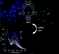
Dipod 1 bearing two pyrenyl tethered benzimidazolium moieties gives a fluorescence spectrum in aqueous medium which reveals a structured emission band between 330–400 nm and a broad emission band centered at 475 nm, respectively due to monomer and excimer emission of the pyrene moieties. The presence of an excimer emission band points to the formation of a pseudocyclic structure. Dipod 1 undergoes highly selective fluorescence quenching of the excimer emission band in the presence of iodide ions, whereas the fluorescence intensity of the monomer emission band remains stable. The ratio of fluorescence intensity I395 nm/I475 nmvs. log [I−] undergoes a linear change over a broad iodide concentration range of 10−9 to 10−5 M with KSV 3.7 × 10⁵ M−1. Dipod 1 can be used to determine iodide ions in urine samples, tap water and sea water conditions with 1 nM iodide as the lowest detection limit. On using paper strips coated with dipod 1, ∼1.7 pg cm−2 iodide ions could be detected. Dipod 1 shows a fluorescence quenching response to 100 nM iodide ions in C6 glioma cells using confocal microscopy. The life time of the dipod 1 shows a linear decrease with log [I−] and points to coordination based recognition of iodide ion.
 Please wait while we load your content...

