Recovering hidden quanta of Cu2+-doped ZnS quantum dots in reductive environment†
Abstract
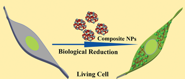
We report that photoluminescence of doped quantum dots (Qdots)—which was otherwise lost in the oxidized form of the dopant—could be recovered in chemical or cellular reducing environment. For example, as-synthesized Cu2+-doped zinc sulfide (ZnS) Qdots in water medium showed weak emission with a peak at 420 nm, following excitation with UV light (320 nm). However, addition of reducing agent led to the appearance of green emission with a peak at 540 nm and with quantum yield as high as 10%, in addition to the weak peak now appearing as a shoulder. The emission disappeared in the presence of an oxidizing agent or with time under ambient conditions. X-Ray photoelectron spectroscopic (XPS) and electron spin resonance (ESR) measurements suggested the presence of Cu2+ in the as-synthesized Qdots, while formation of its reduced form was indicated (by ESR results) following treatment with a reducing agent. Transmission electron microscopy (TEM) and X-ray diffraction (XRD) studies confirmed the formation of ZnS nanocrystals, the size and shape of which did not undergo any change in the presence of a reducing or oxidizing agent. Nanoparticulate forms of the Qdots and chitosan (a biopolymer) composite exhibited similar emission characteristics. Interestingly, when mammalian cancer cells or non-cancerous cells were treated with the composite nanoparticles (NPs), characteristic green fluorescence was observed. Further, the intensity of the fluorescence diminished when the cells were treated later with pyrogallol—a known reactive oxygen species generator. Overall, the results indicated a new way of probing the reducing nature of mammalian cells using the emission properties of the Qdot based on the redox state of its dopant.


 Please wait while we load your content...