Issue 20, 2014

Studying the membrane structure of chicken erythrocytes by in situ atomic force microscopy

Abstract

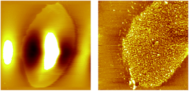
High resolution atomic force microscopy and single molecule force spectroscopy revealed the asymmetric distribution of proteins on both sides of chicken erythrocyte membranes. The cholesterol-enriched domains were directly observed by in situ atomic force microscopy, providing the first direct evidence of lipid rafts in chicken erythrocyte membranes.

Graphical abstract: Studying the membrane structure of chicken erythrocytes by in situ atomic force microscopy

Supplementary files

Article information

Article type
Communication
Submitted
26 May 2014
Accepted
08 Jul 2014
First published
22 Jul 2014

Anal. Methods, 2014,6, 8115-8119

Author version available

Studying the membrane structure of chicken erythrocytes by in situ atomic force microscopy

Y. Tian, M. Cai, H. Xu and H. Wang, Anal. Methods, 2014, 6, 8115 DOI: 10.1039/C4AY01260G

To request permission to reproduce material from this article, please go to the Copyright Clearance Center request page.

If you are an author contributing to an RSC publication, you do not need to request permission provided correct acknowledgement is given.

If you are the author of this article, you do not need to request permission to reproduce figures and diagrams provided correct acknowledgement is given. If you want to reproduce the whole article in a third-party publication (excluding your thesis/dissertation for which permission is not required) please go to the Copyright Clearance Center request page.

Read more about how to correctly acknowledge RSC content.

Social activity

Spotlight

Advertisements