Issue 7, 2012

Analysis of the DNA damage produced by a platinum–acridine antitumor agent and its effects in NCI-H460 lung cancer cells

Abstract
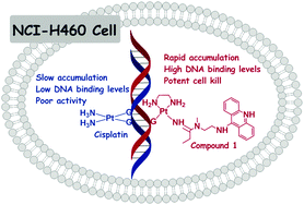
High-performance liquid chromatography in conjunction with electrospray mass spectrometry (LC-ESMS) was used to structurally characterize the adducts formed by the platinum–acridine agent [PtCl(en)(N-(2-(acridin-9-ylamino)ethyl)-N-methylpropionimidamide)](NO3)2 (compound 1) in cell-free DNA. Compound 1 forms monofunctional adducts exclusively with guanine, based on the fragments identified in enzymatic digests (dG*, dGMP*, dApG*, and dTpG*, where the asterisk denotes bound drug). The time course of accumulation and DNA adduct formation of compound 1 and the clinical drug cisplatin in NCI-H460 lung cancer cells at physiologically relevant drug concentrations (0.1 μM) was studied by inductively-coupled plasma mass spectrometry (ICP-MS). Compound 1 accumulates rapidly in cells and reaches intracellular levels of up to 60-fold higher than those determined for cisplatin. The hybrid agent shows unusually high DNA binding levels: while cisplatin adducts form at a maximum frequency of 5 adducts per 106 nucleotides, compound 1 produces 25 adducts per 106 nucleotides after only 3 h of continuous incubation with the lung cancer cells. The high overall levels of compound 1 in the cells and in cellular DNA over the entire 12-h treatment period translate into a rapid decrease in cell viability. Possible implications of these findings for the mechanism of action of compound 1 and the agent's potential to overcome tumor resistance to cisplatin are discussed.

Graphical abstract: Analysis of the DNA damage produced by a platinum–acridine antitumor agent and its effects in NCI-H460 lung cancer cells

Article information

Article type
Paper
Submitted
02 Feb 2012
Accepted
01 Mar 2012
First published
29 Mar 2012

Metallomics, 2012,4, 645-652

Spotlight

Advertisements