Co-cultured endometrial stromal cells and peritoneal mesothelial cells for an in vitro model of endometriosis†
Abstract
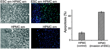
This paper demonstrates an in vitro model to simulate the microenvironment of endometriosis. We used microfluidic channels with cover slips to pattern and release endometrial stromal cells (ESCs) and human peritoneal mesothelial cells (HPMCs) in a way that mimicked the pathophysiology of peritoneal endometriosis. This approach enabled observation in real time interactions between ESCs and HPMCs both in their normal and pathological states. HPMCs from control individuals were able to resist the invasion of ESCs from both control and endometriotic individuals. By contrast, HPMCs from endometriotic individuals were unable to resist the invasion of ESCs from both normal and endometriotic individuals. We further analyzed the dynamics between HPMCs and ESCs from endometriotic individuals. HPMCs from endometriotic individuals relaxed their adhesion to each other at the beginning of invasion of ESCs, lose their adhesion to the substrate and apoptosed when surrounded by ESCs. These data implicate that the peritoneal physiology may play an important role in endometriosis.


 Please wait while we load your content...